13 July 2020
150418
0
3.7 out of 5
Coxarthrosis of the hip joint is a degenerative-dystrophic process that occurs in the articular joint of the head of the femur and the pelvic acetabulum. The disease is more common in middle-aged and elderly people, although it can also occur in young people, including children. Most often, its development is preceded by injuries, as well as a number of pathologies of inflammatory and non-inflammatory nature, and the main signs of the degenerative process in the hip joint are pain and stiffness of movement. In its development, the disease goes through several stages, and if in the early stages it can be dealt with using conservative methods, then in the final stages, treatment of coxarthrosis of the hip joints is effective only through surgery. Otherwise, the pathology will lead to severe impairment or even complete immobilization.
What is coxarthrosis of the hip joint and the mechanism of its development
Coxarthrosis, also called osteoarthrosis and deforming arthrosis, is a complex disease of the hip joints (HJ), accompanied by progressive destruction of cartilage. Over time, this leads to deformation of the surfaces of adjacent bones, as well as the formation of bone growths called osteophytes on them.
According to statistics, coxarthrosis accounts for about 12% of all diseases of the musculoskeletal system. In terms of frequency of occurrence, it is second only to gonarthrosis of the knee joint, but the risks of disability with it are much higher.
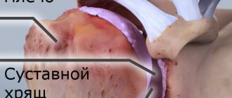
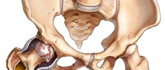
The two hip joints are the largest joints in the body. Each of them is formed by the femur bone and the acetabulum of the pelvis. The femoral head is located in a cup-shaped depression of the pelvic bone and moves freely in different directions. This structure of the joint makes it possible to flex and extend, adduct and abduct, as well as rotate the hip.
To prevent movement from causing discomfort, the surfaces of the bones in contact with each other are covered with an elastic layer called hyaline cartilage. It is this that allows the femoral head to slide easily in the acetabulum. Hyaline cartilage also provides stabilization and shock absorption to the hip joint during movement.
The entire joint is immersed in a kind of case called the articular capsule. It contains a synovial membrane that synthesizes synovial fluid. It is she who lubricates the surface of the cartilage, ensures the flow of water and nutrients into it, i.e., it is responsible for maintaining the normal structure of cartilage tissue.
Above the joint capsule there is a group of femoral and pelvic muscles, with the help of which the joint is moved. The hip joint is also surrounded by a group of ligaments that ensure the stability of its position within physiological boundaries.
Because the hip joint is subject to heavy loads every day, it is prone to rapid wear and tear and injury. The risk of such changes significantly increases the effect of a number of unfavorable factors that are almost inevitable in the modern world, but they will be discussed below. This explains the high prevalence of coxarthrosis.
As a result of the influence of negative factors, the production of synovial fluid is disrupted. Gradually, its quantity decreases, and its qualitative composition also changes: it becomes viscous, thick and is no longer able to fully nourish the cartilage. This leads to acute nutritional deficiency and progressive dehydration of hyaline cartilage. As a result of such changes, the strength and elasticity of cartilage tissue decreases, it delaminates, cracks and decreases in volume. All this prevents the smooth sliding of the femoral head in the acetabulum of the pelvis, which leads to the appearance of signs of coxarthrosis of the hip joint.
Gradually, the interarticular gap narrows, increased friction occurs between the articulating bone surfaces and the pressure of the bones on the hyaline cartilage increases. This leads to even greater injury and abrasion, which cannot but affect the biomechanics of the hip joint and the person’s well-being.
Failure of the hip joint negatively affects not only the biomechanics of the lower extremities, but also the entire locomotor system. This often results in disability.
As pathological changes progress, the hyaline layer gradually completely disappears, which leads to exposure of bone surfaces and a critical increase in the load on the bone joint. During movements, the femoral head is no longer covered by anything and rubs directly against the surface of the pelvic acetabulum. In addition to the fact that this seriously limits mobility and causes unbearable pain, the bones press on each other, simultaneously flattening.
As the bones of the joint deform, bone growths (osteophytes) form on their surface. They can have sharp edges and seriously injure surrounding muscles. This provokes severe pain in the groin area, legs and buttocks. Therefore, the patient unconsciously tries to spare the affected hip joint and avoid movements in it. Lack of adequate stress on the muscles leads to their gradual atrophy, which further aggravates mobility problems. The consequence of this is lameness.
Which specialist should I contact?
Many people, when faced with an illness, have no idea which doctor to go to. The patient is in the dark: should he make an appointment with a general or specialized specialist?
Remember, treatment of arthritis and arthrosis should be carried out under the supervision of different doctors.
Therapist
As soon as you notice signs of illness, visit a therapist. The doctor will conduct an initial examination and ask about the nature of the pain, its duration, and lifestyle. During the examination, the specialist will determine the presence or absence of an inflammatory process.
After diagnosing and making a preliminary diagnosis, the therapist will write a referral to a specialist doctor. His specialization includes examination and observation of not only standard lesions, but also psoriatic arthrosis for pain in the groin area, hip and knee, arthritis of the fingers and palm, forearm, and foot.
Be prepared for the fact that you will need to undergo x-rays and tests to make a diagnosis.
Rheumatologist
The responsibilities of a rheumatologist include examining, interviewing and examining the patient. For an accurate diagnosis, the doctor must prescribe:
- X-ray;
- Ultrasound;
- computed tomography (CT);
- densitometry;
- blood analysis.
This doctor mainly helps in the treatment of rheumatoid and rheumatoid arthritis.
Therapy is based on conservative methods. For example, using Sulfasalazine to lubricate joints.
Arthrologist
Everything related to arthritis and arthrosis is treated by this doctor. Consultation with an arthrologist, especially at an early stage, guarantees recovery. In addition to examination and diagnosis, the doctor can:
- do a therapeutic massage;
- perform manual therapy;
- write out a referral to a rheumatologist or geneticist;
- prescribe special gymnastics (physical therapy) classes.
Also, an arthrologist with experience has the right to give an injection of anti-inflammatory drugs.
To treat arthritis and arthrosis, the doctor prescribes only advanced methods of therapy.
Orthopedist
This is a specialist with a surgical bias. Patients come to see him with the last stage of arthrosis, which is characterized by complete or almost complete destruction of the joint.
An orthopedist's specialization also includes performing operations. In modern medicine, intervention is carried out in two ways:
- Organ-preserving. Doctors preserve the joint, while lost functions are restored, performance increases and pain goes away.
- Endoprosthetics. It is carried out in extreme cases when the damaged joint cannot be saved, but can only be replaced.
Typically, endoprosthetics are prescribed for arthrosis of the hip and knee joints.
Reasons for development
Coxarthrosis of the hip joint can be primary or secondary. In the first case, the reasons for its development cannot be detected, i.e. the disease develops on its own without any apparent reason. Secondary coxarthrosis is the result of a number of changes in the condition of the musculoskeletal system or lifestyle characteristics, in particular:
- injuries of the hip joint, including bone fractures, dislocations, bruises, sprains or ruptures of the surrounding ligaments, chronic microdamages, etc.;
- grueling physical work;
- sedentary lifestyle;
- obesity;
- chronic infectious processes in the body;
- rheumatoid arthritis, gout, tendonitis, bursitis;
- endocrine diseases, metabolic and hormonal disorders, including diabetes;
- congenital anomalies of the hip joint (dislocation, dysplasia);
- aseptic necrosis of the femoral head;
- various types of spinal pathologies;
- genetic predisposition;
- smoking addictions.
In the vast majority of cases, the development of coxarthrosis of the hip joint is caused by inevitable age-related changes, and the presence of other factors from among the above only increases the risk of its occurrence and increases the rate of progression.
Preventive actions
Preventive measures can be used to prevent the occurrence of pathological processes, as well as during remission, preventing the progression of the disease.
Prevention of coxarthrosis involves:
- maintaining a balanced diet;
- systematic sports;
- maintaining a healthy lifestyle, which includes giving up bad habits;
- body weight control;
- systematic preventive examination and timely treatment of various diseases.
Remember that your health is only in your hands. Keep him in optimal condition and enjoy a full life!
Symptoms and degrees
During coxarthrosis, there are 4 degrees of development, of which 1 is the mildest. Initially, the disease may be asymptomatic or manifest with mild pain. More often they occur after intense physical exertion, long walking, or at the end of a busy day. At the first stages of the development of the disease, discomfort is usually attributed to fatigue and is regarded as normal. Therefore, coxarthrosis of the hip joint is extremely rarely diagnosed at stage 1 of development.
Tangible signs of coxarthrosis begin to appear at the 2nd stage of its progression, when the joint space narrows by almost half, and the head of the femur is displaced and deformed. With the transition to the 3rd stage, the pain becomes unbearable and can bother a person even at night; they tend to radiate to the thighs, legs, groin and buttocks. Since the joint space is practically absent, and multiple osteophytes are formed on the bone surfaces, independent movement in such situations is impossible. Therefore, patients are forced to use a cane or crutches.
So, the main symptoms of coxarthrosis of the hip joint are:
- Limitations of mobility - initially, patients may notice difficulties when performing rotational movements of the leg, but over time they are accompanied by morning stiffness and swelling of the hip joint. Because of them, a person needs to warm up for several minutes and, so to speak, walk around in order to achieve restoration of a normal range of movements. Gradually, it becomes more and more difficult for the patient to move his leg.
- A characteristic crunch - occurs when walking, as well as flexion or extension of the hip joint. It is a consequence of friction of bone surfaces against each other and with coxarthrosis it is accompanied by acute or dull pain.
- Pain syndrome – pain initially appears after physical activity and subsides somewhat after a long rest. An acute attack can be provoked by heavy lifting or hypothermia, since coxarthrosis is often complicated by the addition of inflammation of the synovial membrane. As the disease progresses, pain occurs more frequently, persists longer, and becomes more severe.
- Spasm of the thigh muscles is a consequence of pinched nerves and weakening of the ligamentous apparatus, so the muscles spasm compensatoryly in order to hold the head of the femur in the acetabulum. Also, muscle spasm can be provoked by the addition of synovitis.
- Lameness - occurs in the last stages of the development of the disease, since deformation of the bone surfaces provokes the appearance of contracture of the flexor muscles. Therefore, a person cannot fully straighten his leg and maintain it in this position. Also, the patient may involuntarily limp in order to transfer weight to the healthy half of the body, as this helps reduce the intensity of pain.
- Shortening of the leg – observed with coxarthrosis of the 3rd degree. The leg on the side of the affected hip joint can be shortened by 1 cm or more as a result of narrowing of the joint space, decreased muscle tone and flattening of the femoral head.
At the last stage of development, the head of the femur fuses with the acetabulum, which leads to complete immobilization of the leg and disability.
At the same time, degenerative-dystrophic changes can be observed in one hip joint or in both. Accordingly, characteristic symptoms will be observed either on one side or on both sides at once, but in the latter case their severity on the left and right may differ.
Disease prevention
Measures to prevent the development of coxarthrosis include maintaining a healthy, active and active lifestyle, regular exercise to strengthen muscles and joints, and weight control.
As a preventive measure for older people, doctors recommend:
- follow the principles of healthy eating;
- exercise;
- take a course of massage treatments;
- visit the pool;
- do Norwegian walking or skiing;
- undergo sanatorium-resort treatment;
- take chondroprotective drugs once a year.
Diagnostics
The doctor may suspect the presence of coxarthrosis of the hip joint based on the patient’s complaints, external examination and the results of functional tests. During the visual inspection, the length of the legs must be measured. To do this, the patient is asked to stand up and straighten his legs as much as possible. The measurement is taken between the anterior axis of the pelvic bones and any bony structure of the knee, ankle or heel. But if both hip joints are simultaneously affected by coxarthrosis, the data obtained will be uninformative.
But since symptoms typical of coxarthrosis can accompany a number of other inflammatory and non-inflammatory diseases, in order to accurately diagnose the pathology, the patient is required to be prescribed instrumental examination methods. It could be:
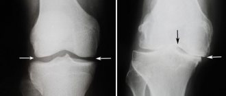
- CT or X-ray of the hip joint - the images show destructive changes in it, narrowing of the joint space, formation of osteophytes and deformation of bone surfaces;
- MRI is the most informative examination method, allowing you to accurately assess the nature of changes in cartilaginous structures, ligaments and the nature of blood circulation in the hip joint area.
Patients are also prescribed laboratory tests to assess their general health and detect diseases that could cause coxarthrosis. This:
- UAC and OAM;
- blood chemistry;
- rheumatic tests;
- puncture of the hip joint with biochemical examination.
The task of diagnosis is to differentiate coxarthrosis of the hip joint with gonarthrosis (damage to the knee joint), as well as radicular syndrome that occurs with osteochondrosis, as well as protrusions and herniations of intervertebral discs. Also, the symptoms of coxarthrosis may resemble manifestations of trochanteric bursitis and the atypical course of ankylosing spondylitis, which requires a full examination in order to determine the true causes of pain and limitations in mobility.
Other specialists
When answering the question of which doctor treats hip joints, one cannot fail to mention an arthrologist. While a rheumatologist and orthopedist treat a wide range of pathologies, an arthrologist specializes in joint diseases – arthrosis and arthritis. He also diagnoses and treats bursitis, tendinitis, periarthritis and other diseases of the periarticular soft tissues. If there is an arthrology clinic in your city, or an arthrologist is seeing you at a multidisciplinary clinic, you should take the opportunity and make an appointment with this specialist. The help of an arthrologist will be useful at any stage of coxarthrosis, since he deals with conservative and surgical treatment.
Who treats coxarthrosis of the hip joint, besides rheumatologists, orthopedists and arthrologists? The treatment of arthrosis combines methods of drug and non-drug therapy, traditional and alternative medicine. Therefore, you may need the help of a number of specialists:
- in manual therapy (the chiropractor performs traction on the joint to reduce pressure on the articular cartilage and restores the normal arrangement of the articulating bones);
- for post-isometric relaxation - stretching muscles and ligaments helps relieve muscle spasms and prepare for manual therapy;
- for therapeutic massage – massage normalizes muscle tone, activates blood supply and nutrition to joints, helps relieve pain;
in physiotherapy – for arthrosis, a variety of physiotherapeutic procedures are indicated, it is important to select their optimal combination and develop a treatment regimen; in physical therapy – exercise therapy is one of the most important components of both complex treatment and postoperative rehabilitation; on reflexology – acupuncture, acupressure, heat puncture;
on hirudotherapy - treatment with leeches.
The treatment of arthrosis, in particular coxarthrosis, is carried out by different specialists. First of all, he is a rheumatologist and orthopedist, as well as a representative of a rarer specialty - arthrologist. A rheumatologist is a general practitioner, and an arthrologist and orthopedist, depending on their specialization, can resort to both conservative and surgical treatment methods.
Non-drug treatment is carried out with the help of massage therapists, physiotherapists, chiropractors, exercise therapy specialists, and rehabilitation specialists. Taking into account the reasons for the development of coxarthrosis and possible complications of this disease, the patient may also require the help of an endocrinologist, nutritionist, phlebologist or angiologist, or vertebrologist.
httpv://www.youtube.com/watch?v=embed/ta-Ch__tME
loading…
Conservative treatment
Conservative treatment of coxarthrosis of the hip joint is effective only in the initial stages of the disease. It is selected individually for each patient and can include a whole range of different methods, each of which will complement the others. Therefore, as part of the treatment of coxarthrosis of the hip joint, patients may be prescribed:
- drug therapy;
- exercise therapy;
- physiotherapy;
- plasmolifting.
For conservative treatment to be effective, patients need to eliminate the effect of a number of factors contributing to the development of coxarthrosis of the hip joint. If you are overweight, it is very important to reduce it as much as possible. This will reduce the load on the affected joint and the risk of progression of the degenerative process.
You should also stop smoking and normalize your physical activity regimen, avoid overload, but also not sit all the time. To prevent further destruction of the hip joint, it is recommended to wear special bandages and orthoses. They provide reliable fixation of the joint and support it during movement.
Drug treatment
The nature of drug therapy is selected strictly individually. In most cases, patients are prescribed:
- NSAIDs are drugs that simultaneously have an analgesic and anti-inflammatory effect (available in the form of tablets, injection solutions and topical agents);
- corticosteroids - drugs with a powerful anti-inflammatory effect, which are prescribed if NSAIDs do not provide a pronounced effect;
- chondroprotectors – help to activate the processes of regeneration of cartilage tissue, but their effectiveness has not been proven;
- muscle relaxants - medications that reduce muscle tone and eliminate spasms, which is necessary when certain muscles or groups are spasming against the background of severe pain;
- drugs to improve blood circulation - most often used in the form of injections and help improve the trophism of the tissues surrounding the joint;
- B vitamins - are indicated to normalize the transmission of nerve impulses, which is especially important when nerves are compressed by deformed bone structures.
For acute pain that cannot be eliminated with tablets, patients can undergo intra-articular or periarticular blockades. They are carried out exclusively by qualified medical professionals in a medical facility and involve the injection of anesthetic solutions with corticosteroids into the joint cavity or directly the area around it.
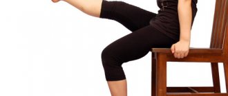
Exercise therapy
Physical therapy is an effective method of combating decreased muscle tone and limited mobility. Thanks to a properly selected set of exercises, it is possible to increase the range of motion and reduce the severity of pain. They also prevent muscle atrophy and help eliminate spasms if coxarthrosis is accompanied by pinched nerve fibers, which reflexively leads to spasms of individual muscles.
Exercise therapy classes can improve blood circulation in the area of the degenerative-dystrophic process. Thanks to this, the quality of trophism of the diseased joint improves and the course of regenerative processes accelerates.
For each patient, a set of exercises should be developed individually by a specialist. In this case, not only the degree of destruction of the hip joint is taken into account, but also the level of physical development of the patient.
Physiotherapy
Physiotherapeutic procedures and massage have an anti-inflammatory, analgesic, tonic, and decongestant effect. Additionally, they help maintain normal tone of the leg muscles, preventing their atony and atrophy.
For coxarthrosis of the hip joint, courses of 10-15 procedures are prescribed:
- ultrasound therapy;
- magnetic therapy;
- laser therapy;
- electrophoresis;
- ultraphonophoresis;
- UHF;
- paraffin treatment.
Many patients are also offered mud therapy. Such procedures have a positive effect only at the 1st stage of development of coxarthrosis of the hip joint or during rehabilitation after surgical treatment. Thanks to therapeutic mud, it is possible to improve the quality of blood circulation and accelerate the restoration of motor capabilities of the affected joint.
Plasmolifering
Plasmolifting or PRP therapy is a procedure that involves the injection of platelet-rich plasma from the patient’s own blood into the cavity of the hip joint. This allows you to activate the processes of restoration of hyaline cartilage.
But, according to some scientists, such a procedure can cause the formation of malignant tumors. This point of view is based on the fact that plasma lifting promotes the formation of a large number of stem cells, the effect of which on the body has not yet been fully studied.
About doctors
Therapist
When pain first occurs, which may be localized not in the hip, but in the knee, contact a therapist or surgeon at a local clinic. Here, primary diagnostics are carried out, including radiography, which makes it possible to make a diagnosis.
Rheumatologist
After making a diagnosis, it is advisable to contact a rheumatologist. It is the rheumatologist in traditional medicine who will prescribe non-steroidal antibiotics and a complex of physiotherapy. If necessary, hormonal medications and other necessary treatment are prescribed.
Surgeon
In complex advanced stages (this is usually the 3rd stage), endoprosthetics will have to be done. Replacement of a damaged joint with an endoprosthesis. This usually helps restore the patient's motor abilities. Since by this time a person can no longer move without crutches or a cane.
Chiropractor
Today, chiropractors have gained wide popularity and are quite successful in treating coxarthrosis. They claim that cartilage tissue can be restored quite successfully.
In any case, everyone must decide for themselves how to treat their body and remember, one of the key components of successful treatment is faith in its effectiveness.
Surgical treatment of coxarthrosis of the hip joint
Despite significant discomfort in the hip joint, many seek medical help too late, when pathological changes in the joint reach grade 3 or even 4 of severity, and functionality is irreversibly depleted.
In case of advanced pathology, surgery is a necessary measure. Only timely surgical intervention will help restore normal mobility and relieve the patient from excruciating pain, i.e., achieve a significant improvement in a person’s quality of life. No medications or physiotherapeutic procedures can restore severely damaged cartilage. At best, painful intra-articular injections and medications can reduce pain. But this will be a temporary phenomenon, after which the pain will return again with the same or even greater force.
Indications for surgery on the hip joint are:
- disappearance of the interarticular space;
- persistent pain in the hip joint that cannot be relieved;
- critical mobility impairment;
- femoral neck fracture.
Depending on the severity of joint destruction and bone deformation, patients may be offered various types of surgical treatment, namely:
- arthrodesis;
- endoprosthetics;
- osteotomy.
Arthrodesis
Arthrodesis is an accessible operation that involves strong fixation of articular bones with metal plates. The result is complete immobilization of the joint. Therefore, with the help of arthrodesis, it is possible to correct only the supporting function of the leg and eliminate pain, but there is no need to talk about restoring mobility or significantly improving the quality of life.
Today, arthrodesis is practically not used, since it deprives a person of the ability to fully move.
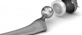
Endoprosthetics
Endoprosthetics with arthroplasty is the only way to radically solve the problem of coxarthrosis of the hip joint with the restoration of all its functions and motor capabilities. This is a high-tech method of solving the problem of coxarthrosis, allowing you to completely forget about it, as well as pain and limitations in mobility, for 15-30 years. Thanks to the use of modern endoprostheses, it is possible to fully restore motor and support functions and provide the patient with a normal life.
The operation involves resection of the femoral head and part of its neck. Surgical preparation of the acetabular bed is also carried out, which involves removing osteophytes, leveling its surface and resection of tissues that have undergone necrosis. Endoprosthetics can even be used to treat elderly patients with coxarthrosis of the hip joint.
The operation is performed under general anesthesia and takes about an hour. Depending on the severity of the degenerative-dystrophic process, the operation can be performed using one of the following methods:
- superficial - involves grinding the acetabulum and head of the femur with further covering them with smooth implants that replace the destroyed hyaline cartilage (the method is rarely used due to the possibility of developing inflammation in the periarticular tissues);
- unipolar - the head of the femur is removed and replaced with an endoprosthesis (used when cartilage is preserved on the surface of the acetabulum and only the femoral head is destroyed);
- bipolar - similar to the previous technique, differing only in the design of the endoprosthesis used, which has a lower coefficient of friction and provides smoother movements in the joint bed;
- total is the most effective and safe method of solving the problem of coxarthrosis of the hip joint, implying complete resection of the femoral head, including part of its neck, as well as the acetabulum, and replacing them with a full-fledged artificial articular joint.
Thus, patients may be recommended to install endoprostheses of various types. Most hip replacements are manufactured in the US and UK. For their manufacture, chemically and biologically inert metals are used: cobalt, chromium, titanium alloys. Ceramics are also often used. Most modern models additionally use polymer gaskets, which provide natural shock-absorbing, stabilizing and sliding properties to the artificial hip joint.
When performing endoprosthetics, the success of the operation is almost 100%.
After surgery, antibiotics are prescribed to prevent the development of infectious complications, and the sutures are removed after 10 days. The size of the postoperative scar is approximately 8 cm. At the same time, the patient is discharged from the clinic. Rehabilitation after endoprosthetics is not complicated, but still requires physiotherapeutic procedures, massage and exercise therapy.
Osteotomy
Osteotomy is a surgical intervention that is a temporary measure before a radical replacement of the hip joint with an artificial endoprosthesis. The essence of the operation is to align the axis of the femur by deliberately breaking it. The resulting fragments are installed in the most appropriate position, thereby slightly unloading the diseased joint. As a result, it is possible to temporarily reduce the severity of pain and improve mobility.
Thus, coxarthrosis of the hip joint is a rather formidable disease that can completely deprive a person of the ability to move independently. It progresses over a long time, and its symptoms, especially in the early stages, are often perceived by patients as a normal condition after physical activity. But this is precisely where the insidiousness of the disease lies, because only at the initial stage of its development can it be dealt with non-surgically. But if the degenerative-dystrophic process has already completely destroyed the hyaline cartilage and led to the bare surfaces of the bones and, even more so, their flattening, only surgery can help the patient. Fortunately, the modern level of medicine and surgery in particular allows us to achieve complete restoration of the normal state of the hip joint and its functions.
Specialists who treat arthrosis of the legs
Before visiting the clinic, you should find out which doctor treats arthrosis there and make an appointment. At the first signs, you should contact a rheumatologist. This doctor treats inflammatory diseases of the bones. He will prescribe a special examination (ultrasound, MRI, densitometry), and based on the results, determine the stage of development of the disease.
After receiving the results of the examination, having determined the symptoms of the disease, the rheumatologist prescribes drug treatment (injections into the joints, intramuscular).
If the development could not be stopped at the initial stage, it is actively progressing, and you cannot do without the help of a surgeon. Surgeons perform two types of operations: joint preservation and prosthetics. Surgeries help the patient return to an active lifestyle, forget about pain in the knee joint and other areas of the lower extremities.
If the joint is not completely destroyed, a specialist surgeon performs the following operations:
If these types of operations cannot be performed, doctors have to resort to radical methods - joint replacement. The destroyed organ is completely replaced with an artificial prosthesis. After such operations, patients lead a normal life and avoid disability.