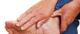
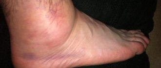
The wrist joint connects the hand to the forearm. It is formed by several bones, two of which - the lunate and scaphoid - are injured most often. Wrist fractures are more common in women, especially older women, due to calcium deficiency and weakened bones. Men have stronger and larger bones, and age-related hormonal changes are less pronounced.
Lack of proper and timely treatment can subsequently cause:
- Pain during functioning;
- Limitations on the volume of movements performed;
- Development of arthrosis;
- Weaknesses of the hand.
Injury to the hand joint is a consequence of increased stress as a result of a fall or sudden emphasis on the hand.
Another reason is osteoporosis, a disease that is accompanied by increased bone fragility.
Stages of rehabilitation after a fracture of the hand
Rehabilitation begins on the second or third day after the fracture and the application of plaster; they will reduce swelling of the hand, accelerate healing and recovery of the damaged area.
To obtain positive results, it is important to follow the entire range of restoration measures. During the period of stay in a cast, the muscles do not receive the necessary load and atrophy, so rehabilitation measures should include massage, physiotherapy and a special set of exercises. It is also necessary to balance your diet.
Manipulations that promote rapid fusion of bone tissue:
- Physiotherapy is the first step to stimulate the performance of the arm muscles.
- Electrophoresis,
- electrical stimulation,
- ultraviolet radiation.
After the cast is removed, more serious exercises begin.
Treatment of a wrist fracture
For fractures of the bones of the wrist joint, conservative or surgical treatment is used, depending on the severity of the disease. Fixation is performed using an orthosis (fixator).
If the patient has a closed fracture without displacement, a plaster or polymer bandage is applied for several weeks until the bones heal completely. In case of a displaced fracture, when fragments are formed, it is important to give them the correct position and fix them in order to exclude arthrosis. In some cases, surgery is performed. At this time, a control x-ray is necessary to avoid repeated displacement.
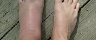
After applying the fixing bandage, painful sensations are possible. In this case, apply cold compresses and use anti-inflammatory drugs.
The plaster should not put pressure on your hand. If your hand becomes numb and your skin becomes pale, you should consult a doctor.
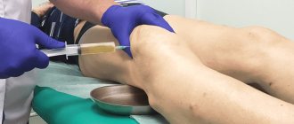
Surgical treatment of radial fractures in a typical location.
Closed reduction and percutaneous fixation with pins.
It can maintain reposition in the sagittal plane and lengthwise while maintaining the cortical plate along the palmar surface. With a comminuted fracture and compression of the bone along the palmar cortex, the reduction cannot be maintained.
The Kapandji technique and the Rayhack technique are used, in which reposition is performed under arthroscopic control.
82-90% excellent results when used according to indications.
External fixation.
If only an external fixator is used, it is impossible to restore normal palmar angulation of the articular surface.
For this reason, it is often used in combination with pins, spokes and in some cases with plates.
The external fixator uses ligamentotaxis to stabilize the fracture. It is better to place external fixator pins on the radius under direct visual control to avoid damage to the superficial branch of the radial nerve. When creating distraction, you should not “overstretch” the joint; the joint space should not be more than 5 mm. Also, you should not excessively retract the hand to the ulnar and palmar side, since in case of splinter damage to the palmar cortex, this will still not allow placing the articular surface at the correct angle.
Immobilization time should not exceed 8 weeks and the external fixator should not interfere with early movement of all fingers.
Complications of external fixator: 1) possible non-union of the fracture due to insufficient stability, 2) stiffness of the wrist joint and decreased grip strength 3) infection at the insertion site of the pins 4) reflex sympathetic dystrophy 5) iatrogenic damage to the superficial radial nerve 6) median nerve neuropathy
Open fracture treatment with internal fixation.
Displacement of the articular surface more than 2 mm, Barton-type fractures. Compression of the cortex along the palmar surface, “depressed” fractures of the articular surface.
Surgical technique.
Currently, volar plates are preferred. Modern plates are thin, pre-curved according to normal anatomy, and lockable screws allow for stable fixation even in severe cases when there is a bone defect in the metaphyseal zone and the epiphysis is represented by a thin strip of bone.
To prevent CRPS (complex regional pain syndrome), the current recommendation is Vitamin C 200 mg daily for 45 days after injury.
Clinical example of treatment of a fracture of the distal metaphysis of the radius using open reduction and osteosynthesis with a plate.
Nuances of recovery programs after fractures
After removing the fixing plaster cast, even with damage to small bones, negative changes occur in the limb. The patient feels discomfort and pain, the muscular-ligamentous apparatus loses its elasticity, the joints lose their elasticity and mobility. The most important goals of resuscitation of such patients are as follows:
- restoration of muscle and joint tissue;
- improvement of blood supply and lymph flow;
- strengthening vascular tissue;
- relief of swelling;
- strengthening of metabolic processes;
- complete return to joints of their former mobility.
In case of large-scale and severe fractures and the need for subsequent long-term immobilization, the recovery procedure is lengthy and requires a comprehensive approach. The return to normal life is especially long and difficult for elderly patients, weakened people suffering from pathologies associated with metabolic disorders, hematopoietic functions and poor vascular patency.
Early postoperative period
Wound care. Typically, postoperative sutures do not require removal, and the wound does not require special care. The doctor may recommend that you treat the wound with brilliant green for several days, in most cases this is enough. However, you need to protect the wound from injury, avoid rubbing the wound with clothing, and avoid hypothermia.
Shower. If sutures are not required or are removed, you can shower. Hot showers are not recommended. The wound is washed with warm water without using a washcloth. After washing, you need to blot the wound area with a towel, do not rub! After the wound has dried, you can treat it with brilliant green.
Mode. During the first month after surgery, a gentle regime is necessary. The wound must heal, and the body “get used” to the new features of the blood supply. Therefore, physical activity, baths, saunas, hypothermia are not recommended, and alcohol intake should be completely avoided.
Medicines. After the operation, you will be prescribed medications. Strictly follow the regimen and dosage. More information about medications will be discussed below.
What to do to avoid repeated operations?
Most people know what a healthy lifestyle is, that smoking, overeating and lack of exercise are harmful to health. Following simple rules (see the article “Prevention of Atherosclerosis”), which should become a habit, will help not only to avoid repeated operations, but to maintain health for many years.
Lifestyle
An active lifestyle is very important, not only for the prevention of atherosclerosis, but for maintaining health in general.
It is important for post-surgery patients to choose the right type of exercise. This is primarily walking, doing housework, gardening, or climbing stairs. You can work out in the gym (physical therapy), swim. But it is important to remember that exercise associated with heavy sweating (when you sweat a lot) is undesirable. If they are unavoidable, always keep a water bottle with you.
Diet
It is necessary to form the habit of healthy eating. Changing your diet is essential to normalize your weight and reduce your cholesterol levels, which play a huge role in plaque formation.
Medications
Taking medications after arterial surgery is extremely important. This should become a daily habit. In order not to forget to take a pill, you can use, for example, a reminder on your phone.
The main medications that need to be taken after artery surgery are “blood-thinning” drugs (antiplatelet agents and anticoagulants). Taking these medications will help avoid thrombosis of the reconstruction area. Therefore, strictly follow the dosage regimen and dosage. Under no circumstances should you stop taking these medications on your own. However, taking “blood-thinning” drugs can provoke various bleedings (nasal bleeding, menstrual bleeding, bleeding gums, spontaneous formation of hematomas, and so on). If signs of bleeding appear, you should immediately consult a doctor. If you see another doctor, such as a dentist or gynecologist, be warned about taking “blood-thinning” medications.
To prevent further development of atherosclerosis, drugs are prescribed to lower cholesterol levels ().
Bath, sauna
The steam room is not recommended for patients after arterial surgery, especially in combination with alcohol. The fact is that in the steam room a person sweats and loses a lot of fluid, which leads to “thickening” of the blood and can provoke the formation of a blood clot, especially in the operation area. The same applies to any situations in which increased sweating occurs (physical activity, hot weather, etc.). If such situations are unavoidable, always keep a water bottle with you.
Alcohol
Excessive alcohol consumption is a risk factor for the development of thrombosis of various locations, even in a healthy person. As for the patient after arterial surgery, alcohol abuse greatly increases the risk of developing thrombosis of the arterial reconstruction zone. Most patients with atherosclerosis after artery surgery completely refuse to drink alcoholic beverages.
Visit doctor
Regular visits to the doctor and additional research methods (blood tests, ultrasound, etc.) will help avoid unfavorable situations.
Don't neglect your health.
Visit your doctor regularly and follow all his recommendations.
Fresh fracture of the radius
Home > Fracture of the radius
It's no secret that a person has a radius bone in their hand, and in that radius bone there is a typical place where it breaks.
It would seem that the fragment is very small and the fracture is insignificant
This image also shows a noticeable displacement along the articular surface, which, despite the apparent insignificance of the fragment, requires further examination
Indeed, when falling on the hand, the radius bone in the area of the distal metaepiphysis is often broken (this is the correct name for that “typical place”).
Computed tomography clearly shows the defect of the articular surface
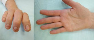
Due to damage to the articular surface of the radius, subluxation of the lunate bone occurred
There are different types of fractures depending on the displacement of the fragments: there is a Colles, Smith, Thompson fracture. There is also an AO classification, in which each type of fracture is assigned an alphanumeric code depending on the complexity. In any case, these nuances are for specialists, and this article is more popular than scientific.
Closed manual reduction (reduction of fragments) is a basic skill of any traumatologist. A fracture of the radius in a typical location is just such an injury for which this manipulation is very effective. There are several ways to reduce fragments of the distal end of the radius - this is manual traction along the axis, and hanging the hand by the fingers and reposition due to muscle relaxation, and holding the wires in a special way (Kapandzhi osteosynthesis).
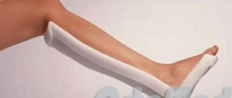
After placing the fragments in place, they are fixed with a hard bandage made of plaster or plastic. Immobilization of the elbow for fractures of the radius in a typical location can be considered a “relic of the tsarist regime”; most traumatologists have not done this for a long time, although classic Soviet textbooks recommend it. Unfortunately, I happened to meet a patient who had plaster applied to her fingertips, and this was a grave mistake.
After 7-10 days, it is necessary to perform control radiographs of the wrist joint, because During this time, swelling in the fracture area decreases and secondary displacement of fragments may occur.
In modern orthopedics, surgery for fractures of the radius is becoming more popular due to the refinement of our knowledge about the anatomy of this area and the publication of the results of various treatment methods. Mandatory for restoration are the length of the radius, a continuous smooth articular surface, and its angles of inclination. To clarify the position of fragments in complex fractures of the distal ray, it is necessary to perform computed tomography for high-quality preoperative planning.
Even for such a small fragment you have to use a full-fledged special plate
The lateral view shows that the subluxation of the lunate has been corrected.
After a correctly performed operation or after 6 weeks in a comfortable and individual hand orthosis, a person has every chance to return to normal activities without fear for his hand.
You can discuss your case in more detail during a personal meeting.
The estimated cost of surgical treatment of this pathology is 90,000 - 120,000 rubles, depending on the volume and complexity of the intervention
You might be interested in:
- Mallet finger injury
- Skier's finger
- Scaphoid fracture
Recovery after a fracture at the Innovative Medical Center
To prevent atrophy and degenerative changes in muscles, our center successfully uses kinesitherapy for rehabilitation after a fracture. Dosed, individually developed complexes allow you to begin the recovery process even with an immobilizing bandage:
- when performing exercises, only muscle tissue is involved;
- exercises on multifunctional decompression simulators eliminate the load on bones and joints;
- adapted force effects improve trophic processes, which eliminates pain and discomfort during rehabilitation after a fracture.
The training program is drawn up taking into account the location of the damage, the degree of damage and the patient’s condition. The correctness of the exercises is monitored by kinesiotherapists and instructors of the center. Constant monitoring ensures maximum effectiveness of classes and makes it possible to carefully monitor the dynamics of the rehabilitation process after a fracture, making adjustments to the recovery complex.
Activation of blood circulation and strengthening of the muscle frame eliminate the development of post-traumatic complications and stagnant processes in tissues. And effective and painless rehabilitation after a fracture allows for complete restoration of motor functions without long-term drug treatment.
Our center’s kinesiotherapists will help you return to a full, active life, using the body’s own regenerative resources - sign up for a course of 12 classes at the phone numbers indicated on the website and feel how your condition is improving.
Recovery period after fractures
With various bone injuries, the important task for the patient is to improve the health of the body, complete socialization and a full return to their previous life and physical activity. The time required for bone tissue to heal varies from person to person and depends on a number of factors. The scale of damage also plays a big role:
- Mild degree. Complete healing occurs in 25-30 days. This includes injuries to the ribs, hands and fingers.
- Medium severity. It takes about 3 months for bones to heal.
- Severe fractures. They almost always require surgical intervention; healing time can reach 1-1.5 years.
For the fastest possible recovery, the patient is required to strictly follow all prescribed procedures and doctor’s recommendations. Including advice related to daily routine and nutrition. The most important thing is to complete the prescribed course of health procedures. And don’t forget to do simple exercises at home, so as not to complicate rehabilitation and not provoke the development of stagnant processes.