Last Updated on 06/23/2017 by Perelomanet
Almost all bones in the human body grow together quite quickly and correctly. But it also happens that the bones are restored slowly or not at all correctly, becoming deformed. This happens when a person has poor health or poor circulation. What to do if a fracture does not heal? To do this, you need to understand the mechanism of bone fusion and understand what factors influence the acceleration of this process.
Stages of bone regeneration
In medical practice, the following regeneration stages have been identified:
- Catabolism of tissue structures and cellular infiltration. After damage, the tissue dies, cells disintegrate into elements, and hematomas appear.
- Stage of cell differentiation. This stage is characterized by primary bone fusion. With good blood supply, fusion occurs according to the type of primary osteogenesis. The process takes 10-15 days.
- Stage of primary osteon formation. Callus begins to form on the damaged area. Primary fusion takes place. The tissue breaks through with capillaries, its protein base hardens. A chaotic network of bone trabeculae grows, and they, connecting, form the primary osteon.
- Stage of callus spongiosis. This stage is characterized by the appearance of plastic bone cover, the cortical substance appears, and the damaged structure is restored. Depending on the severity of the damage, this stage can last several months or up to 3 years.
A prerequisite for a normally healing fracture is that the recovery stages proceed without disturbances or complications.
Fracture healing rate in adults
The process of bone fusion is complex and takes a long time. With a closed fracture in one place of the limb, the healing rate is high and ranges from 9 to 14 days. Multiple injuries heal on average in about 1 month. An open fracture is considered the most dangerous and takes the longest to recover; the healing period in such cases exceeds 2 months. When the bones are displaced relative to each other, the duration of the regeneration process increases even more.
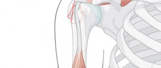
Healing of fractures of the upper extremities occurs slowly, but they pose less danger to humans than injuries to the lower extremities. They heal in the following periods:
- phalanges of fingers - 22 days;
- wrist bones - 29 days;
- radius - 29-36 days;
- ulna - 61-76 days;
- forearm bones - 70-85 days;
- humerus - 42-59 days.
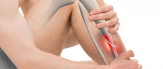
Healing time for fractures of the lower extremities:
- calcaneus - 35-42 days;
- metatarsal bone - 21-42 days;
- ankle - 45-60 days;
- patella - 30 days;
- femur - 60-120 days;
- pelvic bones - 30 days.
The reasons for the low healing rate may be improper treatment, excessive stress on the broken limb, or insufficient calcium levels in the body.
Treatment.
When bone healing is slow, there are two treatment options – conservative and surgical.
- Conservative treatment is an extension of the period of immobilization of the fracture sufficient for its healing. A non-union fracture is treated by administering drugs that stimulate the process of bone formation; it is possible to inject the patient’s blood into the gap between the fragments, as well as strengthening and tonic injections.
- If the consolidation time increases by more than 1.5-2 months, then the patient is prescribed surgical treatment. In the traumatology department, the patient will undergo one of the necessary operations according to indications: extrafocal compression osteosynthesis, bone grafting, Beck tunneling or sliding graft surgery according to Khakhutov.
Healing rate of childhood fractures
In a child, fracture treatment occurs 30% faster than in adults. This is due to the high content of ossein and protein in the children's skeleton. The periosteum is thicker and has excellent blood supply. The skeleton of children is constantly increasing, and the presence of growth plates accelerates bone fusion even more. In children from six to twelve years of age, with damaged bone tissue, correction of fragments is observed without surgical intervention, and therefore, in most situations, specialists make do only with the application of plaster.
A little about fractures
A fracture is a disruption of bone tissue due to increased mechanical stress. If the damage enters the consolidation stage, this means that the bones begin to grow together without pathological deformations. This most often happens in childhood, when the body is still young and strong enough to easily cope with the problem.
Sometimes, especially in adulthood, the situation develops differently - with an unfavorable prognosis. In the case of an unconsolidated fracture, the patient needs immediate hospitalization and surgical intervention - one in-person or, especially, an absentee consultation with a surgeon is not enough.
First aid for a fracture
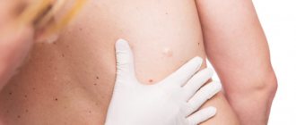
The rate of healing of broken bones is greatly influenced by the provision of first aid for fractures. If it is an open fracture, it is very important that the wound does not get infected in order to avoid inflammation and suppuration in this area. Therefore, the damaged area needs to be disinfected; for this, the circumference of the wound should be treated with an antiseptic and covered with a sterile napkin until the medical team arrives.
To transport the victim to a medical facility, it is necessary to organize immobilization of the limb. To complete the task, they use available means - plywood, flat boards, tree branches, which are secured to the injured limb with a cloth or bandage. If a person has a spinal injury, then a solid stretcher or improvised means, such as flat boards, on which the patient must be carefully laid, are used for transportation.
The timing of consolidation of fractures directly depends on the provision of first aid and emergency transportation of the victim to the hospital.
Healing mechanism
Healing of fractures begins immediately after injury. Fusion can be of two types:
- Primary fusion. If the bones are reliably connected, there is no need to build up a callus on the broken area; the fracture heals easily and with good blood circulation.
- Secondary fusion. In this case, it is necessary to build up the callus due to the active movement of bone fragments.
The mechanism of fracture healing is very complex, and therefore is divided into certain stages:
- The first stage involves the formation of a clot formed from the blood surrounding the damaged area. After some time, they transform into new tissue for bone building. This clot forms within a few days after the injury.
- At the second stage, this clot is filled with osteoblasts and osteoclasts. They greatly support healing and restoration. By filling the clot around the fracture, they smooth out and align the bone fragments, after which a granular bridge is created. It is he who will hold the edges of the bone to prevent displacement.
- The third stage is characterized by the appearance of callus. After a few weeks (2-3) from injury, the granular bridge turns into bone tissue. During this period of time, it is still very fragile and differs from ordinary bone tissue. This area is called callus. To prevent damage, it is important that the fracture is securely immobilized.
- During the fourth stage, complete healing of the fracture occurs. After a certain time after the incident, depending on its severity and area (3-10 weeks), blood circulation in this place is completely normalized, which helps strengthen the bone. The tissue takes a little longer to recover (6-12 months).
At the end of all stages, the fused bone regains its strength and is able to withstand various loads.
Features of the period of consolidation of various fractures
The period of fusion can take place in different ways. If a rib, phalanx or macula is damaged, it begins after about three weeks, in the case of the collarbone - no earlier than the fourth. The fusion of the bones of the upper extremities takes 2-3 months, and the lower ones – even longer, up to four. Consolidation of the femur occurs only after six months, so such injuries are extremely dangerous.
Violation of these deadlines indicates problems in the body, such as exhaustion, vitamin deficiency, diabetes mellitus, purulent infection, poor circulation or other pathology. In this case, additional examination and urgent measures are necessary, otherwise the patient may face repeated surgery.
Factors influencing the rate of bone fusion
The healing of a broken bone depends on a number of factors that either speed it up or hinder it. The regeneration process itself is individual for each patient.
First aid is critical to the speed of healing. With an open fracture, it is important to prevent infection from entering the wound, because inflammation and suppuration will slow down the regeneration process.
Healing occurs faster when small bones are broken.
In open fractures, callus forms much longer if a wound infection develops, which is accompanied by bone sequestration and post-traumatic osteomyelitis. That is why, if the fracture is treated incorrectly, the formation of callus slows down or does not occur at all. In such situations, fractures that do not heal for a long time, characterized by slow consolidation, as well as false joints appear:
- If patients suffer from hypovitaminosis and vitamin deficiency (osteomalacia in pregnant women, rickets, scurvy).
- If there are disturbances in the activity of the parathyroid glands (decreased calcium concentration in the blood) and adrenal hyperfunction.
- The presence of concomitant diseases occurring in the chronic stage, as well as inflammatory processes. Any pathological processes in the body significantly delay the recovery period after a fracture.
- The presence of excess body weight negatively affects the healing process of bone tissue.
- Metabolic disorder.
- Failure to comply with the terms of wearing a plaster cast. Many cases of bone tissue taking too long to heal are due to the fact that a person does not want to walk in a cast for a long time and removes it before the time prescribed by the doctor. The fused area of the bone is under pressure and displacement occurs.
How quickly bones heal depends on factors such as the need to install an implant. This occurs in cases where there are too many bone fragments, they are very small, and it is not possible to reassemble them.
Fracture (ICD-10: M84 Violation of bone integrity) - a violation of bone continuity, can be caused by a single strong mechanical damage, as well as repeated damage or stress (fatigue or stress fracture). A simple (closed) fracture - there is no communication between the broken bone and the external environment, an open (complex) fracture - there is such a communication.
The healing process of a fracture can be divided into 3 overlapping phases: inflammation, repair, and remodeling.
In the inflammatory phase immediately after the fracture, rupture of the vessels of the medullary canal, cortical plate, periosteum and adjacent soft tissues is noted; formation of a hematoma between the ends of the damaged bone (12 hours); acute traumatic inflammation with migration of polymorphonuclear leukocytes and monocytes to the site of damage (24–48 hours); organization of hematoma, proliferation of fibroblast-like elements and endothelial cells, formation of granulation tissue (48–72 hours).
During the repair phase, morphogenetic proteins released from damaged bone promote the differentiation of pluripotent granulation tissue cells into osteoblasts and chondroblasts. These cells produce a mixture of cartilage, rough fibrous bone and fibrous tissue that form the primary callus. The latter fills the gap between the ends of the damaged bone, connects and stabilizes the fracture, and reaches its maximum size by the end of 2–3 weeks (for uncomplicated fractures).
Primary (temporary) callus is an initially formed coarse fibrous bone that fills the gap between the ends of the damaged bone, connects and stabilizes the fracture, and reaches its maximum size by the end of 2-3 weeks. Callus is largely derived from osteoblast precursors migrating from the inner layer of periosteum and endosteum.
Secondary callus is formed by resorption of primitive and formation of lamellar bone, which contributes to a strong bone connection at the fracture site (after 6 weeks).
In calluses, according to localization, 3 zones are distinguished: endosteal callus - formed in the bone marrow cavity, intermediary callus - connects the ends of the cortical plates and periosteal callus - around the opposing ends of bone fragments. Periosteal and endosteal callus formations are temporary; they only provide fixation of fragments necessary for the fusion process. The fusion of fragments along the fracture line occurs due to the intermediary callus, which is formed later than the periosteal and endosteal callus. The least pronounced is endosteal callus, in which bone formation occurs, as a rule, according to the desmal type, without a preliminary cartilaginous stage.
Osteogenic cells, in the presence of a good blood supply, differentiate into osteoblasts that form new bone trabeculae (desmal or intramembrane osteogenesis); in the absence or insufficient development of blood vessels (outer part of the callus), pluripotent cells differentiate into chondroblasts, which form cartilage (enchondral osteogenesis). Mechanical stress (eg, a poorly immobilized fracture) also promotes the formation of cartilage in the callus.
During the remodeling phase of fracture healing, the strength characteristics and structure of the bone are restored, while through the resorption of primitive and the formation of lamellar bone, which promotes a strong bone connection at the fracture site (after the 6th week), a secondary callus is formed. The cartilaginous callus undergoes enchondral ossification and is replaced by coarse fibrous bone trabeculae.
Osteoclasts, the number of which increases at this stage, resorb mineralized dead bone, as well as excess trabecular coarse fibrous bone of the primary callus. At the same time, proliferating osteogenic cells differentiate into osteoblasts, which form new bone trabeculae oriented according to the lines of mechanical stress to which the callus is exposed. The rough fibrous bone of the primary callus is gradually removed and replaced by lamellar bone. In this case, Haversian systems are formed in which newly formed osteons of one fragment cross the fracture line and are embedded in another fragment.
During the process of remodeling, the periosteal callus almost completely disappears, the intermediary callus gradually turns into compact bone with a characteristic osteon structure, and the endosteal callus is rebuilt into a medullary cavity with elements of cancellous bone.
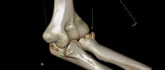
A strong connection of fragments occurs only after the entire primary callus (surrounding the fracture area) is reconstructed with the formation of new havers systems, which, passing through the fracture line, fasten the fragments together. At the end of this process, the original configuration of the bone can be completely restored, while the location of the fracture cannot be determined on the x-ray.
Primary healing of a fracture is healing (especially of the cortical bone), carried out without the formation of massive periosteal (fibrous-cartilaginous) callus, under conditions of stable osteosynthesis; characterized by rapid restoration (up to 5 weeks) of the normal structure and function of the damaged bone.
Secondary fracture healing is healing that occurs through the formation phase of a fibrocartilaginous callus; observed when fragments are mobile and not tightly attached.