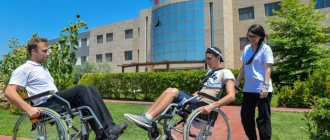
Physiotherapy uses natural factors such as currents, magnets, water, light and heat to treat diseases. How does the power of natural elements serve to restore human health?
Margarita Manasyan
Therapist with over 30 years of experience
Ask a Question
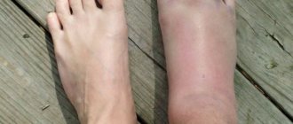
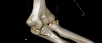
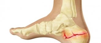
A fracture is a serious injury that is characterized by a violation of the integrity of the bone and adjacent tissues.
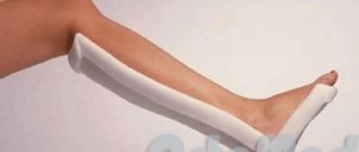
The main task in case of a fracture is to restore the anatomically correct location of the bone. For this purpose, plaster is applied to the fracture site and, if necessary, knitting needles are installed.
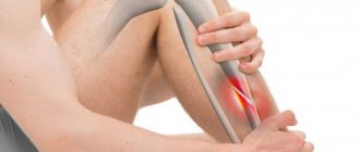
Next, the patient faces a long period of healing and rehabilitation. At this time, the patient experiences great discomfort and is constrained in his movements. Pain is a faithful companion to fractures.
Physiotherapy helps shorten the rehabilitation period after a fracture, relieves pain and swelling, and promotes rapid healing of tissues. Physiotherapeutic procedures can be used within 3-4 days after injury.
How does physical therapy help with fractures?
The main goal of physical therapy for fractures is the speedy restoration of the injured bone and its function without complications and pain.
Physiotherapy after a fracture helps:
- relieving pain during the “development” of atrophied muscles;
- activation of blood circulation in the extremities;
- elimination of contracture (limited joint mobility);
- tissue restoration (muscles, ligaments);
- acceleration of bone fusion;
- relieving swelling.
First stage of rehabilitation
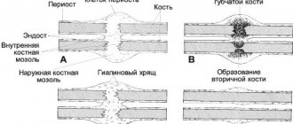
At the first stage, the following procedures reduce pain, have a bactericidal effect, improve venous and lymphatic drainage, and stimulate the growth of callus.
Namely, it is carried out:
- UHF therapy to provide a warming effect, starting 2 days after the fracture. During the session, which lasts up to 15 minutes, the vessels dilate, blood flow is activated, which reduces pain and swelling;
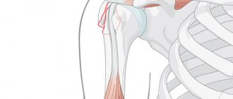
- Magnetic therapy for a shoulder fracture to accelerate the process of healing of fragments, stimulation of calcium and phosphorus metabolism. Using a constant or alternating field, blast cells are differentiated into osteocytes. Course – 10-15 procedures of 10-15 minutes each are carried out directly over the plaster cast. Metal implants do not interfere with this method of physiotherapy;
- electrophoresis to reduce pain, swelling of tissues using solutions of lidocaine or novocaine. Electrodes are installed parallel on both sides of the fault line. Course – 10-15 procedures, session duration – 20 minutes;
- exposure to interference currents to activate microcirculation and metabolic processes in the problem area, reduce pain and swelling of tissues. A total of 10-15 procedures lasting 15 minutes are required. 4 electrodes are applied to the fracture site and pulses of a certain rhythmic frequency are applied;
- massage of the limb above and below the plaster cast and healthy limb to provide a warming and therapeutic effect;
- Exercise therapy after 3-4 days for the joints located below the fracture and plaster and for a healthy limb. Perform feasible exercises to activate blood circulation, eliminate congestion and strengthen muscle and other soft tissue.
Electrophoresis: installation of electrodes on the shoulder area after removal of the plaster
How is UHF performed?
The patient is seated on a wooden chair without removing a T-shirt, shirt or sweater, as the waves penetrate the clothing and the cast. Prepare capacitor plates (electrodes) according to the size of the affected area, fix them in holders and wipe them with an alcohol-containing solution, then bring them to the painful area. The electrodes are installed transversely or longitudinally.
With the transverse method, the electrodes are placed opposite each other, i.e. on both sides of the shoulder at a distance of at least 2 cm from the body. Then the electromagnetic field will penetrate the entire body and have a general effect on the body.
With the longitudinal method, electrodes are installed only to the line of fracture or crack in the bone, since the waves will penetrate shallowly. Then the electrode should be no further than 1 cm from the body.
It is important to know. The closer the plate is to the body, the stronger the thermal effect. Incorrect placement of the plates leads to burns. The required UHF dose of the required power is set by a special regulator located on the generator panel.
Set the following dose of heat sensation:
- thermal - with a power of 100-150 W - for pronounced thermal sensations with a provocative purpose;
- oligothermic - with a power of 40-100 W - for blood circulation and improvement of cellular nutrition, metabolic processes, the patient feels a slight warmth;
- athermic - with a power of 15-40 W - for anti-inflammatory effects, the heat may not be felt.
If the plates (electrodes) are placed incorrectly and safety rules are not followed, the following may occur:
- thermal injuries or burns if a metal plate touches the skin or a damp cloth pad is used;
- bleeding if UHF is used before surgery. The electromagnetic field heats the tissue, causing hyperemia at the fracture site, which subsequently causes microbleeding;
- scars in the connective tissue that protect the wound site from infection. Therefore, before surgery to install plates or wires at the site of a bone fracture, UHF is not performed;
- electric shocks when the patient’s body comes into contact with exposed parts of the device that are energized.
How is electrophoresis performed?
During electrophoresis, medicinal substances are introduced into the patient's body through intact skin and mucous membranes using direct current. The procedure relieves inflammation, pain, resolves and has an antibacterial effect.
In addition to lidocaine and novocaine, drugs with the same charge are used to enhance the effects of each other or enzymes (Trypsin, Chemotrypsin), with organic acids and substances (for example, with mumiyo). To improve the phoretic ability of the skin, electrophoresis is carried out after ultrasound, paraffin and ozokerite applications, baths, sollux and massage.
Drug electrophoresis is carried out from a mixture:
- For 100 ml of distilled water - 0.2 g of Novocaine, 0.1 g of Diphenhydramine, 1 ml of 0.1% solution of Adrenaline.
- For 100 ml of distilled water - 0.02 g of Sovcaine, Lidocaine and Dicaine, 0.1 g of Tritecaine, 2 ml of Adrenaline solution (0.1%).
- Novocaine solution (5%) – 500 ml, 0.5 g Diphenhydramine, 0.06 g Platyfillin.
- Novocaine solution (0.5%) – 100 ml, 1.2 ml Adrenaline.
What physical therapy methods are used to treat fractures?
The most effective and modern physiotherapeutic means used now are:
- Electrotherapy . A procedure aimed at relieving pain, stimulating muscle tissue and nerve endings.
- Magnetotherapy . A low-frequency magnetic field helps get rid of swelling, pain, inflammation, and improves blood microcirculation. The procedure is especially effective for chronic pain in the spine, joints and muscles.
- Laser therapy . Has an analgesic, absorbable, anti-inflammatory effect. Under the influence of this procedure, regenerative processes are accelerated and the transmission of nerve impulses is activated.
- Electro- and phonophoresis . A non-invasive procedure, the purpose of which is to painlessly introduce anti-inflammatory and antibacterial drugs under the upper layers of the dermis. Stimulates the healing process of bone and muscle tissue.
It has been proven that physical therapy after fractures helps reduce the rehabilitation period by half and avoids complications.
Cosmetic procedures are contraindicated during pregnancy and lactation.
Second stage of rehabilitation
During the second stage, the growth and strengthening of the callus is activated, muscle atrophy and stiffness in the shoulder joint are prevented by the following procedures:
- interference currents of different frequencies;
- ultraviolet (general suberythemal) irradiation with a minimum dose to expand capillaries and supply tissues with nutrition and oxygen due to the warming effect and local erythemal effect;
- electrophoresis with calcium ions to strengthen the structure of callus and cell differentiation. 2 electrodes are used: one each for the damaged and healthy limb. Course – 10-15 procedures lasting 15-20 minutes;
- massage above and below the injury to improve blood flow, reduce muscle spasms, and prevent muscle atrophy.
Compression fracture of the spine in children: treatment
For uncomplicated forms of spinal compression fracture in children , treatment is functional. All necessary conditions are created to relieve the load from the affected area of the back. Special designs are used for this:
- Delbe ring;
- Gleason loop;
- In the supine position, bolsters are placed under the lumbar and cervical regions.
The patient is prescribed painkillers, which are selected depending on age. The most important condition is that the child must maintain bed rest; the bed needs to be hard, raised at the head by 30°. As recovery progresses, courses of exercise therapy, massages, electrophoresis and other methods of physiotherapy begin.
Treatment of spinal compression fractures in the elderly
In older people, bones become fragile and regeneration processes are slowed down. Treatment of spinal compression fractures in the elderly involves following a complex set of measures:
- NVPS group drugs are used to relieve pain and reduce the inflammatory process;
- calcium supplements and vitamin complexes are introduced to stimulate the restoration of bone tissue;
- complete physical rest and bed rest are ensured;
- the patient should lie on a hard surface, cushions are placed under the lower back and neck;
- fixing elements and rigid corsets are used;
The restoration of motor activity occurs gradually, exercise therapy classes are carried out only with an instructor, and the corset is not removed. Courses of massages and physiotherapy (UHF, UV) are required.
Compression fracture of the lumbar spine: treatment
The lumbar region is the most mobile and bears the greatest load. For these reasons, the bone structures in this area are considered the most vulnerable. For a compression fracture of the lumbar spine, treatment with conservative methods is as follows:
- for 3-4 months the patient is advised to regularly wear a lumbar corset or a rigid bandage;
- to prevent decompensation of the spinal cord, traction of the lumbosacral region is provided;
- Bed rest is maintained to remove the load from the damaged area, a cushion is placed;
- analgesic drugs, NSAIDs, chondroprotectors and vitamin-mineral complexes are administered;
- physiotherapy is performed.
When, based on the results of additional diagnostics, the patient is ready to get up, massages begin and a system of exercise therapy is developed to restore motor activity and subsequently strengthen the back muscle corset.
Compression fracture of the thoracic spine: treatment
For a compression fracture of the thoracic spine, treatment is similar to a similar pathology in the lumbar region. The differences are minor:
- a corset is selected, the design of which fixes the thoracic spine;
- in most cases, breathing exercises are required during the rehabilitation process.
In case of a compression fracture of the spine, treatment and rehabilitation are carried out by qualified specialists. In such cases, an integrated approach is important; one cannot rush; each stage of therapy is approached gradually. Surgical interventions are carried out only in severe cases, if there are appropriate indications.
Basic methods of rehabilitation and its need after fractures
The body of an elderly person is considered “worn out”, and it is more difficult for it to recover than a young person. Therefore, treatment after a fracture must begin immediately. It can be conservative, and in severe cases surgery is required. The next important stage is rehabilitation. It is necessary so that the patient can return to his normal lifestyle and avoid future injury as much as possible. You can recover either in a hospital or at home, but under the mandatory supervision of a doctor.
The most popular methods include:
- Physiotherapy . If there are no contraindications, you can visit the pool, as swimming strengthens the spine. Simple walking tours are also shown.
- Physiotherapy.
- Massage and manual therapy.
- Diet . A proper diet will make up for the deficiency of vitamins and microelements necessary for the body, for example, calcium, protein, zinc, vitamin D, etc.
- Treatment of osteoporosis . By strengthening your bones, you can avoid relapses.
With timely treatment and proper rehabilitation, an elderly patient will be able to return to their normal lifestyle in 1.5-2 years.