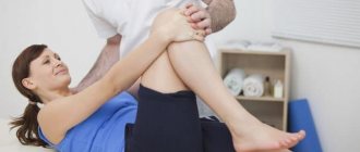
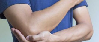
Among other types of injuries that damage ligaments, this injury is the least common. However, a sprained elbow cannot be classified as a purely sports injury. You can get damage to the elbow joint both in everyday life and from a strong fall.
The conditional classification of elbow injury is specific. Thus, damage to the joint (its joints) can manifest itself externally and internally. In this case, trauma is divided into 3 types:
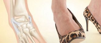
- stretching of the internal epicondyle, which is often called the golfer’s joint, since the specific position of the elbow when playing golf “favors” injury;
- “throwing” sprain or baseball player’s elbow joint (injury occurs during sudden throwing movements, often involves tearing the fibers of the ligaments);
- so-called tennis elbow – damage to the ligaments located on the inside of the elbow joint.
In all cases, pain becomes a pronounced symptom of sprain. In the case of the elbow set of ligaments, it manifests itself acutely. Only the location of the pain differs: on the inside, just above the joint if damaged during throwing, in the elbow cavity from the inside, if it is a golfer’s elbow, or on the outside if the external epicondyle is injured.
In addition to pain, sprains are characterized by swelling of the affected ligaments, subcutaneous hemorrhages, and stiffness of movement. The painful effect makes itself felt when moving, can be reflected in the hand, shoulder, and is often felt when kneading the fingers.
Clinically Relevant Anatomy
The elbow joint is stabilized by three ligaments:
- Ulnar collateral ligament (ulnar ligament).
- Radial collateral ligament (radial ligament).
- Annular ligament of the radius.
The ulnar and radial collateral ligaments provide valgus and varus stability, as well as the ability to safely rotate the elbow joint. The annular ligament wraps around the head of the radius, stabilizing it in the radial notch. Each of these ligaments can be damaged by overuse or injury to the elbow.
Diagnostic studies for injuries to the articular tissue of the elbow
Elbow on x-ray during diagnosis.
If an injury occurs, the patient must contact a traumatologist for an examination, which includes:
- Collect an oral history of a sprain.
- Counting the pulse, examining the skin for hyperemia or cyanosis. Skin manifestations occur when blood flow is disrupted.
- Assessment of motor activity of the damaged joint.
- Inspect the victim's body for other injuries.
After completing the history taking and palpation of the injured arm, the doctor prescribes a hardware examination:
- X-ray.
- Ultrasonic.
- Computed tomography.
- Magnetic resonance imaging.
Important! Laboratory research methods are carried out when there is serious damage to the joint tissue and the patient is hospitalized. They include taking a general blood test and passing urine for OAM.
Taking blood from a vein during hospitalization of a patient with damage to the fibers of the elbow ligaments.
Clinical picture
Although ligamentous injuries are rare, patients may present with varus or valgus instability due to overuse or injury. Radial knee injury is usually caused by trauma or intense force in the varus direction. These injuries usually involve a fracture or subluxation of the elbow joint. A tear or sprain of the ulnar joint can occur with excessive valgus loading or stressful movements due to pitching or throwing a ball. Commonly seen in young baseball pitchers, a tear or sprain of the ulnar CL of the elbow can also be found in athletes involved in activities with high repetitive overhead activities (such as playing tennis or volleyball).
Symptomatic features of sprained ligament fibers in the elbow
Pain when stretching depends on the number of damaged fibers.
Symptoms appear from the first minutes of injury. At the initial stage, pain, hyperemia of the skin occurs, and swelling of the soft tissue gradually appears. After some time, the pain intensifies, the joint becomes inactive.
The development of symptoms depending on the stage of stretching:
- Stage 1. When several fibers break, minor pain occurs. The joint is amenable to movement. The skin is clean, without pronounced hyperemia. There is no swelling of the soft tissues.
- Stage 2. Sharp pain. Soft tissue swelling occurs. The joint is painful when moving. Possible hemorrhage.
- Stage 3. Severe pain syndrome. Severe swelling of soft tissues with hyperemia of the skin, which is replaced by cyanosis. The joint is fixed in one position, its movement is limited.
The picture shows elbow ligament disorders depending on the degree of damage.
Diagnostic procedures
- Varus stress test, tests for radial joint weakness of the elbow joint.
- Valgus stress test, tests for ulnar joint weakness of the elbow joint.
- Valgus stress test in motion, tests for chronic strain or ulnar joint rupture due to overuse (sensitivity: 100%, specificity: 0.75).
- Modified milking maneuver, tests for ulnar joint tension or rupture due to overuse.
- Palpation.
In order not to miss anything interesting, subscribe to our Telegram channel.
Diagnostics
You should visit a doctor immediately after an injury. The specialist will collect anamnesis and palpate the damaged area. It is impossible to make a correct diagnosis based on external manifestations and complaints, so an instrumental examination must be carried out.
The traumatologist needs to tell in detail what happened, how the injury occurred, how severe it was. In some cases, falls can lead not only to sprained elbow joints, but also to damage to the spine, which is extremely dangerous.
Diagnostic techniques for elbow sprains:
- Radiography. Allows you to determine damage to bones and joints, identify fractures or dislocations.
- Ultrasound. It is more informative during stretching, as it makes it possible to see the condition of the ligamentous apparatus. Read more about ultrasound of joints→
- CT or MRI. The most informative, but expensive examination technique. Using tomography, you can not only assess the condition of soft tissues, but also determine the presence of damage to blood vessels and nerve endings, and correctly determine the severity of the sprain. Read more about MRI of joints→
- Electromyography. Rarely used to assess the passage of nerve impulses.
Until the diagnosis is known, it is necessary to limit physical activity and not self-medicate.
Criteria for evaluation
For elbow ligament injuries, three generally accepted methods are used to assess patient-reported outcomes:
- Disabilities of the Arm, Shoulder and Hand (DASH) is a 30-question questionnaire with a score ranging from 0 to 100, with 0 being no limitation. The DASH is well studied and valid with minimal clinically significant differences; either a 15-point clinically minimally important difference (MCID) scale or a 12.7-point minimally significant difference (MCD) scale.
- The Quick DASH test is usually used instead of DASH. The patient chooses the answer that is most characteristic of him (1-5 points for each question). Instructions for recording responses are listed below the questionnaire form, but Quick DASH does not have MCIDs studied like DASH does.
- The patient's personal functional scale is a scale in which the patient selects 5 tasks that are difficult to perform and evaluates these tasks from 0 to 10 points, where 0 is inability to perform and 10 is execution. The MCID for an average of 5 tasks is 2, while for one task the MCID is 3 points.
Providing first aid to the patient and subsequent treatment of the elbow joint
Examination and palpation of the elbow by a traumatologist.
Treatment of a sprained elbow joint begins with providing assistance to the victim at the scene of injury or during transportation to a medical facility.
It includes:
- Protecting the patient from receiving other injuries, providing him with peace.
- Fixing the joint with a scarf.
- Apply a cold compress for no more than 20 minutes. with an interval of 15 minutes.
- Supply of an analgesic and, in stressful situations, a mild sedative.
Applying an ice pack at the prehospital level of care.
The damaged elbow joint can be completely restored if the patient contacts a traumatologist within the first 48 hours after the incident. During hospitalization, the patient is given an elastic bandage or the injured elbow is placed in a supporting immobilizing bandage.
The use of a bandage for injury to the elbow joint.
Drug treatment is carried out in the form of a blockade of novocaine with the addition of vitamin B12 and an analgesic to the injection. The patient is prescribed physiotherapy in the form of ascorbic acid electrophoresis.
Introduction of blockade by injection in the treatment of elbow ligaments in the hospital department.
Restoring the functioning of a damaged joint consists of three levels:
- Level 1. During immobilization, the patient is taught to tense the muscles without moving. Such exercises prevent the joint from completely atrophying and keep it in constant tone.
- Level 2. During this recovery period, the bandage or immobilization splint is removed from the patient's arm. Restoration of hand movement and joint function is carried out by performing light physical exercises. Patients are prescribed swimming in the pool and light arm swings.
- Level 3. At the final stage of restoration of the affected fibers, exercises are performed with increasing physical activity and adding electrical stimulation of muscle tissue.
The product includes instructions that describe in detail how to apply the gel to relieve pain in the elbow. The price of the product is available for purchase to every patient.
Differential diagnosis
- Heterotopic ossification: significant loss of passive range of motion without loss of muscle strength.
- Malignant neoplasms: acute progressive pain not associated with movement.
- Inflammatory arthritis: abnormal systemic signs.
- Fracture: Trauma history, elbow extension test (specificity: 0.69, sensitivity: 0.97), noted limitations in range of motion, and hematoma.
- Dislocation: increased bony prominence, joint effusion, or the appearance of elongation of the forearm, which may affect neurovascular status.
- Inflammatory process: sudden swelling without injury.
- Damage to the vascular system: numbness, tingling, abnormal pulsation.
- Radiating pain from the neck.
- Referring pain from the shoulder.
Symptoms of elbow ligament rupture
The signs are clearly expressed, and it is impossible to confuse them with others. Sensations occur on the left or right side of the forearm, on the bend. In medical practice they are classified into three degrees. With the first and second, you can do without qualified help. The third degree of ligament rupture is dynamic and involves immobilization, completion of a full therapeutic course, and rehabilitation. In mild to moderate injuries, symptoms may take time to appear, but after an elbow muscle rupture, piercing pain spreads throughout the entire arm.
Characteristic symptoms:
- swelling;
- increasing symptoms;
- extensive hematoma;
- functional limitation or abnormal mobility.
Treatment
Due to the paucity of high-quality literature describing these conditions, management guidelines based on a “deterioration score” approach are recommended.
- Nonsteroidal anti-inflammatory drugs (NSAIDs) are commonly used to relieve pain and inflammation. If the pain is severe, corticosteroid medications may be prescribed.
- Common injuries to evaluate during examination may include decreased range of motion of the elbow or shoulder, joint effusion, and decreased muscle strength of the upper extremity. Internal rotation deficits at the glenohumeral joint are commonly seen in pitchers and athletes who frequently perform repetitive overhead activities. Research has shown a direct relationship with decreased internal rotation and excessive external rotation in baseball pitchers and elbow joint injuries.
- It is recommended that patients with these conditions remain active while avoiding stress on the ligaments. Activities that increase symptoms should be minimized initially to allow the ligaments to heal. The activity level can then gradually increase.
- Pain and swelling can be relieved by periodically applying ice during flare-ups.
- Ulnar CL surgery is indicated for cases of complete ligament rupture and for athletes who wish to maintain previous activity levels. The most common surgical procedure, Tommy John surgery, is the replacement of the elbow joint with a tendon from elsewhere in the body (often from the patient's forearm, hamstrings, or foot). This procedure is most often performed on athletes involved in certain sports, especially baseball.
- Surgery to repair a radial knee joint alone is rare and is usually associated with a fracture, dislocation, or subluxation of the elbow joint. Due to decreased structural stability of the joint, the surgeon may decide to perform open reduction with internal fixation (ORIF).
Elbow biceps tendon rupture: treatment
Conservative
Treating a biceps tendon tear conservatively will include resting the injured arm and avoiding heavy lifting or activities that could aggravate the injury (such as weight training). Applying cold and ice can help relieve swelling and general pain. You can also take anti-inflammatory medications and non-steroidal drugs to relieve pain. Special physiotherapeutic exercises aimed at developing flexibility and strength also help.
Triceps stretching
Triceps stretches are often overlooked when it comes to arm training. Triceps stretching is beneficial for preventing delayed onset muscle soreness (DOMS)
- Place your hand on your upper back with your elbow bent.
- Apply gentle pressure to your elbow with your other hand, lowering it as low as possible.
- Hold for 10-30 seconds.
Muscles involved: Triceps brachii
Video with hand stretching exercises performed:
Prevention
Having suffered a sprain once, there is a high probability of repeated injury of this kind. For prevention, injury to the elbow joint should be avoided. You need to choose comfortable shoes and watch your step to avoid falls. If you are into cycling, rollerblading or skateboarding, be sure to wear elbow pads.
A sprain is a fairly serious type of injury. If the elbow joint is treated incorrectly or untimely, pain will accompany any movement of the elbow for years.
Author: Oksana Belokur, doctor, especially for Ortopediya.pro
Possible complications
If you do not consult a doctor in time or self-medicate, then even mild damage to the elbow ligaments can lead to:
- arthrosis – damage to articular cartilage;
- damage to the nerves in the forearm or shoulder;
- permanent limitation of mobility, etc.
Any complications will make it difficult to treat the underlying damage, and the consequences of the above diseases will remain for a long time. Therefore, it is strongly recommended to consult a doctor at the first symptoms indicating damage to the ligaments of the elbow joint.
Three degrees of pathology and their characteristic symptoms
There are 3 degrees of elbow ligament sprain. Symptoms depend on the volume of damaged fibers:
| Stretch Rate | Signs |
| 1st degree – damage to a small number of fibers | Sudden but tolerable pain that subsides within a week Minor swelling, possible bruising, minor mobility restrictions Unpleasant sensations intensify with movements of the elbow, rotation of the hand and forearm, but usually do not cause severe discomfort and subside a few days after the injury. |
| 2nd degree – tear of a large number of fibers | Combined with other injuries (soft tissue bruises, rupture of blood vessels) The joint hurts a lot, sharp pain, swelling, hemorrhage in the joint area, local temperature rises Movements are limited (bending and unbending at the elbow, moving a limb and leaning on it is painful) A patient with a 2nd degree injury takes care of his elbow, trying to relieve unpleasant symptoms Such injuries are usually accompanied by bruises of soft tissues and rupture of the walls of blood vessels, so a person develops a hematoma. |
| 3rd degree – violation of the integrity of the entire bundle of ligament fibers | All symptoms are pronounced (intense pain, swelling, hematoma, hot skin) With concomitant tearing of large nerves, pain radiates to the shoulder, forearm and hand Any movement causes acute pain, it is impossible to lean on the arm, the elbow becomes excessively mobile (the torn ligament no longer strengthens it). This is especially noticeable when trying to straighten, rotate inward or outward the injured arm with the healthy arm (span and range of motion at the elbow exceed the norm). |
What consequences are possible after stretching:
- Small scars at the site of the tear make the elbow ligaments less resistant to repeated injury. In the future, they can be damaged even under the slightest load.
- In 10% of cases, after some time, a person develops stabbing sensations and aching pain in the area of injury.
- If left untreated, the tissue may heal incorrectly and elbow stiffness may develop.
- A severe complication of sprain is periarthritis (inflammation of the periarticular tissues).
Three degrees of sprain. Click on photo to enlarge
Stretching the flexor radialis
Stretching the flexor muscles can be difficult to perform, but a wall or any vertical, stable surface will help enhance the effect of the exercise.
- Face the wall and place your palm on its surface, fingers up.
- Slowly try to move the palm of your hand, without lifting it from the wall, clockwise with your fingers downwards as far as possible.
- Make sure your body remains straight and motionless. Stay in this position for 10-30 seconds.
Variations: You can perform this exercise on all fours, turning the palms of your hands towards you.
Muscles involved: Flexor radialis Flexor ulnaris Flexor digitorum superficialis Biceps brachii