Home > Hand injuries
Fractures of the hand bones are the most common fractures among all the bones of the human skeleton. Such fractures occur in the workplace or at home as a result of being hit by or about heavy objects, during a fall, as well as when playing various sports.
Most of these fractures are treated conservatively without surgery, however, some fractures: open, intra-articular, unstable, with angular or rotational displacement, require surgical treatment, that is, fixation of fragments using pins, screws or plates. If left untreated, fractures of the metacarpal bones and phalanges of the fingers heal incorrectly and can lead to limited function and cosmetic appearance of the hand. Improperly healed intra-articular fractures cause post-traumatic osteoarthritis, which can cause pain and limitation of movement in the damaged joint.
It should be remembered that not all displaced fractures cause dysfunction of the hand or finger. Also, in parallel with an unevenly healed fracture, there may be other problems that are not related to the injury.
A malunited fracture of a finger is a more complex problem than any other malunited fracture. After all, good hand function also depends on joint mobility, sensitivity, skin coverage, blood supply, and gliding of flexor and extensor tendons. Any pathology of these structures may limit the options for surgical reconstruction of the finger.
If a broken finger has healed incorrectly, you must first weigh all the possible risks and benefits of surgical intervention, discuss the goals and possibilities of treatment with a hand surgeon.
How to diagnose?
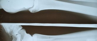
It is usually not difficult to identify dysfunction of the hand associated with improper healing of a fracture. There are often external signs, such as pathological rotation, a hump on the back of the hand, angular deformation of the finger, and others. To clarify the nature of the bone deformation, an x-ray is usually sufficient. It is very important to photograph each finger separately in a clear frontal and lateral view.
Rotational displacement is assessed when clenching the fingers into a fist: whether there is crossing or not. And here difficulties may arise, for example, we assume that the finger is simply not yet developed and therefore does not bend, but in fact there is a slight rotational displacement, which makes it difficult to bend.
So for a complete diagnosis, both analysis of radiographs and examination by a specialist are important. After all, tendon mobility can only be assessed during examination; ultrasound or MRI are not very helpful in assessing the condition of soft tissues when it comes to fingers.
In the case of intra-articular fractures, a computed tomography scan may be ordered to assess displacement.
Causes
A fall on the upper limb is the most common precipitating factor. Also, do not overlook the powerful physical impact on the phalanx - blow or pressure. In any case, diagnosis must be timely, otherwise complications during treatment are guaranteed.
It is important to mention that damage affects the phalanges of the little finger and the joints equally. Diagnosis of such herbs is extremely difficult, since visual symptoms are not always present.
In such situations, it is worth paying attention to the presence of pain and discomfort, as well as directly to the level of functionality of the finger.
Treatment
So what to do if your finger is not fused correctly?
It is worth contacting a specialist in the treatment of hand pathologies. A regular emergency room or traumatology department in a hospital is unlikely to take on the treatment of the consequences of a wrist fracture, and they will be right.
First, it is necessary to find out whether the patient’s complaints are a consequence of malunion or a lack of rehabilitation. Post-traumatic contractures of the hand joints are almost always first treated by a hand therapist using dynamic orthosis and other techniques for non-surgical joint development.
If the bone of the finger on the hand has really fused incorrectly, then the surgeon, together with the patient, decides on the timing and methods of correcting this deformity. Most fractures of the phalanges and metacarpals can be repaired and fixed with pins.
I prefer to perform such operations with local anesthesia because it is safe and convenient. Directly on the operating table, the patient moves the newly fixed fingers, which makes it possible to verify the correction of the deformity and the stability of the fixation. In the postoperative period, this gives confidence to allow early development of movements.
Usually I don't leave the knitting needles sticking out, but rather bite them under the skin. They are removed after 5-6 weeks through small punctures, also under local anesthesia.
While the fracture is healing after surgery, an individual orthosis is made from thermoplastic, which is much lighter and more comfortable than conventional plaster.
At home
When treated at home, the injured hand is constantly exposed to external influences. The plaster cast can become wet and deform; in winter, the material quickly cools, which negatively affects the healing process.
Important! Clinics are increasingly using moisture-resistant polymers instead of traditional plaster bandages. They are light in weight, with low thermal conductivity.
After removing the plaster, rehabilitation includes a course of massage to normalize blood circulation, lymph outflow, relieve joint stiffness, eliminate pain, and discomfort when working with the hand.
Drug treatment includes anti-inflammatory and antiseptic ointments; for pain, non-hormonal painkillers (NSAIDs) for oral administration (once a day, but not more than 7 days). If pain persists for more than a week, you should see a doctor for additional diagnostics.
On average, a broken finger heals in 21 days. For cracks, healing occurs within 2 weeks. Restoration of the hand's functionality occurs within a month. Injury with displacement of bone fragments requires longer immobilization - 1-1.5 months, rehabilitation takes up to 2.5 months. With multiple, open, compression fractures, the recovery period lasts up to 90 days, subject to successful treatment and compliance with all doctor’s recommendations.
Case study No. 1
A girl whose phalanx of her finger fused incorrectly described her complaints like this:
“If the hand is at rest, there is a pulling sensation in the finger somewhere from the middle of the palm, as if it is heavier than the rest, or as if stiff, this does not bother you, it is just noticeable. When vibrating, shaking a finger, it hurts from slightly to very much, for example, if you touch something with your hand (not hit, but lightly) - get caught on clothes, hand in hand, furniture - it already hurts, I wince, but tolerable. My husband suddenly took my hand - I screamed, there was sharp pain. Holding a weighty object (salad bowl, book) in your hand (4 fingers at the bottom, big at the top) hurts, I immediately grab it with my other hand. I unclench my fist after I bring the bag from the store and have to endure it for a couple of minutes until the pain subsides - although the load seems to be greater on the other fingers, and it didn’t hurt to carry it. At the same time, it hurts to bend and unbend a finger only if you try to do it with effort, further than he can do” (the author’s spelling and punctuation have been preserved).
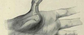
The image clearly shows a displacement along the articular surface of the middle phalanx, which was not eliminated during treatment of the fracture.
In this case, corrective osteotomy is impossible and plastic surgery was performed with an osteochondral graft from the hamate bone (hemi-hamate arthroplasty).
In this operation, a piece of bone with cartilage from the hamate is taken, which is ideal for replacing a defect in the articular surface of the base of the middle phalanx.
This is what the finger looks like after surgery.
Three months later, the girl wrote about him in a completely different way:
“I perform all the actions that I couldn’t do with my hand before the operation because of pain in the finger without any problems, the only slightly painful sensations are when I try to fully bend it and immediately straighten it completely and vice versa, but the pain is not sharp and goes away immediately.
The set of rehabilitation exercises that the doctor showed me at the handclinic began slowly - now I perform it much more actively, the strength in the finger is almost on par with the rest, plus the steering wheel (a good compression trainer), plus briefcases - I use my hand actively, even the muscle of the left forearm is already aligned with the right, otherwise I compared them in August by accident, I was quite surprised at how weak my left arm was in six months after the fracture.
As far as I understand your forecasts, things with the finger are, in principle, as expected.”
Symptoms
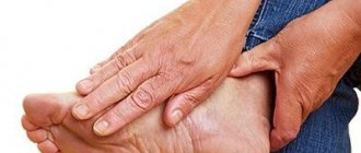
Signs of a fracture of the little finger on the hand may not be very pronounced visually, so diagnosis is difficult. If the symptoms of a fracture of the little finger on the hand are noticeably expressed, they can be determined by the following changes.
- Inability to move a finger as usual;
- Formation of a hematoma, characterized by redness or blueness;
- Acute, sharp pain that increases with manipulation of the injured finger;
- Gradual increase in swelling.
Fracture of the phalanx of the finger
Our fingers make very fine, coordinated movements and disruption of these movements can have a huge impact on daily and professional activities. To maintain full hand function, it is important that all finger fractures are evaluated by a physician to determine appropriate treatment. If you think that a broken finger is a minor injury, then you are seriously mistaken. Without proper treatment, a fracture of the finger can cause serious problems: limited flexion of the finger (contracture), pain with minor loads, decreased grip of the hand, whether it is a fracture of the nail or the main phalanx of the finger.
Anatomy of hand bones
The human hand is formed by 27 bones:
- 8 carpal bones;
- 5 metacarpal bones;
- The 14 bones that form the fingers are called phalanges. The first finger has only two phalanges: proximal and distal. Unlike the rest of the fingers, which consist of three phalanges: proximal, middle and distal.
Fractures of the metacarpal bones of the hand account for 30% of all hand fractures in adults.
Types of finger fracture
Because of
- Traumatic fractures are damage to the finger bone due to trauma.
- Pathological fracture - a fracture of a finger in the area of pathological restructuring (affected by any disease - osteoporosis, tumor, osteomyelitis, etc.) Osteoporosis is the most common cause of a pathological fracture.
The nature
- Closed fractures (without breaking the skin)
— Incomplete
— Complete
- Open fractures (with skin damage)
— Primary open
— Secondary open
Based on the presence of offset:
- Fractures without displacement of fragments
- Displaced fractures.
Rehabilitation
The rehabilitation period begins approximately one and a half months after the injury. The purpose of the exercises is to activate the processes involved in bone fusion, as well as to fully restore the functionality of the finger. In addition to the physiotherapeutic procedures prescribed by the doctor, the following exercises are allowed.
- Take grains of different types of cereals into your hand and distribute them on different plates;
- Work with the expander regularly, performing exercises 20 times a day;
- Sit in front of the table and place your hand on a flat surface, palm down (try raising your little finger up without opening your palm);
- Three times a week, make a hand bath with sea salt and your favorite essential oils.
First aid
First prehospital care consists of a number of activities:
- Anesthesia. It is necessary to take NSAIDs, for example, Ketorol, Ibuprofen, Analgin.
- Limb immobilization. In case of closed damage, it is enough to place the brush on a hard surface. It is better not to apply a splint, as if there are fragments, this may cause them to shift.
- Cold. Cold compresses can be used to reduce swelling and hematoma.
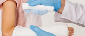
- In case of an open fracture, an aseptic bandage is applied to stop bleeding. Only the edges of the wound are treated with an antiseptic. A bandage is applied around the fragment.
- Delivery to hospital. This is done with caution; the hand must be immobilized. If this is not possible, it is better to wait for the team at the scene of the injury.
Signs
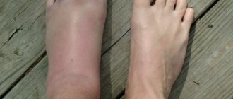
Some of the signs of a finger fracture are its atypical appearance, change in shape, and pain.
Let's look at the symptoms of a fracture separately for the fingers and toes.
It must be taken into account that in both cases there are both relative and absolute signs.
Relative signs of finger injury include:
- swelling and redness in the injured area;
- the inflamed area becomes warmer;
- mobility is impaired;
- if you try to put pressure on the injured area, the pain intensifies.
There are also absolute signs:
- palpation clearly reveals the presence of a crack;
- change in finger shape;
- crepitus on palpation;
- the presence of pathological mobility;
- shortening of the injured phalanx.
Parents should understand that checking for reliable symptoms of a fracture will be accompanied by severe pain and may also cause progression of the injury. It must be borne in mind that in the absence of the necessary education, the risk of damage to tendons, nerves and blood vessels increases significantly.
Let's look at a broken toe in a child, relative signs:
- motor dysfunction;
- bruising, severe pain in the foot;
- a fracture in the area of the distal phalanx is characterized by the presence of aching pain, in the main one - acute (painful sensations will be brighter, because in this place there is an articulation of the main phalanx with the bony frame of the leg);
- Quite large vessels run along the main phalanx; in case of a fracture, there will be a strong bruise under the skin, as well as under the nails;
- if the fracture is observed in the third, second and fourth fingers, then the child may not feel anything.
If we consider a fracture of the thumb, then we need to take into account its features:
- there are only two phalanges;
- superiority of its size over other fingers;
- tendency to intra-articular fractures and disorders.
This injury is characterized by:
- very painful sensations;
- inability to step on a limb;
- the entire leg below the knee may swell;
- a bluish tint may appear.
The absolute symptoms of a toe injury include:
- abnormal mobility at the site of the suspected fracture;
- when pressed, crepitus is observed as a result of friction of fragments;
- unnatural position of the injured finger.
Recovery period
Activities for the rehabilitation period are selected by the doctor individually for each patient. As a rule, the rehabilitation course consists of several stages. 7-10 days after the plaster is removed, the patient is prescribed UV treatment and interference currents.
Terms of complete healing in complicated fractures.
Fact! The duration of immobilization for a finger fracture is 2-4 weeks. The doctor will be able to determine more precise timing of fusion after an accurate examination of the X-ray image and assessment of the patient’s general vital signs.
If positive dynamics after removing the plaster cast are observed after 20-30 days, proceed to the second section of the course of rehabilitation procedures:
- UHF;
- electrophoresis with hydrocortisone;
- massage;
- medicinal baths.
It is quite difficult to accurately determine the likely time frame for complete restoration of finger mobility.
Likely consequences
How to prevent the consequences of a fracture.
The consequences may be different:
- The bone heals very slowly - the only treatment option is prolonged wearing of a cast. To speed up the process, it is necessary to fill the diet with vitamins.
- Incorrect splicing. In this case, the fracture is repaired and repositioned again.
- No splicing. To identify this pathology, radiography is carried out for 2-3 weeks. If positive dynamics are not observed, surgical intervention is performed.
- Stiffness. To restore normal functioning of the phalanx, constant physical treatment is necessary.
- Pain syndrome - many people know about this complication. Indeed, the fracture site reacts more acutely to any changes in climatic conditions and atmospheric pressure.
Impaired mobility of the little finger after removal of the cast is observed in many patients. This complication is the result of prolonged immobilization of the joint.
The process of restoring the mobility of the limb (subject to successful fusion) will not take much time, however, the patient should pay attention to the doctor’s recommendations regarding the rehabilitation period.