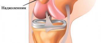
Anatomy of the structure
The knee joints in the human body have an important musculoskeletal function. The structure of each knee joint includes the patella or the so-called oval bone. The function of the patella is to prevent direct damage to the musculo-ligamentous apparatus of the joint itself, thus it seems to cover the soft tissue. The stability of the attachment and stability of the patella is provided by ligaments at the attachment points of the quadriceps femoris muscle, the muscles of the lower leg, as well as its own ligament. All these elements together provide sufficient strength to the quadriceps muscle to ensure movement of the limb. If a person receives a knee injury, including in the area of the patella, it can lead to both limited mobility of the joint and result in various complications.
Classification of patellar dislocations
- Congenital.
This pathological process is rare and basically implies, as a rule, underdevelopment of the connective tissue structures included in the structure of the knee joint. It rarely occurs separately and is combined with other types of dislocations. - Acquired or traumatic.
This pathology occurs due to direct traumatic effects. If more than a year does not pass between dislocations, then such dislocations are called habitual. Dislocations are also differentiated depending on how long ago they occurred: acute and chronic.
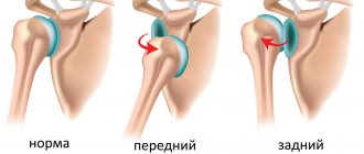
There is a classification based on the direction of bone displacement of the patella.
- Rotation
is when the patella changes its position relative to its axis. - Lateral -
usually appears due to a direct injury to the knee when the lower leg is extended. - Vertical is
an extremely rare type of dislocation, in which the patella in the horizontal plane enters the joint cavity.
According to the severity of the process
- Mild
- slight discomfort may be felt and a diagnosis can only be made after a thorough examination. - Medium degree
- leads to changes in gait and noticeable pain. - Severe degree
- entails the appearance of severe pain and often to the point of complete immobilization of the limb.
First stage
It begins a few days after reduction and application of a fixing bandage. It is important to train all leg muscles, from the toes to the thigh. This will help normalize blood circulation and maintain muscle tone, which will speed up the healing process.
Exercise therapy for the first rehabilitation period:
- Move your toes: bend and straighten.
- Grasp small objects laid out on the floor with your fingers.
- In a sitting or lying position, move your foot, abduct and adduct it.
- Rhythmically squeeze and unclench your thigh muscles. Perform from 5 to 20 times.
- Squeeze the muscles of the buttocks.
At this stage, it is recommended to do breathing exercises. Proper deep breathing allows you to saturate the body with oxygen, normalize blood circulation and improve the flow of blood with nutrients to the injured area.
Breathing exercise:
- Place one hand on the solar plexus, the other on the chest.
- Take a breath, inflating your stomach.
- As you exhale, slowly deflate your stomach.
- When performing gymnastics, the chest must be controlled with your hand so that it remains motionless.
A healthy limb can do any movement, both passive and active. If the traumatic injury is not accompanied by complications, you can bend and straighten the knee. This exercise is performed at a slow pace; the squat should not be deep. But it can be done only after the permission of the attending physician, very carefully and after removing the splint.
It is recommended to contract muscles rhythmically as often as possible, devoting a few minutes to this activity every hour.
Diagnosis of patellar dislocation
If a patellar dislocation is suspected, it is necessary to conduct a clear and detailed examination and differentiate this pathology from a patellar fracture or any other disease of the knee joint. The list of diagnostic studies includes:
- Collecting the patient's medical history, examining him with palpation of the affected area.
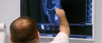
- Next, fluoroscopy should be done, preferably in several projections and simultaneously with a healthy joint for comparison.
- The most informative procedure for diagnosis is MRI, which makes it possible to most broadly assess the pathological process and subsequently prescribe the most correct and effective treatment.
- If fluoroscopy and MRI do not make it possible to make a diagnosis, the doctor may often prescribe a knee arthroscopy procedure, which gives not only a diagnostic result, but also a therapeutic effect.
- Subsequently, the patient undergoes a course of therapy and rehabilitation prescribed as part of the diagnosis.
Third stage
This stage can be started only after the fixing bandage has been removed and provided that the patella is restored correctly and there are no complications. By this time, the patient should be able to walk without the support of crutches.
During exercise, it is imperative to protect the patella from stress by wearing an orthosis or fixing it with an elastic bandage. Outside of physical activity, bandages are not needed. By the time this stage begins, the knee is already sufficiently developed, so you can increase the intensity and range of movements. Despite this, the load should still be moderate and the pace should be leisurely, although not as slow as before.
Before starting a set of workouts, you need to stretch your knee well and tone the muscles. You cannot exercise without first warming up. It is recommended to start by supporting your leg with your hands and slowly pulling it towards your body. You can perform self-massage to speed up blood circulation.
Other warm-up exercises are walking in place, without lifting your feet high from the floor, and slightly lifting your shins back. Gymnastics:
- The healthy leg is the supporting leg; it needs to be bent and the other leg raised slightly. After a few days, you can support the injured knee, but not for long. If pain occurs, the exercise should be stopped immediately.
- Squats are slow, not deep. At this time, you need to hold on to any support with your hands. Every day, gradually increase the number of squats, as well as the depth.
- Lunges forward and backward.
- Place the injured limb on a low bench and squat on the supporting leg. Then change places.
- Walking up the steps. You need to rise with emphasis on the damaged kneecap, and descend on your healthy leg. Gradually, the exercise should be complicated, stepping through several steps.
- The starting position is to kneel down, placing your palms on the floor. From this position, perform squats using your buttocks, your hands should not leave the floor. The number of repetitions is 10-15, after which lift your hands off the floor, raising your torso, touch your buttocks to your heels.
- Get on all fours. Bend your leg, pulling it towards your stomach. Freeze in this position for a few seconds, return to the starting position, repeat with the other.
- Exercise on the gymnastic wall. First, place your feet on each bar, then step over several bars.
Treatment methods for patellar luxation
Conservative treatment of dislocation
If an injury occurs, the best option is to immediately consult a doctor; if this is not possible, the injured limb is immobilized, and cold is applied to the area of injury to reduce swelling, pain and reduce the likelihood of bleeding. Next, as soon as possible, you still need to contact a traumatologist or a trauma center. The doctor’s tactics should be aimed at anesthetizing the damaged area in order to painlessly and effectively realign the patella to avoid possible complications. Then I apply a plaster cast or a fixing bandage to the limb, the duration of which, as a rule, is at least 6 weeks. During this time, it is advisable for patients to undergo a course of physiotherapy using UHF. After removing the plaster, the patient needs to undergo repeated fluoroscopy and, if the result of treatment is positive, undergo a course of rehabilitation to maximize the restoration of joint mobility.
Patella dislocation surgery
Surgical intervention is resorted to only in case of a fracture of the patella, or if conservative treatment has not brought the desired result. The tactics of surgical intervention in each individual case are selected individually and depend on the severity of the pathological process. However, recovery usually requires a longer period, at least 9 weeks. The variety of surgical interventions is wide, but the most commonly used are:
- arthroscopy,
- plastic surgery of the medial ligament using open access,
- transposition of the distal attachment of the ligament.
If the intervention was carried out correctly and on time, then it is possible not only to suture and fix the joint capsule, but also to prevent the development of complications such as hemarthrosis, damage to joint cartilage. If a dislocation of the patella is combined with a rupture of the ligaments, then it is not possible to sew them together. In some cases, there is a need to restore joint mobility and ensure its function using artificial or donor tissue.
Bibliography
1. D. L. Egmond, R. Schuitemaker. De knieregio. In: A. J. F. Mink, H. J. ter Veer, J. A. C. Th. Vorselaars. Extremiteiten manuele therapy in enge en ruime zin. 1e uitgave. Houten. Bohn Stafleu Van Loghum bv. 2006. p. 559 – 628 Level of evidence: D 2. Omer Matthijs, Didi van Paridon-Edauw, Dos winkel. Hoofdstuk 2 knie. Manuele therapie van de perifere gewrichten. 1e uitgave. Houten. Bohn Stafleu Van Loghum bv. 2004. p. 220 – 235 Level of evidence: D 3. G.VD. Bijl Jr., C. G. De Graaf, P. A. De Ridder. “hoofdstuk”. Actief en passief bewegen in de gewrichten der extremiteiten. De tijdsstroom. 1975. p. 126 Level of evidence: A1 4. R. meeusen. 2011. Praktijkgids knieletsels. Cursus. Vrije universiteit brussel. Level of evidence: D 5. Harry B. SKinner, Robert L. Barrack, Michael S. Bedmar, George D. Clarson et al. Sports medicine. In: Shelley Reinhardt, Isabel Nogueira, Peter J. Boyle. Current diagnosis en treatment in orthopedics. 2e edition. United states of America. McGraw-Hill. 2000. p. 125 – 175 Level of evidence: A2 7. Karen S. Beeton. The knee. Manual therapy masterclass: the peripheral joints. Churchill Livingstone. Elsevier. 2003. p. 54 – 55 Level of evidence: A2 8. Pierre-paul Castelyn. “hoofdstuk”. Acute knee injuries, diagnostic and treatment management proposals. Vub University press. 2001. p. 42-43 Level of evidence: A1 9. Smith TO., Davies L., Chester R., Clark A., Donell ST. Clinical outcomes of rehabilitation for patients following lateral patellar dislocation: a systematic review. Phystiotherapy. Volume 96. Issue 4. December 2010. p. 269 – 281 Level of evidence: A1 13. Journal of otrhopaedic surgery and research.Primary traumatic patellar dislocation. Chun-Hao Tsai. https://www.ncbi.nlm.nih.gov/pmc/articles/PMC3511801 14. Atkin DM, Fithian DC, Marangi KS, Stone ML, Dobson BE, Mendelsohn C. Characteristics of patients with primary acute lateral patellar dislocation and their recovery within the first 6 months of injury. Am J Sports Med. 2000;28:472–479. [PubMed] 15. Atkin DM, Fithian DC, Marangi KS, Stone ML, Dobson BE, Mendelsohn C. Characteristics of patients with primary acute lateral patellar dislocation and their recovery within the first 6 months of injury. Am J Sports Med. 2000;28:472–479. [PubMed] 16. Kirsch MD, Fitzgerald SW, Friedman H, Rogers LF. Transient lateral patellar dislocation: diagnosis with MR imaging. AJR Am J Roentgenol. 1993;161:109–113. [PubMed] 17. https://www.physio-pedia.com/Quadriceps_tendon_tear/Differential diagnosis 18. Hohlweck J., Diagnostic and therapeutic management of primary and recurrent patellar dislocations - analysis of a national survey and the current literature. Zeitschrift für Orthopädie und Unfallchirurgie. 2013 Aug;151(4):380-8. [Pubmed] 19. Picture from: www.slideshare.net/bhavinj/mri-of-patellar-disorders (Bhavin Jankharia, Doctor at Jankharia Imaging, Mumbai, India) 20. Nikku R, Nietosvaara Y, Aalto K, Kallio PE. Operative treatment of primary patellar dislocation does not improve medium-term outcome: A 7-year follow-up report and risk analysis of 127 randomized patients. Acta Orthop. 2005;76:699–704. doi: 10.1080/17453670510041790. [PubMed] (level of evidence 1B) 21. Arendt EA, Fithian DC, Cohen E. Current concepts of lateral patella dislocation. Clin Sports Med. 2002;21:499–519. doi: 10.1016/S0278-5919(02)00031-5. [PubMed] (level of evidence 2C) 22. Buchner M, Baudendistel B, Sabo D, Schmitt H. Acute traumatic primary patellar dislocation: long-term results comparing conservative and surgical treatment. Clin J Sport Med. 2005;15:62–66. doi: 10.1097/01.jsm.0000157315.10756.14. [PubMed] (level of evidence 2B) 23. Fithian DC, Paxton EW, Cohen AB. Indications in the treatment of patellar instability. J Knee Surg. 2004;17:47–56. [PubMed] (level of evidence 1C) 24. Koskinen SK, Rantanen JP, Nelimarkka OI, Kujala UM. Effect of Elmslie-Trillat and Roux-Goldthwait procedures on patellofemoral relationships and symptoms in patients with patellar dislocations. Am J Knee Surg. 1998;11:167–173. [PubMed] (level of evidence 1C) 25. Hohlweck J., Diagnostic and therapeutic management of primary and recurrent patellar dislocations—analysis of a nationwide survey and the current literature. Zeitschrift für Orthopädie und Unfallchirurgie. 2013 Aug;151(4):380-8. [Pubmed] (level of evidence 1C) 26. Milan Apostolovic, Boris Vukomanovic, Nemanja Slavkovic, Vladimir Vuckovic, Miodrag Vukcevic, Goran Djuricic, and Nikola Kocev; [PubMed] 27. Akkie Rood, Harm Boons, Joris Ploegmakers, William van der Stappen, Sander Koëter; Tape versus cast for non-operative treatment of primary patellar dislocation: a randomized controlled trial; Archives of Orthopedic and Trauma Surgery; 2012 [PubMed] (level of evidence 1B) 28. Stefancin JJ, Parker RD. First-time traumatic patellar dislocation: a systematic review. Clin Orthop Relat Res. 2007;455:93–101. [PubMed] (level of evidence 2A) 29. Sillanpaa PJ, Mattila VM, Maenpaa H, Kiuru M, Visuri T, Pihlajamaki H. Treatment with and without initial stabilizing surgery for primary traumatic patellar dislocation. A prospective randomized study. J Bone Joint Surg Am. 2009;91:263–273. doi: 10.2106/JBJS.G.01449. [PubMed] (level of evidence 1B) 30. van Gemert et al.. Patellar dislocation: cylinder cast, splint or brace? An evidence-based review of the literature. International Journal of Emergency Medicine 2012 5.45 31. White BJ et al. Patellofemoral instability: bulletin of the NYU Hospital for Joint Diseases 2009; 67 32. David Drez, T.Bradley Edwards; Claude S. Williams: Results of medial patellofemoral ligament reconstruction in the treatment of patellar dislocation; March 2001; Arthroscopy: The Journal of Arthroscopic and Related Surgery (level of evidence 4) 33. Peter R. Miller, Roger M. Klein. Robert A. Teitge: Medial dislocation of the patella; August 1991, Volume 20; Issue 6; pp 429-431 34. Elizabeth W. Paxton, Donald C. Fithian, Mary Lou Stone and Patricia Silva: The Reliability and Validity of Knee-Specific and General Health Instruments in Assessing Acute Patellar Dislocation Outcomes; July 2003 35. Mizuno, Y., Kumagai, M., Mattessich, SM, Elias, JJ, Ramrattan, N., Cosgarea, AJ and Chao, EYS (2001), Q-angle influences tibiofemoral and patellofemoral kinematics. J. Orthop. Res., 19: 834–840. doi: 10.1016/S0736-0266(01)00008-0 36. Smith TO, The reliability and validity of the Q-angle: a systematic review, Knee Surg Sports Traumatol Arthrosc. 2008 Dec;16(12):1068-79. doi:10.1007/s00167-008-0643-6. Epub 2008 Oct 8. 37. Emami MJ, Q-angle: an invaluable parameter for evaluation of anterior knee pain, Arch Iran Med. 2007 Jan;10(1):24-6. 38. https://ukhealthcare.uky.edu/uploadedFiles/UKHC-SportsMed-Medial-Patellof... 39. Chris S et al; Femoral Neuropathy due to patellar dislocation in a theatical and jazz dancer: a case report; Arch Phys Med Rehabil Vol 86, June 2005. 40. Neel P. Jain, MD, Najeeb Khan, MD, and Donald C. Fithian, MD; A Treatment Algorithm for Primary Patellar Dislocations; Sports Health. March 2011
Rehabilitation after injury
To fully and quickly restore joint mobility, it is extremely important to undergo a high-quality course of rehabilitation treatment. The process itself must be carried out under the careful supervision of the attending physician. The rehabilitation course includes not only physical activity to develop the joint, but also massage and physiotherapeutic procedures. A set of physical exercises is selected for each patient individually, depending on his age, condition and severity of injury. The purpose of the prescribed loads is not only to restore the function of the knee joint, but also to achieve the previous amplitude and strength of movements. During the entire rehabilitation period, the patient uses a fixing bandage, because immediately after recovery it is impossible to put a full load on the joint, and they also do not allow it to fully bend and unbend, which can lead to re-displacement of the patella.
Reasons for the development of the disease
There can be several reasons for a dislocation. Most often, the injury is provoked by a sharp mechanical force in the area of the patella (impact). Less commonly, pathology is caused by a sharp contraction of a muscle, for example, during a fall from a height, an accident, or during sports training. In addition, specialists from Dr. Glazkov’s clinics emphasize that there are other factors whose presence predisposes to the development of the disease. This is a congenital or acquired anomaly that provokes a violation of the structure of the musculoskeletal system. If we are talking about a congenital defect, it should be noted that the defect manifests itself differently in boys and girls, therefore the therapeutic methods prescribed by specialists are also different.
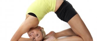
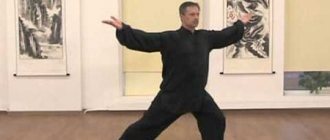
Prevention
In order to avoid recurrences of patellar dislocation, it is necessary to perform a set of physical activities daily; they will help the patient strengthen the musculo-ligamentous apparatus of the knee, which will not allow the patella to move later. At the same time, it is better not to do heavy loads and sudden movements, so you should avoid extreme sports, as well as heavy physical activity. Patellar dislocation must be treated without fail; this condition cannot be cured on its own. If this is not done, it can subsequently lead to serious and dangerous complications.
Symptoms
Symptoms are represented by the following manifestations:
- Acute painful sensations.
- Knee deformity due to displacement.
- Swelling, presence of hematoma.
- Stiffness, limited amplitude when flexing/extending a limb or complete loss of the ability to move in a joint (the knee is in a bent position).
- Abnormal location of the patella, which is felt upon palpation.
It is better to transport such a patient to a medical facility by immobilizing the leg in the position in which it is located and applying cold to relieve pain and reduce swelling.
Complications of patellar dislocation
Chronic dislocation of the patella can lead to the development of gonarthrosis. If you do not undergo a full course of rehabilitation, then perhaps the dislocation will become habitual. And even with light loads, and sometimes even with simple walking, the patella will shift. At the same time, therapy for this type of pathology will require more radical treatment. When the patella dislocates, the patient experiences discomfort in the affected limb and tries to protect it as much as possible, which over time can lead to weakening of the muscles, ligaments and cartilage tissue and result in their degeneration and even atrophy.
Arthroscopic techniques
Arthroscopic techniques
- Injury to the patellar ligament, accompanied by its incomplete rupture, requires conservative therapy. For complete rupture, surgical treatment is used.
- A non-displaced fracture involves immobilizing the bone using a cast. In case of slight displacement, the fragments are first repositioned. For a comminuted fracture, surgery with osteosynthesis is performed.
- In case of dislocation, it is reduced using anesthesia and subsequent immobilization.
Arthroscopic surgical techniques are used for combined injuries.