General information
What is instability of the C3-C4 cervical vertebrae? Instability of the cervical spine is a condition in which the vertebrae of the cervical spine are unable to maintain the correct anatomical position and, at the same time, the range of motion is greater than the physiological norm (pathological mobility). Spinal instability is a fairly common phenomenon and most of this pathology occurs in the cervical spine, which is due to its high activity and load (tilting/turning the head). As a result, against the background of increased mobility of the vertebrae, various structures suffer: the ligamentous apparatus, roots, spinal cord, vertebral arteries, which take part in the blood supply to the spinal cord.
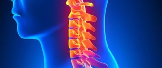
Features of the structure and function of the cervical spine
Atlas (C1) and axis (C2) - the first/second cervical vertebrae connect the base of the skull with the spinal column (atlantoaxial-occipital complex). At the same time, the Atlas has a specific structure (there is no vertebral body), and on its upper surface there are concave articular processes connected to the condyles of the occipital bone. The axis has a body that passes into the odontoid process, which, protruding upward, articulates with the surface of the anterior arch of Atlas (Fig. below).
The first and second vertebrae form three joints: one between the arch of the atlas and the odontoid process of the axis and two paired ones between C1 and C2, which are functionally combined into a combined joint, providing about half of the movements in the cervical region.
Rotational/axial loads in this region occur on the intervertebral discs of the vertebral body. They are responsible for axial pressure, shock load and maintaining the vertical position of the human body. The load on the spine is distributed by the nucleus pulposus . Intervertebral joints located in the articular capsules do not bear axial load. Important importance is given to the ligamentous apparatus, which fixes the intervertebral discs/vertebrae to each other and determines the range of motion of the spine.
The biomechanics of movements in the cervical spine occurs around three axes:
- Flexion/extension around the transverse axis.
- Circular movements around the longitudinal axis.
- Lateral bends around the sagittal axis.
Other features of the cervical spine include the narrowness of the spinal canal, the weakness of the muscular corset of this area, and a well-branched neurovascular network, which, with instability of the cervical spine, contributes to the development of neurological symptoms.
The spinal column combines the properties of mobility and stability:
- Mobility is determined primarily by the structural features of the vertebrae, mechanical strength and size of the vertebral structures and intervertebral disc.
- The leading element in stabilizing the spine is the fibrous/pulpous nucleus ligament of the intervertebral disc and the capsule of the intervertebral joints. The stability of the vertebrate is ensured by the stability of all its segments and protects it from deformation under conditions of physiological stress.
Of particular importance is the fact that cervical pathologies often accompany serious consequences, in particular paresis/paralysis of the upper limbs. Advanced instability can provoke cerebral circulatory disorders, respiratory failure, and impaired vision/hearing. Therefore, treatment of this pathology must begin as early as possible.
Unstable vertebra - is it dangerous?
The main danger of the disease is that at the initial stage it manifests itself, as a rule, only by pain during excessive load, sudden movement or minor injury. In such cases, usually no one even thinks about contacting a neurologist, preferring to “wait it out” until the unpleasant sensations go away on their own.
They can indeed disappear for a while (sometimes even for years), but soon they will remind themselves again, because an unstable vertebra, due to its mobility, constantly injures the nerve endings and the spinal cord. This can lead to the most tragic consequences, such as:
- osteochondrosis,
- intervertebral disc herniation,
- spinal cord injury
- compression of spinal vessels,
- paralysis.
Pathogenesis
The pathogenesis of instability of the cervical spine is based on several anatomical anomalies:
- destruction of the disc/disturbance of its structure, which contributes to the appearance of excessive translational movement in the posterior (dorsolateral) direction;
- the inability of the disc to perform the function of stabilization and transfer of the center of gravity to neighboring structures (discs, ligaments);
- increase in the neutral zone;
- formation of a center of pathological movement (rotation around the longitudinal axis).
Classification
There are several types of instability of the cervical spine.
Post-traumatic instability. The most common type. Is a consequence:
- Providing obstetric assistance during childbirth (application of forceps, squeezing out the baby).
- Injuries (road accidents, sports injuries, falls from a height) in the form of dislocations / fractures of the vertebrae, resulting from compression, flexion/extension, flexion-rotation mechanisms of injury. Complications in the form of post-traumatic instability develop in 10-15% of cases of vertebral fractures/dislocations, and unstable is the segment of the spine in which the height of the intervertebral discs is reduced, the disc is damaged or the ligaments are torn. As a rule, with post-traumatic instability, severe spinal/radicular symptoms develop.
Degenerative instability . Develops with osteochondrosis of the spine due to the disintegration of the fibrous ring/fragmentation of the disc tissue, which reduces its ability to fixate. The cause may be both a violation of the metabolism of cartilage tissue and a violation of the statics of the spine. When a load is placed on a degenerative disc of the spine, pathological mobility is formed, often with displacement of the vertebrae (degenerative spondylolisthesis). Displacement of the vertebra contributes to overload of the posterior supporting complex, which gradually leads to the development of degenerative spondyloarthrosis. In most cases, this type of instability occurs at the C3–C4, C4–C5, or C5–C6 vertebral levels.
Dysplastic instability.
Develops as a consequence of dysplastic syndrome. Manifestations of dysplasia include changes in the structure of collagen fibers, narrowing of the intervertebral disc, incorrect position of the nucleus pulposus, wedge-shaped vertebral bodies, and disruption of the integrity of the endplates. This contributes to the development of disruption of the mechanical properties of the disc, the relationship between the fibrous ring and the nucleus pulposus, which reduces the rigidity of fixation of the vertebrae, and can occur at all levels of the cervical spine (C1-C7).
Signs of dysplasia can be found in various structures of the spinal column - in the vertebral body, intervertebral disc/joints, and spinal ligaments. This kind of instability is caused mainly by congenital inferiority of the intervertebral disc, less often by asymmetry of the intervertebral joints, changes in the position/size of the articular facets, and underdevelopment of the articular processes.
Postoperative instability.
It is a consequence of surgical interventions in which it is necessary to remove/resect facets, significantly disrupting the integrity of the supporting complexes and ligamentous apparatus, which leads to a significant increase in the load on the vertebrae/intervertebral joints.
It is customary to distinguish several stages of instability:
- The first stage is mechanical (pathological mobility develops at the level of the destabilized spinal motion segment). It is often not detected x-ray. Develops mainly at a young age (up to 20 years). It manifests itself as pain in the neck near the spine, less often as radicular pain.
- The second stage is neurological, accompanied by damage to the spinal structures and has pronounced neurological symptoms. Develops in people 20-60 years old.
- The third stage is combined (manifestations of the first and second stages are present simultaneously). It develops mainly in people over 60 years of age.
Prevention
If we talk about pathology that arises as a result of labor, then the health of the mother plays an important role in prevention. Women who lead a healthy lifestyle, play sports, are active, take care of their health and maintain their immunity, endure childbirth more easily.
It is also important to control your weight and not gain more than the permissible norms during pregnancy. A large fetus is less active and more difficult to pass through the birth canal
At an older age, children are recommended to play sports. Swimming has a good effect on the condition of the spine. In addition to everything you need:
- Provide your child with a comfortable sleep by choosing the correct height pillow and orthopedic mattress.
- Monitor correct posture when sitting at a desk. If necessary, use an orthopedic corset.
- Avoid games with possible neck injuries.
- Use massage techniques to strengthen your neck muscles.
- Treat inflammatory and infectious diseases promptly and completely.
- Do not overuse physical activity, especially during the period of growth and skeletal formation.
- Treat osteochondrosis, osteoporosis at the first signs.
Timely detection of the problem gives a good chance of cure and prevention of complications. In severe cases, therapy contributes to a significant improvement in quality of life. The final result depends on the patient’s organization or the mother’s responsible approach to solving the problem.
source
Causes
The main causes of instability of the cervical spine include:
- dysplasia .
- Spinal injuries involving the cervical spine (domestic, road, birth, sports).
- Anomalies of spinal structures.
- Disorders of bone tissue mineralization processes.
- Osteochondrosis in the cervical region.
- Degenerative changes in the vertebral discs ( hernias / protrusions ).
- Involvement of bone tissue in the infectious process ( osteomyelitis / tuberculosis ).
- Systemic inflammatory diseases ( systemic lupus erythematosus / rheumatoid arthritis ).
- Spine operations (ligaments, discs, facets).
Symptoms
Symptoms of cervical spine instability can vary significantly depending on the severity of the instability. The main manifestations of instability of the cervical vertebrae are:
- Pain syndrome, including headache . Appears mainly against the background of physical activity or immediately after it, as well as during prolonged stay in an uncomfortable position (sitting with the head tilted down/in front, flexion/extension of the head).
- Muscle symptoms. It manifests itself as a feeling of fatigue, tension in the neck muscles.
- Neurological focal symptoms. Characterized by numbness/weakness of the upper extremities, shooting pains, and pain on palpation of paravertebral points.
- Vestibulo-cochlear disorders. Dizziness , tinnitus, less often - visual disturbances. Appear when the vertebral artery is compressed.
- Hypertension syndrome. Increased intracranial / blood pressure , which increases dizziness and headache.
- Sleep disorders. It is observed against the background of chronic pain. Feeling of constant discomfort, inability to sleep, interrupted sleep.
- Spinal deformity. Changes in the shape of the neck (increased kyphosis).
Also common manifestations of instability include decreased muscle tone in the neck, soreness of the skin, numbness, weakness in the limbs, and less commonly, arrows.
How to treat vertebral instability
The key to success in the treatment of any spinal disease is an integrated approach. This is the only way to achieve a good and lasting effect. However, there is not and cannot be a single complex suitable for everyone. It all depends on the degree of displacement of the vertebra and symptoms (discomfort, pain, muscle weakness below the displacement zone, surges in blood pressure, etc.).
Among the methods successfully used in our clinic to restore vertebral stability:
- Osteopathy helps to gently return a displaced vertebra to its place for both adults and children. The doctor's hands cause absolutely no pain.
- Manual therapy - restores muscle balance in the body, normalizes muscle tone, and removes excessive stress from the spine and joints.
- Plasma therapy promotes the accumulation of platelets isolated from the patient’s own blood around the site of the disease. Platelets are natural stimulators of body tissue regeneration. This method is perfectly combined with almost any other treatment methods.
- Isometric kinesiotherapy - promotes spinal traction, due to which the vertebrae themselves assume a normal position, changing movement patterns to the correct ones.
- Acupuncture - relieves muscle spasms and pain, restores innervation and blood circulation.
- Hirudotherapy - thins the blood and promotes its flow with nutrients and oxygen to the source of pain.
And the most important thing is professionalism, not only of the attending physician, but also of the doctors performing the treatment procedures. Under no circumstances should you trust the treatment of vertebral instability to inexperienced massage therapists or chiropractors. These should be competent and experienced specialists working in the same team with the attending physician and other procedural doctors.
Trust your health only to trusted specialists!
Tests and diagnostics
The diagnosis is made based on:
- Collecting complaints and studying the history of the disease (characteristics of the pain syndrome, localization, presence of concomitant symptoms - dizziness, headache, unsteadiness while walking, etc.).
- Neurological examination data (soreness of the cervical muscles/paravertebral points of the spine, range of motion of the spinal joints/condition of the muscular corset, presence of radiating pain, sensory disturbances, etc.).
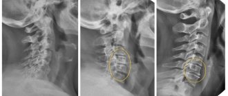
- X-ray examination/CT/MRI of the cervical spine. Allows you to determine the condition of the bone structures of the spine, the width of the spinal canal, and the displacement of the vertebrae.
Treatment methods
The effectiveness of treatment is achieved through a complex of measures. The course of treatment consists of a number of procedures. Drug treatment consists of taking vasodilators and sedatives. For headaches, painkillers are prescribed. If there is a subluxation of a vertebra, it needs to be put back in place. This can be done with the help of osteopathy.
Relaxation of overly tense cervical muscles is achieved by classical massage. At the same time, atrophied muscles are strengthened and restored. At the same time, doctors recommend undergoing physiotherapy sessions. Electrophoresis improves the condition of blood vessels and stimulates cerebral circulation.
Exercise therapy consolidates the results of previous procedures. Exercises should be done constantly. An infant does gymnastics with his mother every day. This will strengthen the neck muscles and will no longer return to a painful state. A good prevention of SHOP violations is visiting the pool. If treatment is started in a timely manner, instability is completely eliminated.
How is transportation performed for a spinal fracture?
Consequences and complications
The most common complications of cervical spine instability are:
- Vertebral artery syndrome (compression of the vertebral artery), which may be accompanied by the appearance of cerebral/vestibular symptoms ( dizziness , headache , noise effects). In cases of acute compression, an acute attack may develop, manifested by severe dizziness with nausea / vomiting , and loss of coordination. When the vertebral artery is compressed over a long period of time, it can lead to chronic impairment of spinal/cerebral circulation.
- Severe pain syndrome , often becoming chronic due to weakening of muscle tone.
- Disorder of sensitivity/motor function due to compression of the nerves located in the intermuscular spaces.
- Spinal stenosis . Clinically manifested by paralysis/paresis of the upper and lower extremities
Useful properties of exercise therapy
Exercises for spondylolisthesis of the lumbar region or any other region can achieve a lot of positive results. Namely:
- strengthening the back muscles;
- improvement of blood circulation in the tissues adjacent to the affected vertebra;
- elimination of pinched nerve processes;
- elimination of pain, inflammation and swelling in the sacrum, lower back or other part of the back;
- surge of vitality.
Such results can be counted on only if you follow the training technique and their regularity.
List of sources
- Kremer Jurgen. Diseases of the intervertebral discs. Per. from English; under general ed. prof. V.A. Shirokova. – M.: MEDpress-inform, 2013. – 472 p.: ill.
- Travell and Simons. Myofascial Pain and Dysfunction: A Guide to Trigger Points. In 2 volumes. T.1. // Simons D.G., Travell J.G., Simons L.S.: Trans. from English – 2nd ed., revised and expanded. – M.: Medicine, 2005. – 1192 p.
- Neck pain: differential diagnosis and basic approaches to treatment / Shostak N.A., Pravdyuk N.G. // General Medicine - 2009 - No. 2.
- Mumentaler Marco. Differential diagnosis in neurology. Guide to the assessment, classification and differential diagnosis of neurological symptoms / Marco Mumenthaler, Claudio Basseti, Christoph Detwiler; lane with him. – 3rd ed. – M.: MEDpress-inform, 2012. – 360 p.
- Popelyansky Ya.Yu. Orthopedic neurology (vertebroneurology): a guide for doctors / Ya.Yu. Popelyansky. – 5th ed. – M.: MEDpress-inform, 2011. – 672 p.
Stabilizing operations
In advanced cases, hypermobility of the spine will have to be treated surgically.
Indications for stabilizing surgery:
- Conservative methods do not give effect for 2 months or more.
- Partial displacement of the vertebral body.
- Presence of neurological symptoms.
- Frequent exacerbation of pathology.
To cure NSOP by stabilizing the affected segment, the following surgical techniques are used:
- Arthrodesis is the fusion of 2 or more vertebrae using metal structures (metal plates, screws).
- Vertebroplasty - a defect in the vertebra is filled with durable bone cement, which restores its integrity and strength.
- Transplantation – the resulting defect is filled with fragments of the patient’s bone tissue.
- Endoprosthesis replacement is the replacement of a damaged intervertebral disc or vertebra with an endoprosthesis made of plastic, ceramic or metal.
Reference. Most often, arthrodesis is performed after injuries, operations to remove affected fragments of the cartilage pad or vertebra. But the most effective is endoprosthetics, which allows you to restore stability and normal mobility of the affected segment. An artificial prosthesis lasts 15–20 years.