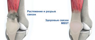
An ankle sprain is a nasty sports injury in which a ligament is partially or completely torn. Such injuries are most often the result of traumatic effects on the ligamentous apparatus of the ankle joint. The damage may involve several ligaments at once, not just one.
The ArthroMedCenter Joint and Spine Clinic has been treating sports injuries for many years, using an innovative technique - MBST therapy - to restore damaged tissue.
The increased degree of injury to the ankle ligaments is due to the fact that the ankle joint is an extremely mobile joint, with a large degree of freedom of movement. The lower leg is constantly subjected to a load equal to the body weight (and when performing physical exercises, the weight increases even more). This injury is very common among athletes: if during training a person did not follow safety precautions and exercise technique, then he can damage the ligaments. However, this injury often occurs in everyday life, for example, due to simple carelessness: you can fall unsuccessfully.
MBST therapy
The ArthroMedCenter clinic offers a progressive treatment method aimed at restoring tissue after an ankle sprain - MBST therapy. MBST is a therapeutic magnetic resonance therapy used in clinical practice in the mode of several programs encoded on a chip card. Programs are designed to treat injuries and illnesses.
For ankle sprains, the course of treatment is 7 or 10 procedures, 1 hour per day. At this time, the patient lies on the device and “receives” the incoming energy without feeling discomfort.
There are 3-day programs prescribed to athletes after intense competitions and training, as well as to prevent damage to cartilage tissue. Magnetic resonance therapy uses energy that is produced by hydrogen atoms. In some way, sports injuries are treated with bioenergy, and its use is not associated with consequences for the body and the risk of complications. The procedure can be repeated.
First aid to the victim
The timing of restoration of the active functioning of the joint is influenced by timely first aid. In case of injury, apply a cold compress to the ankle area as quickly as possible. It causes a reflex narrowing of blood and lymphatic vessels. Swelling and post-traumatic inflammation are relieved, and the severity of pain is reduced. What can be used for compress:
- a bag of ice cubes;
- packaging with frozen vegetable mixture;
- frozen meat or fish.
A bag of cubes or frozen foods is wrapped in several layers of thick fabric and applied to the joint for 10 minutes. Then take a break for 20-30 minutes to prevent tissue frostbite. Such treatment procedures are indicated for patients in the first days of treatment.
“Doctors are hiding the truth!”
Even “advanced” joint problems can be cured at home! Just remember to apply this once a day...
>
An important part of the therapy is fixing the injured limb with an elastic bandage and holding it in an elevated position. After diagnosis, long-term immobilization is often required using a plaster cast, splint, semi-rigid or rigid orthosis.
How does MBST work?
The principle of operation of this therapeutic method is based on the fact that tissue metabolism is regulated by electric and magnetic fields. And if in a healthy organ or system the processes of regeneration, that is, renewal, occur independently, then in case of dysfunction they require impulse influence from the outside. In MBST therapy, magnetic resonance is used as such an impulse, which sends signals to diseased tissues that reproduce signals from a healthy body system. Normalized metabolism allows you to resume the regeneration process in the cells of cartilage, bone tissue, tendons, etc. This allows you to eliminate the cause of disorders of the musculoskeletal system, and not only relieve the patient from the unpleasant symptoms of the disease.
Based on research, physicists and engineers were able to design equipment for the non-surgical treatment of sports injuries and diseases of the musculoskeletal system. Scientific and clinical studies have fully confirmed the restoration of cartilage using this method, and the method itself has been patented under the name MBST. This is the first and so far only therapeutic method that uses magnetic resonance for chronic and osteoarticular diseases. It has been officially approved for the treatment of osteoarthritis and osteoporosis and has been used for more than 15 years in Germany, Switzerland, Austria, Israel, Spain, Great Britain, Italy, Croatia, Romania, Slovenia, Czech Republic, and more recently in Turkey, Malaysia, the Philippines , in the Netherlands, in China and in India.
What other injuries can be treated with MBST?
Indications for using the MBST method:
- Achilles tendon injuries.
- Tendon injuries, partial tear of the medial collateral ligament of the knee, or tendinitis due to patellar tendon syndrome.
- Hip (muscle injuries).
- Knee joints (jumper's knee, cartilage and meniscus damage).
- As a conservative addition to cartilage wear/degradation and in cases after arthroscopy.
- Bone contusion/Bone swelling, osteochondritis dissecans or bone necrosis, pseudarthrosis, fractures and stress fractures.
Why does ankle sprain occur?
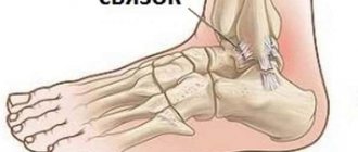
The ligamentous apparatus of the ankle can withstand quite serious loads, so in order to injure it, it is necessary to make an effort. An injury occurs at a time when the load is distributed incorrectly, that is, it is distributed not over several ligaments, but into one. As a result of overstrain, it stretches and breaks. Increased stress on the ankle ligaments can occur:
- when tucking the outer edge of the foot, the entire body weight will be distributed precisely over this area. In this case, injury occurs due to excessive supination of the leg;
- with maximum extension of the leg (while fixing the foot). In this case, injury occurs to the anterior talofibular and interfibular ligaments;
- in situations where the body weight is transferred to the forefoot (when it is fixed), and the lower leg is bent. In this case we are talking about an injury to the Achilles tendon.
- when the foot rotates (externally or internally), the foot is fixed. Ligaments are injured depending on the direction of the applied load - these can be internal or external ligaments, tendons of the peroneus brevis and longus muscles, etc. If we are talking about excessive internal rotation, the tendons of the tibialis posterior muscle can be damaged.
Types and degrees of ankle sprains
There are several types of injuries:
- turning the foot inward (inversion);
- turning the stoop outward (eversion);
- sprain of the upper ankle.
An ankle sprain can have several degrees of severity, which are distinguished based on the amount of damage to the ankle ligaments: The first degree is minor trauma. This type of injury is characterized by tearing of the fibers, but more than half of them remain intact. Treatment for such an injury does not last long, as does the rehabilitation period after it.
The second degree is a severe ankle sprain. Treatment in this case is more labor-intensive and lengthy than with the first degree of sprain. The second degree of severity is characterized by rupture of a significant part of the collagen fibers (about half). You can observe swelling of the injured area, as well as displacement of the articular elements.
Third degree - complete rupture of the ankle ligaments. There is pathological mobility in the joint, very pronounced swelling and pain in the injured area. It is important that immediately after receiving such an injury, first aid for an ankle sprain is provided, and then the person is taken to the clinic. This degree of severity is characterized by longer and more serious treatment. The rehabilitation process in this case also slows down.
Clinical picture
grade 1 rupture of the ankle ligaments The victim continues to lead his previous lifestyle without limiting his physical activity. But post-traumatic inflammation progresses. Hematoma and swelling occur, often spreading to the entire ankle. Now, while walking, severe pain occurs, localized in the ankle. To reduce its intensity, a person tries not to lean on his leg and begins to noticeably limp. Symptoms of grade 2 and 3 ligamentous injuries are much more pronounced. What signs indicate ligament rupture or complete separation from the bone:
- pain . Occurs immediately at the moment of injury. Its intensity often exceeds the pain syndrome associated with fractures. As long as extensive swelling has not developed, the victim can move independently. With more serious injuries, any load on the leg causes such severe pain that the person cannot even lean on the injured limb;
- swelling _ The main symptom by which a traumatologist determines ligament rupture. Swelling can form on both the lateral and medial sides of the ankle. With a complete separation, it spreads to the foot, but this condition is diagnosed extremely rarely. Severe swelling persists for 5-7 days and then gradually disappears. Since the accumulation of exudate is always associated with damage to the capillaries, an extensive bruise forms at the site of the edema;
- hematoma _ A bruise is only an indirect sign of ligament rupture. A few days after the injury, it is localized on the injured side of the ankle. After about 2-3 weeks, the hematoma moves down to the foot. In damaged tissues, due to the inflammatory process, a gradual breakdown of blood cells occurs. This is visualized by a change in the color of the hematoma. At first it is intensely dark blue, even purple. Gradually, a greenish tint begins to predominate in the color scheme, and then yellow.
A day after grade 2 and 3 injuries, the victim cannot fully lean on the injured leg and move. The occurrence of edema causes increased severity of pain. This occurs as a result of compression of the sensitive nerve endings by the accumulating fluid.
In severe ligament injuries with rupture of the joint capsule, traumatic hemarthrosis occurs. This is the name for hemorrhage into the joint cavity as a result of rupture of the vessels that supply blood to the internal joint structures. Hemarthrosis can provoke the development of destructive and degenerative tissue changes. To extract accumulated blood, a puncture is performed, followed by treatment of the cavity with antiseptics.
First aid for sprains
When spraining the ankle joint, timely first aid greatly influences the restoration of the tissues of the injured ligament and determines the time for complete restoration of joint function.
- First of all, apply ice wrapped in a towel (to prevent frostbite) to the injured ankle for 10-15 minutes, repeat the procedure after 15 minutes. Such manipulations will help not only relieve pain, but also stop the spread of edema (under the influence of cold, blood vessels reflexively narrow).
- The injured ankle must be kept at rest, and until an accurate diagnosis is made, you should not lean on the foot. Using a homemade or transport splint, you need to secure your leg. Fixation of the area should be maintained until examined by a traumatologist or surgeon.
- Place a bolster under your leg to give it an elevated position.
- Introduce an analgesic into the body (orally or by injection) if possible.
Damage classification
“An effective and affordable remedy for joint pain exists...” ...
For ease of diagnosis and choice of treatment methods, injuries are divided into certain groups. The main criteria are the degree of tissue damage and clinical manifestations. Ligamentous rupture is classified as follows:
- 1st degree . Minor tearing of individual fibers or bundles formed from them. The damaged ankle joint can be easily felt through the skin, the range of motion is slightly impaired or completely preserved. The patient is able to rest on his foot for a short time without feeling significant pain;
- 2nd degree . A tear of a large number of connective tissue fibers is diagnosed. On palpation, the victim complains of pain, and the joint itself can hardly be felt due to increasing swelling. The symptoms are much more pronounced. All attempts by the victim to lean on the injured leg cause severe pain, similar to that arising from dislocations or fractures;
- 3rd degree. With such damage, a complete separation of one, and in some cases several ligaments, occurs from the bone base. The sensations after a ruptured ankle ligament resemble those of a bone fracture. Extensive swelling and hematoma quickly form. The functional activity of the foot is so reduced that emphasis on it is impossible for several reasons. Firstly, it is severe pain. It is so intense that the victim may lose consciousness. Secondly, the anatomical relationship of the articular elements is seriously disturbed.
Regardless of the severity of symptoms, differential diagnosis is indicated for the patient. Its results will help to most informatively assess the degree of damage and the number of complications that have developed.
Symptoms of an ankle sprain
Symptoms of ankle sprains appear depending on the extent of the injury and the number of tendons involved in the injury mechanism. A complete rupture of the ankle ligaments is only caused by a sprain. This condition is characterized by excessive mobility in all projections of the joint. The main symptoms of an ankle sprain are:
- severe pain that occurs at the time of injury (fall, blow, etc.);
- swelling around the injured area (around the medial or lateral malleolus);
- bruising;
- extensive hematomas;
- local increase in temperature (in the injured area).
When trying to make a movement, a person also continues to experience pain.
Repeated ankle sprains lead to complete rupture of the tendon. More long-term and complex as well as longer rehabilitation will be required. That is why specialists at the ArthroMedCenter clinic recommend that you consult a doctor at the first sign of a sprain. To exclude repeated injuries, as well as the development of any other pathologies, it is worth carrying out a full course of treatment, including rehabilitation.
Complete or partial rupture of ligaments. Treatment methods
Ligament rupture is one of many injuries to the musculoskeletal system. They can be complete or partial. Represent damage to the integrity of the ligament. Appear as a result of injury during movements, especially sudden ones.
How can this happen?
Ligaments are an auxiliary element of the joint; they help connect the bones that form the joint. They differ from tendons in that they help hold muscles and bones together. Ligaments consist of fibrous tissue and have a certain amount of collagen and elastic fibers. Their strength depends on collagen fibers, and their elongation depends on elastic fibers. But ligaments often tear due to the fact that they do not stretch well.
Each joint has a strictly limited range of motion. The bony articular surfaces and ligaments that strengthen and fix the joint are the limiting factors. When a person commits actions that exceed the acceptable level, a rupture occurs. Cases of such injury due to a fall or blow are very common.
This type of damage varies in severity. Let's list them:
- A microfracture is what is often mistakenly called a tensile fracture. But you must always remember that sprains are
almost non-existent. At the same time, the overall integrity is not broken; we observe the rupture of individual fibers. - A partial rupture is also called a tear; a rupture of a certain area occurs during such damage, and a complete violation of the transverse is not observed.
- The rupture is complete - this means that the integrity in the cross section has already been completely broken. The ligament is torn into two parts, proximal and distal, with such an injury. One part is closer to the center, the second - to the periphery.
There are cases of a combination of rupture with injuries that are considered more complex.
Signs of damage
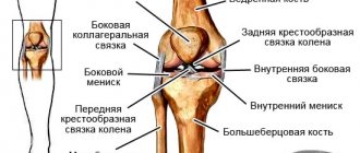
In practice, the knee and ankle joints of the lower extremities, the shoulder and wrist joints of the upper extremities localize the most common ruptures of the ligamentous apparatus. Although in theory, ruptures occur in any joints.
Based on certain signs, it is possible to determine the nature of the injury, namely:
- Edema. Excessive accumulation of fluid occurs in nearby tissues when there is a joint defect. Thus, damage leads to an inflammatory process with damage to small blood vessels, which leads to swelling.
- Pain. Since there are a large number of nerve endings in the tissues of the ligaments, any rupture is felt in the body by sharp pain. Ligamentin then develops in the injured ligament.
- Hematoma. When a ligament rupture leads to hemorrhage, the injured area expands in size, the skin becomes hot and turns blue.
- Limitation of mobility. Pain and bruises reduce the number of movements.
It is imperative to highlight the fact that you should never simply rely on pain symptoms without a detailed diagnosis. They are only primary and play an additional role, and instrumental research should be carried out. I would like to note that an x-ray cannot reflect the degree of rupture; it can only diagnose the existence of dislocations and fractures. Computed tomography, as well as MRI, are types of diagnostics in which it is possible to determine the degree of severity.
Stages in treatment
The speed of recovery, timely and correct approach to first aid are interconnected. It is advisable to treat ligaments immediately after an injury occurs. First we talk about immobilization, in other words, immobilization. It must be understood that the degree of the gap and its location guide the choice of means of rest.
A pressure bandage on the joint is a remedy that is used for mild micro-tears. An elastic bandage is also convenient for fixing ligaments. And it is necessary to use, for example, plaster splints in case of a complete rupture with the presence of hemarthrosis.
In order to relieve inflammation, relieve pain and swelling, local cold is required. You can apply ice to the affected area in the first one or two days.
The next stage in treatment for long-term fixation of the joint is the use of anti-inflammatory ointments and warming. Ointments can relieve pain and remove inflammation. To improve blood circulation in certain areas, locally irritating ointments are used. But such ointments can be applied only after local blood flow has been eliminated and swelling has subsided.
To improve local blood circulation, it is effective to use a semi-alcohol compress. This is a mixture of alcohol and clean water in equal proportions. Gauze soaked in this solution is applied to the area, then the area is covered with a plastic bag, a layer of cotton wool is placed on top, and then secured with a bandage. Ointments containing horse chestnut are excellent for relieving swelling. The rehabilitation period can take from three weeks, and sometimes up to two months.
Recovery is the next stage in treatment. Physiotherapy is applicable here. Therapeutic gymnastics will also have a positive effect on the ligaments. Loads that can cause pain are unacceptable; they must be performed gently and smoothly. A scar forms at the site of the rupture, and gradually they grow together provided that the treatment method is correctly selected.
Folk remedies for mild rupture
Very often, when providing assistance at home in case of injury, we resort to folk remedies. Here are some of them:
- You can use a cabbage leaf, which is applied to the bruised area.
- Potato compress is also used - a mixture of grated potatoes, onions and cabbage. Everything is mixed into a homogeneous mass and fixed on the damaged area.
These simple recipes will help you at home.
Author: K.M.N., Academician of the Russian Academy of Medical Sciences M.A. Bobyr
Diagnosis of ankle sprains
The diagnosis of sprain is made based on the existing symptoms, as well as data from such studies as:
- magnetic resonance imaging (MRI);
- ultrasonic examination (ultrasound) of the joint;
- arthroscopy.
X-rays are not used to make a diagnosis, since the ligaments are a soft tissue formation, i.e. they are not visible on an X-ray. However, the patient may be referred for an x-ray to rule out a fracture. The symptoms of fractures and sprains are very similar, in addition, some symptoms can be combined with each other.
Ankle exercises
The ankle joint is a complex joint. Recovering from an injury will require a person to focus on four factors such as:
- range of motion
- force
- flexibility
- balance
Each of these functions is critical to a healthy ankle. Various exercises will focus on one or more of these factors.
Physical therapy is an integral part of the recovery process. The exercise helps strengthen the ankle joint and prevent recurring sprains or other problems.
A 2021 study suggests that failure to perform exercises may lead to the development of chronic ankle instability, which may require surgery.
Although a person may experience slight discomfort while performing these exercises, they should not cause pain. If a person feels pain during exercise, he should stop and rest his foot.
Preventing ankle sprains
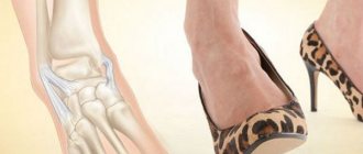
Ankle sprains can occur in anyone if they are not careful during exercise and outdoor activities. If you want to play sports, do it in suitable shoes and clothing. Walk carefully in high-heeled shoes, avoid potholes and potholes, and watch your step when walking somewhere. Try to fight excess weight, as obesity of any degree puts excessive stress on the joints. Lead an active lifestyle, play sports, eat well. Moderate physical activity strengthens.
How to treat a sprained ankle?
Physiotherapy
- The use of ultrasound - this procedure not only improves microcirculation of the damaged area, but also accelerates the process of lymph outflow. After this procedure, the ointments are absorbed much better, and the drug accumulates in the tissues.
- UHF is used to reduce local inflammation and accelerate reparative processes in tissues. Vasodilation helps improve the trophism of the affected area.
- Paraffin therapy is one of the main activities. The main effect of this therapy is to reduce pain and eliminate inflammatory processes. Can be used in any period after injury.
- Magnetic therapy – promotes the outflow of blood and lymph, enhances the absorption of local medications, and relieves inflammation.
- Electrophoresis with non-steroidal anti-inflammatory drugs or novocaine. Pain relief is achieved by dilating blood vessels.
Gymnastics
Any gymnastic exercises to strengthen the ankle joint can only be performed in the long term (about 1-3 months, depending on the degree of injury).
- Development of motor skills of the toes.
- Rolling a bottle with your foot.
- Walking on your heels.
- Running on small pebbles or sand.
- Circular rotations of the foot.
- Extension and flexion of the leg at the ankle joint.
- Walking on the outside and inside of the foot.
- Walk on your toes for a few minutes every day.