Disorders of calcium metabolism are called calcification, calcification, or calcification. It is based on the deposition of calcium salts in cells and intercellular space. In this regard, intracellular and extracellular calcification are distinguished, and according to prevalence - systemic (general) and local.
Areas of calcification may take the form of tiny grains, detectable only under a microscope, or foci that are clearly visible to the naked eye. Calcified tissue becomes dense and brittle and resembles stone. Bone formation may occur in areas of calcification. Inflammation appears around the calcium deposits with the proliferation of connective tissue elements, the accumulation of giant cells of foreign bodies and the development of the capsule.
According to the mechanism of development, calcification is classified into metastatic, dystrophic and metabolic.
Development mechanism
This process is local, that is, it affects a specific area.
The main cause of calcification is tissue changes that provoke increased absorption of calcium (lime) from tissue fluid and blood. The main factor in the development of this process is the alkalization of the environment, as well as an increase in the activity of enzymes that are released from dead tissue. Petrification occurs in:
- chronic inflammatory foci;
- tuberculous necrotic foci;
- places of cell death;
- gummah;
- heart attacks.
In case of petrification, “armored lungs” are observed on the pleura, and “armored heart” is observed on the pericardium.
Dystrophic calcification
Dystrophic calcification, or petrification (from the Greek petra - rock, stone), is characterized by local deposition of calcium salts in problem areas. These can be dead or deeply degenerated tissues, tissues with reduced metabolism, which include cartilage and tendons.
With dystrophic calcification, hypercalcemia is absent, and the adsorption of calcium salts from the blood and tissue fluid is caused by physicochemical changes in tissues undergoing necrosis or degeneration.
Petrificates are found in various organs and tissues, are white in color, and have a rocky density. Most often, petrificates are found in areas of chronic inflammation, heart attacks, arterial walls with atherosclerosis, in scar tissue, such as heart valves with defects, cartilage, and venous blood clots.
Classification
1. According to etiology:
- traumatic;
- degenerative;
- inflammatory.
2. By localization:
- brain calcification;
- petrification of joints, ligaments;
- calcification of blood vessels and so on.
3. In accordance with the location of petrification in a particular system (part) of the body:
- calcifications in the tissues/organs of the heart and vascular system (circulatory and lymphatic);
- petrification in organs/tissues of the nervous system;
- respiratory organs;
- musculoskeletal system;
- genitourinary system;
- Gastrointestinal tract and glands;
- hematopoietic system and intrasecretory organs;
- other calcifications.
4. According to the x-ray picture:
- in the form of massive regional formations, which more often occupy part of the organ (calcification of the pericardium or pleura) or (less often) multiple petrifications (with ossifying progressive myositis);
- individual foci that can be multiple or single, large or small (calcified pulmonary tuberculosis foci, calcified lymph nodes, and so on);
- petrificates in the form of stones (pancreatic, bile, salivary, etc.)
It is worth noting that both regional and focal calcifications can be organ (that is, located in one organ) or systemic (that is, present throughout the entire system).
5. In addition, calcification can be:
- physiological, that is, developing due to aging (involution);
- pathological, developing in places of various neoplasms.
Calcification of the pineal gland
Calcification is (as mentioned above) the formation of accumulations of undissolved calcifications in various organs or tissues in which such salts should not normally be contained.
The cause of pineal calcification can be congenital pathologies, various infections and metabolic disorders. Physiological calcification of the pineal gland is most often (40%) found in patients under 20 years of age. In this case, compact neoplasms with a diameter of up to 1 cm are formed in the organ.
In cases where calcifications are large in size, it is worth studying them in detail, as they can become the basis for malignant neoplasms. Dystrophic (pathological) calcification in the pineal gland occurs as a result of trauma, chemotherapy, ischemia, and so on, and is characterized by the deposition of cholesterol and lime in neoplasms.
Calcification of the pineal gland is accompanied by dysfunction of the latter, which can provoke the development of cancer, multiple sclerosis and schizophrenia due to blockade of melatonin synthesis. Filling of the pineal gland (calcification) with calcifications increases the likelihood of developing nervous exhaustion, anxiety, depression and gastrointestinal pathologies.
Metastatic calcification
Metastatic calcification (calcareous metastases) is systemic in nature and is accompanied by the deposition of calcium salts throughout the body in various organs and tissues. The cause of its development is hypercalcemia. The reasons for the increase in the concentration of calcium in the blood plasma may be increased leaching of calcium from the bones, decreased excretion of calcium from the body, or a violation of the hormonal regulation of calcium metabolism. For example, with hyperproduction of parathyroid hormone or lack of calcitonin, hypervitaminosis D.
An inflammatory reaction occurs around the deposits of calcium salts, and sometimes foreign body granulomas are formed.
Ligamentous calcification
Calcification of ligaments is a fairly common occurrence associated with age-related changes in the body, injury and inflammation. Ligamentous calcification is often asymptomatic and discovered incidentally during X-ray examinations.
Such involutive processes in cartilage and ligaments during calcification of joints are accompanied by a loss of shock-absorbing properties, plasticity and elasticity in the joints.
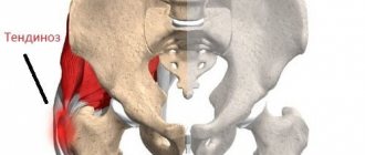
Most often, tendon calcification develops in the spine (cervical/lumbar spondylosis deformans), due to tears in the area where the annulus fibrosus and longitudinal vertebral ligament attach to the edge of the vertebra, as a result of which the intervertebral disc is displaced, tearing the ligament away from the vertebra. This is where calcifications/ossifications develop.
In addition, similar processes are often found in the spinal-costal joints (ribs 9-10), hip and phalangeal joints (Eberden and Bouchard nodes), being a local demonstration of the aging of the body.
How to recognize pathology: signs and symptoms
Diagnosis of pineal gland calcification is a difficult task, complicated by the lack of characteristic symptoms. Most manifestations are common symptoms of other diseases.
Doctors include indirect signs of pathology:
- frequent headaches of moderate intensity and often diffuse in nature;
- feeling of heaviness in the head;
- constant feeling of anxiety;
- neurological disorders;
- tendency to depression.
In some cases, pathology can cause disruptions in the functioning of the gastrointestinal tract, so frequent digestive disorders, stool disorders, heartburn not related to eating habits and behavior can also be a reason for undergoing an in-depth examination to determine the functioning of the pineal gland.
Spurs
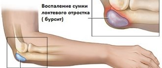
Calcification of tendons at the points of their attachment to bones, which look like spikes and points, are called spurs. Similar formations occur in the pelvic, ulnar, occipital, and calcaneal bones.
The cause of calcification in this case is inflammatory processes, physical activity and age-related changes. The most common diagnosis is a heel spur (at the insertion of the Achilles tendon).
The formation of spurs is often accompanied by pain and limitation of movement; radiographs show deformities of the foot, replacement of soft tissues with fatty tissues and transformation of tendons into bone tissue.
Calcification of heart valves
- Calcification of the aortic valve. The cause of this disease is rheumatic valvulitis, leading to degenerative changes in tissues. In this case, the valve flaps are deformed and soldered together. At the same time, calcifications form on them, which block the aortic ostium. In some cases, the process extends to the interventricular septum, the mitral valve leaflet and the wall of the ventricle (left). As a result, aortic insufficiency develops.
- Calcification of the mitral valve. It is quite difficult to diagnose such a pathology, due to the fact that its symptoms are similar to the clinical picture of cardiosclerosis, hypertension and rheumatism. More often this disease is detected in elderly patients.
Vascular calcification
- Calcification of the aorta. Develops in patients over 60 years of age. The clinical picture of the disease depends on the level of vessel damage.
- Calcification of cerebral vessels. Calcification is in this case a synonym for atherosclerosis. Due to the accumulation of lipids on the walls, insufficiency of blood circulation in the brain occurs, which is fraught with the development of strokes, dementia, and so on.
- Calcification of the coronary arteries. In this case, cholesterol and fats settle on the walls of these vessels, that is, atherosclerotic plaques are formed, which leads to a loss of elasticity and a change in the shape of the vessel, as a result of which the blood supply to the myocardium is disrupted, and in the case of complete blockage of the lumen, tissue necrosis.
And a little about secrets...
If you have ever tried to study the problem of thyroid diseases, you have probably encountered the following difficulties:
- drug treatment prescribed by doctors, solving one problem creates others;
- replacement therapy drugs that enter the body from the outside help only for the duration of use;
- medications used to treat hormonal disorders cost a lot of money;
- medications taken orally disrupt the gastrointestinal tract;
- Constant fluctuations in hormonal levels spoil your mood and prevent you from enjoying life. Published by econet.ru. If you have any questions on this topic, ask them to the experts and readers of our project here .
PS And remember, just by changing your consumption, we are changing the world together! © econet
Brain calcification
Calcinosis can affect various brain structures:
- cerebral cortex;
- cerebral vessels;
- hard shell.
Such changes develop due to various reasons, the main of which are:
- Past or existing infections (tuberculosis, cysticercosis, HIV).
- Intrauterine (congenital) infections (TORCH).
- Injuries.
- Atherosclerosis.
- Inflammation.
- Tumors.
- Metabolic, endocrine disorders.
What is the pineal gland?
The pineal gland is a part of the brain that has nerve cells (neurons) in its structure and is responsible for the production of serotonin, melanin and other hormones.
The laying and formation of the organ begins in the fifth week of gestation, so expectant mothers during this period need to be especially attentive to their own health, take vitamins and medications prescribed by the doctor, and avoid consuming harmful drinks and foods, the main one of which is alcohol.
The shape of the pineal gland is not constant and changes throughout a person’s life. In infancy, it is usually a ball, but as you grow older, the pineal gland becomes elongated and slightly flattened on the sides. The average size of the lateral surface in an adult is 4.5-5 mm.
The main growth of the organ occurs during a period of increased hormonal activity in adolescents. This most often occurs when the child reaches puberty.
In order to avoid problems associated with improper functioning of the pineal gland in the future, it is important to eat properly during this period and avoid increased mental and physical stress.
The best publications in the Econet.ru Telegram channel. Subscribe!
Calcification: treatment
Therapy for calcification depends on the location and extent of the process, as well as the severity of symptoms and the age of the patient.
- To normalize calcium metabolism, it is recommended to restore the balance of calcium and magnesium in the blood. Magnesium controls the flow of calcium and dissolves calcifications, and also promotes the removal of excess microelements and its proper absorption. Therefore, in addition to diuretics, the patient is recommended to take magnesium supplements.
- Dieting. The patient should avoid foods that are fortified with calcium (vegetables, milk, etc.) and vitamin D.
- In the case of massive foci of calcification (in particular on the skin and subcutaneous tissue), surgical treatment is recommended.
- Early detection of pathology promotes a speedy recovery and is the prevention of serious complications. As mentioned above, therapy depends on the location of the calcification focus.
- In some cases, patients are recommended to use folk remedies, but such therapy should be carried out strictly under the supervision of the treating doctor.
- Treatment of mitral valve calcification is carried out using mitral commissurotomy, as well as prescribing preventive drug treatment. Thanks to these methods, cardiac activity is restored and the patient can lead a normal active lifestyle.
- To slow down calcification of the aorta, therapy with drugs based on statins, nicotonic acid, and so on is used. In the case of an advanced process, surgical interventions are used.
The structure of the pineal gland and its role
The main part of the pineal gland is the pinealocyte, which is why the pineal gland is also called the pineal gland. Pineal cells contain lipoid acid and pigment inclusions, which are responsible for the basic functions of the organs.
It has been proven that the pineal gland is responsible for the production of hormones necessary to ensure human biological rhythms, the functioning of internal organs and the functioning of the brain.
Doctors identify several important functions of the pineal gland:
- regulation of the synthesis of sex hormones;
- normalization of the functioning of the ovaries and other organs of the female reproductive system;
- production of biologically active substances to maintain the vital functions of the body;
- ensuring circadian rhythms;
- decreased blood glucose levels (due to penialin produced by pineal cells);
- maintaining normal water-salt balance.
The speed of falling asleep, duration and quality of sleep depend on the functioning of the pineal gland. During normal functioning of the organ, the activity of the brain appendages at night is blocked, which ensures proper rest and the production of hormones and biologically active substances in quantities sufficient to maintain the healthy functioning of organs and systems.
Important! The pineal gland restrains the functioning of the reproductive system until the child reaches puberty.