How is it formed?
A growth with a rod forms in response to constant friction and pressure of the skin area. With a long-term mechanical impact of the same type on the skin, the keratinized particles of the epidermis do not have time to exfoliate. They layer on top of each other and form a compaction. Keratinized zones or dead cells of the epidermis appear quickly, accumulate, and the mechanism of their renewal is disrupted.
Callus formation begins in the top layer of skin in a place where tight-fitting shoes or other factors put pressure on the cells, injure them and prevent them from sloughing off. To protect the lower layers of the dermis from traumatic effects, the skin in this area becomes harder. At the first stage of callus formation, you may notice that the skin has become dry. If nothing is done at this moment, the provoking factor is not removed, then the growth will begin to go deeper and form a hard internal core that will affect the lower layers of the dermis.
At the same time, as the callus grows deeper, its area gradually decreases and its shape becomes cone-shaped. When pressing precisely on its top, a person feels severe pain, because the hard, sharp rod at this moment presses on the soft tissue of the dermis surrounding it.
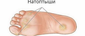
The formation may appear on the legs and arms. The core callus occurs on the little toe, on the bony protrusions, on the pads of the fingers, in the third and fourth interdigital spaces on the foot, in the center of the heel.
What does a callus look like?
How can you tell if a callus has a core?
It is easy to recognize such a formation by its appearance. It is a rounded, whitish, yellow or yellowish-gray compaction slightly raised above the skin. In the center there is a small depression or “pit” that resembles a cork. This is the top of the stem, which can be compared to the head of a carnation.
The rod can grow deep evenly, perpendicular to the surface of the skin, or at an angle. Depending on the severity, its length ranges from several millimeters to centimeters. There are often cases when the rod reaches the subcutaneous fat and even the bones of the foot.
How the core callus passes under the skin, look at the photo:
At rest, the callus is not felt between the toes, on the heel or anywhere else. But it causes intense pain while walking, often causing forced gait disturbance and lameness. Pain occurs due to contact of the rod with nerve endings that penetrate the deep layers of the dermis. Many patients compare the unpleasant sensations arising from a callus to the feeling of a splinter being stuck into the foot.
You can independently suspect a growth based on the following signs:
- the formation is small in size, has a rounded clear outline, smooth edges;
- The color of the callus is yellowish, the structure feels dense to the touch;
- change in skin pattern;
- dull pain or discomfort when walking if the callus is located on the foot;
- increased pain when pressed.
This type of compaction develops slowly. In the early stages, its formation can be detected by slight redness of the injured area, itching, and tingling. If a rounded, convex area with a dimple in the center appears on the skin, this already indicates that the callus has a core.
1.What is a bone spur?
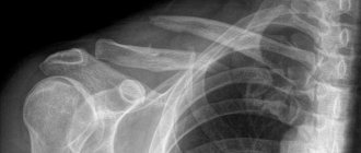
Bone spur or osteophyte
is a growth that forms on top of normal bone. The specific name of this phenomenon misleads many people who mistakenly believe that a bone spur is something sharp, but it is soft to the touch, a completely normal bone tumor.
Most often, osteophytes are located
on the lower or posterior surface of the heel bone, less often they are found on the shoulders, elbows, hips or knees.
As a rule, their presence is not accompanied by painful phenomena, but when they come into contact with soft tissues, bones and tendons, they can cause excruciating and severe pain.
A huge role is played by
the localization of the bone spur
. If a bone spur is located, for example, on the olecranon, then its owner will not feel discomfort or other painful manifestations, and, therefore, there is no need for its treatment. But if a bone spur has formed on the heel bone, which is used when a person walks, then over time it will cause increasingly stronger and longer-lasting pain.
Another common location for a bone spur, or osteophyte, is the shoulder area.
. The complex structure of the shoulder joint determines its versatility, the ability to move the shoulder and arm in various directions. Over time, the tendons, muscles, ligaments and bones that make up the shoulder joint begin to wear out. This process primarily affects the tendons that attach the muscles to the upper arm.
When moving, the tendons can come into contact with the bones, causing the formation of bone spurs with subsequent inflammation and pain in the affected area. This pathology is typical for older people and athletes, as well as for those who, due to their professional activities, are forced to hold their hands above their heads.
A must read! Help with treatment and hospitalization!
Reasons for appearance
Dermatologists consider callus as a complication of dry normal callus that was not removed in time. The main reason for its formation is prolonged physical impact on the skin. Therefore, most often the appearance of calluses on the toes and other parts of the foot is provoked by uncomfortable shoes. This includes shoes with narrow toes, thin soles, shoes, boots, and high-heeled sandals. If you wear such shoes at work from morning to evening, you expose certain areas of your foot to increased stress. This causes calluses to form near the little finger and under the pads of the fingers.
It is important that the shoes fit correctly and are made from soft, natural materials. If it is tight, the legs will be squeezed. And if it is too loose, then the foot will slide and rub in it. In shoes made of synthetic materials, feet will sweat, which will also create favorable conditions for the formation of calluses, especially in combination with friction.
You need to be careful with new shoes. The leg should get used to it gradually. Prolonged physical activity or sports, running, aerobics, football, basketball in new sneakers can cause roughening of the skin in areas of greatest pressure.
Other factors that lead to the formation of a seal with a rod are:
- rough seams of socks and tights, squeezing the skin;
- increased loads on the legs of professional athletes;
- climbing stairs every day;
- diseases of the foot joints (arthrosis, arthritis), heel spurs;
- uneven distribution of body weight on the foot with flat feet, clubfoot, hallux valgus, varus deformity;
- frequent walking barefoot, which may be accompanied by a splinter or other small foreign body getting under the skin;
- nail fungus (onychomycosis) and foot skin;
- psoriasis;
- chronic pathologies that affect a person’s gait (diabetes, cerebral palsy, Parkinson’s disease);
- the presence of old dry calluses in which the core may begin to grow;
- excess weight.
Calluses form on the fingers much less frequently than on the feet.
They appear as a result:
- working with bare hands with household tools and gardening equipment;
- certain professions (blacksmiths, carpenters, joiners);
- constant cooking, working with a knife, rolling pin;
- practicing some sports (lifting weights, exercises on horizontal bars, uneven bars, rings, tennis, badminton);
- playing musical instruments (guitar, violin, viola).
People with poor blood circulation in the extremities (diabetic foot, vascular diseases), weakened immunity, bad habits (smoking), and chronic dermatological pathologies are more likely to get this type of callus. Risk factors include old age.
The formation of a seal with a rod is facilitated by frequent walking barefoot or in open shoes. This increases the risk of splinters, pieces of glass, grains of sand or other foreign particles getting under the skin or in shoes, which will injure the skin for a long time and accelerate the protective mechanism of keratinization. Most often, formation appears in the summer, when we put on sandals, flip-flops, sandals on bare feet, and walk barefoot in an apartment, on the beach or in a summer cottage. The skin becomes less protected and more easily injured, which contributes to the formation of calluses.
Treatment of callus with medications
It is immediately worth noting that drug treatment of calluses has the least therapeutic effectiveness.
This phenomenon is associated with the inability of drugs to penetrate deep into tissues, eliminating the root of the formation.
The products used for core calluses often contain keratolytic components and can be effective at the beginning of callus development.
As a rule, products based on salicylic acid are used.
Salicylic acid is a keratoplastic and keratolytic agent.
In addition, it has an antibacterial and antifungal effect.
Salicylic acid works by softening keratin, a natural protein that is part of the skin's structure and produced by the body.
The drug has a softening effect on the stratum corneum of the dermis, which facilitates its removal.
Salicylic acid is often used in combination with other substances.
Allows for a higher therapeutic effect.
When using salicylic acid preparations, the simultaneous use of the following drugs is not recommended:
- Alcohol-containing medications.
- Any other local medications that contain dibenzoyl peroxide or retinoid.
- Soapy substances that have a drying effect.
- Cosmetics that have an exfoliating effect.
Diagnostic features
In appearance, an ingrown callus can easily be confused with a plantar wart, which is caused by the human papillomavirus (HPV). Unlike a callus, a wart is the result of HPV infection, which can occur when walking barefoot on dirty surfaces, the ground, in public showers, and gyms. When the wart is opened, small black dots will be visible under the layer of keratinized skin. These are thrombosed capillaries that bleed when pressed. Plantar warts can occur anywhere on the foot, not necessarily in areas of greatest pressure.
A dermatologist or podologist can easily distinguish a callus from a plantar wart and make the correct diagnosis. For a detailed examination, he may use a dermatoscope. This is a device that magnifies the image of the surface layer of the skin 10 times. If necessary, the doctor will take a scraping of epithelial cells from the seal to determine the nature of this formation. Since, in addition to plantar warts, differential diagnosis with oncological skin diseases is sometimes important.
What is the difference between a plantar wart and a callus with a core?
- An ingrown callus does not bleed, even with strong pressure.
- Warts usually appear in groups rather than as single individuals.
- There is a depression in the central part of the callus with a core, and the wart is covered on top with small dark-colored dots.
- With a plantar wart, the papillary pattern or raised lines on the skin bypass the wart, but with a core callus they persist and pass through it. This is due to the fact that when infected with HPV, the DNA of the epidermal cells changes, and the skin pattern at the site of the lesion is no longer formed.
- Plantar warts cause pain when pressing from the sides when forming a skin fold, and ingrown calluses cause pain when pressing from above in a vertical direction.
It is important to understand the reasons that caused the callus. A doctor will help here. If necessary, he will order tests, ask about lifestyle, evaluate the correct choice of shoes and foot care, recommend a consultation with an orthopedist, prescribe additional examinations, blood tests for sugar, HPV.
Methods for removing callus
How to get rid of a painful formation? Treatment for a callus on the foot involves its complete removal. To do this, you need to remove or destroy the rod. It is recommended to start treatment as early as possible in order to quickly get rid of discomfort and pain when walking. The shorter the rod, the easier it will be to get rid of it and the faster the wound will heal and the skin will heal.
By itself, such a callus will not disappear. To solve the problem you will need medical help. Be sure to consult a podiatrist if there are circulatory problems in the lower extremities, inflammation develops at the site of rough skin (redness, purulent discharge), or a fungal infection.
The main problem when removing such calluses is the core that grows deeply into the skin. If you remove only the top part, it will do nothing. The pain will remain, and the removed keratinized skin will quickly recover. It is not always possible to get rid of a problem in one procedure. This depends on the depth of ingrowth of the rod, the chosen removal method, skin characteristics, age, health status of the patient, and the presence of concomitant foot problems.
Previously, surgical methods were used to remove callus. But now there are many minimally invasive, painless procedures that quickly and painlessly solve this problem. Therefore, you should not be afraid or put off visiting a podiatrist.
Due to the large number of disadvantages, surgical removal of callus is used extremely rarely, only in advanced stages with very large callus sizes and deep ingrowth of the core. This operation involves excision of the keratinized area and is performed under local anesthesia. The doctor uses special scissors to cut out the layers of the growth and remove the rod. After removal is completed, the resulting wound is treated with antibiotic ointment. Given the high level of development of alternative methods of combating calluses, surgical excision is today considered an outdated method.
Its serious disadvantages include:
- soreness;
- the need to use local anesthesia;
- traumatic, bleeding;
- risk of infection;
- long healing period;
- large postoperative scars.
What methods are used now?
To remove a callus on the leg along with the stratum corneum and root, the following modern low-traumatic methods are used:
- hardware pedicure;
- laser exposure;
- cryodestruction;
- radio wave therapy;
- electrocoagulation.
Each of the listed methods has its own indications, advantages and disadvantages. Sometimes some of them are used in combination. Let's look at them in more detail below.
Hardware pedicure
Hardware pedicure is an effective and affordable way to remove callus. The rough skin and shaft are drilled out with special diamond or ceramic cutters; their length and diameter are selected depending on the size and depth of the root.
Hardware pedicure does not cause pain, only a slight burning and tingling sensation is possible due to the increase in temperature in the affected area. After drilling the rod is completed, a depression remains on the skin, into which antibacterial, antifungal or antiseptic ointment is placed to prevent infectious complications.
The procedure is performed under 100% sterility conditions. The replaceable attachments used for the device can be easily sterilized. The feet are treated with an antiseptic.
If necessary, keratolytics or substances that soften rough skin are first applied to the keratinized areas. They may contain salicylic or lactic acid. Keratolytics act only on dead skin cells, without affecting young skin.
For ingrown calluses, first remove the upper stratum corneum. Then they start drilling the rod. They begin to work with cutters of larger diameter, moving into the skin, using smaller attachments. The size of the cutter is selected according to the diameter of the rod. If it exceeds the diameter of the growth, the risk of damage to healthy tissue will increase. High precision and experience in performing manipulations are important here so as not to damage surrounding tissues. At the end, a medicinal ointment is placed into the resulting cavity and a bandage is applied.
Depending on the size of the callus, it may take from 1 to 5 sessions to completely remove it.
Hardware pedicure is the safest and most comfortable for the patient. All manipulations are carried out only on the affected areas without affecting healthy tissue. The depression formed after drilling the rod is usually healed within 2 days.
Laser method
Laser therapy is widely used for many medical, dermatological and cosmetic problems. Calluses with a core also respond well to treatment with this method, even in advanced stages.
Two types of lasers are used to remove it:
- erbium with a beam penetration depth of up to 20 microns;
- carbon dioxide with a beam penetration depth of up to 100 microns.
How does a laser remove a callus? Under the action of a laser beam, the keratinized upper layer of skin is heated in a targeted manner, the structure of its cells changes, and they evaporate. The hardened seal is removed layer by layer. The depth of exposure is controlled by a doctor, so healthy tissues are not injured. The procedure takes 5–10 minutes. At the end, the specialist treats the skin with disinfectant and medicinal compounds that will speed up healing and prevent infectious complications.
In place of the former callus, a crust forms, under which young dermal cells gradually appear. Complete healing occurs within 10 – 14 days. The mark on the skin after laser treatment is almost invisible.
The laser beam additionally has an antiseptic effect and destroys pathogenic microorganisms present on the skin, which eliminates infection during the procedure. Evaporation of tissue is accompanied by simultaneous cauterization of blood vessels, so there is no even minimal blood loss.
The disadvantages of laser treatment of callus include high cost and the need for injections of local anesthetics for pain relief.
This method cannot be used for people who have:
- diabetes;
- dermatological pathologies of any nature;
- disruptions in the immune system;
- mental disorders;
- oncological diseases.
Laser treatment can be used at any stage of growth with a rod, but in practice this method is more often used in advanced cases, with old formations.
Cryodestruction
Cryodestruction is a method of removing ingrown calluses with liquid nitrogen. When the procedure is performed under the influence of low temperatures (-196 °C), the keratinized skin freezes and dies. This process is called cryonecrosis. Nitrogen is odorless and colorless and looks like a clear liquid.
It is applied only to the problem area, so nearby healthy tissue is practically not affected. When the procedure is performed by an experienced doctor, the risk of injury is minimal. The diameter of the nozzles from which nitrogen is supplied to the skin is selected in accordance with the size of the callus.
In terms of time, removing one callus takes from 30 seconds to 2 minutes, depending on the depth of the ingrowth of the rod.
The procedure does not cause pain or risk of infection. Only mild discomfort due to exposure to cold is possible. During cryodestruction, cold acts as an anesthetic. If the callus is too deep, then local anesthesia with lidocaine is used.
Rejection of dead tissue begins a few days after treatment. Immediately after the manipulation, the keratinized skin turns pale, and a water bubble appears in its place. After a few days, it dries out and becomes covered with a crust, under which a new soft layer of epidermis forms. Under no circumstances should you puncture the bladder or tear off the crust, this will provoke inflammation and lead to the formation of an unsightly scar. After 2 weeks, the crust will fall off on its own, and underneath there will be new smooth skin. Until the skin is completely healed, a person may experience discomfort at the site of exposure.
The disadvantages of this method include:
- long rehabilitation period, which takes at least 14 days;
- impossibility of spot application;
- the impossibility of simultaneous removal of several closely located calluses, since this creates a large wound surface that will take a very long time to heal;
- risk of damage to healthy tissue;
- risks of secondary infection due to the formation of areas of wet necrosis;
- low effectiveness for large calluses with a deep core.
Removal of calluses with liquid nitrogen is permitted only after consultation with a doctor. Cryodestruction can be performed on both adults and children. However, it is recommended only for ingrown rods of short length, since it is difficult to control the depth of tissue freezing. When treating deep calluses, the risk of damage to healthy skin increases.
Electrocoagulation
Electrocoagulation is a method of cauterizing an ingrown callus using an electric current. Under the influence of high temperature (80°C), proteins in keratinized skin are destroyed and cell death occurs. After the procedure, a protective crust forms at the site of the growth; it disappears after 7–12 days.
Electrocoagulation takes about two minutes and requires preliminary anesthesia. A specialist acts on the seal with a special needle from which a high-frequency current is supplied.
The advantages of the procedure include low cost, simplicity, and the ability to regulate the depth of exposure to tissue. During electrocoagulation, blood clotting occurs in the treatment area, which eliminates bleeding and infection. The method makes it possible to conduct a histological analysis of removed tissues, if it turns out that this is necessary.
Disadvantages include pain after the procedure, the likelihood of damage to healthy tissue surrounding the callus, the formation of a scar during healing if the growth is deep, and the risk of relapse if treatment is insufficient.
Electrocoagulation has many contraindications and is not suitable for everyone.
It cannot be done in the following cases:
- intolerance to electrical procedures;
- blood clotting disorders;
- presence of a pacemaker;
- systemic blood diseases;
- diabetes mellitus;
- increased sensitivity of the skin to light (photodermatosis).
Radio wave therapy
Radio wave therapy is based on the use of high-frequency radio waves, when a thermal effect is exerted on the affected areas of the skin. The procedure is performed on a device whose operating principle is similar to that of a microwave oven. This means that when removing calluses, the device itself does not heat up, but only the tissues at the site of exposure to radio waves heat up.
The method is non-contact, the electrode does not touch the skin, the destruction of the callus occurs at the cellular level. Radio waves cause the evaporation of liquid inside the keratinized cells, while the healthy cells surrounding the callus do not react to it and are not damaged.
The procedure is painless, does not require anesthesia, and does not cause bleeding. It only takes a few minutes. After its completion, a thin brown crust forms on the treated area, which disappears on its own after a few days. The recovery period takes up to two weeks.
Radio wave therapy has an antiseptic effect, destroys pathogenic microorganisms, which prevents infection and inflammation. Does not leave scars or scars on the skin.
Compared to laser radiation, electrocoagulation, and cryodestruction, radio wave therapy has a minimum of contraindications and has the least damaging effect. However, its main disadvantage is its high cost.
Traditional medicine in the treatment of calluses
Folk remedies cannot affect the development of callus and are not an effective treatment method.
The treatment and removal of callus must be carried out by a qualified specialist.
This will minimize negative consequences and complications.
It is necessary to begin treatment of callus in the early stages of its development.
When there is still a chance to get rid of the problem without resorting to surgical methods.
As it progresses, the root of the callus will penetrate into the deeper layers of the epidermis.
Then it may be necessary to use more radical and expensive methods of therapy.
Skin care after callus removal
At the site of the removed callus, a small wound remains on the skin.
To speed up its healing and prevent complications, it is important to organize proper care in the first days:
- do not wet;
- prevent dirt from getting in;
- limit the load on the leg;
- do not apply regular foot skin care creams to the site of the removed callus;
- treat with an antiseptic and special medicinal healing compounds, which will be recommended by the doctor who performed the procedure;
- Do not peel off the crust that forms at the site of the wound, otherwise the rehabilitation will be very delayed.
During the healing period, the doctor can place a special unloading corrector in the place where there was a callus before. It will reduce pressure on the sore area while walking and protect against irritation and rubbing.
It is very important, together with a podologist or orthopedist, to find out the reason why the pathological compaction occurred. If the provoking factor is not removed, the formation will appear again. You can start by reviewing your shoes. It is recommended to avoid high heels. Shoes should be comfortable and fit.
If it turns out that the cause is flat feet or other structural disorders of the foot, then it is necessary to treat them. Do special exercises to strengthen the muscles that support the arches, wear corrective orthopedic insoles, and select shoes in accordance with the recommendations of the orthopedist.
About treatment at home
You won't be able to deal with callus on your own. This type of keratinized growths is considered the most difficult to remove. The problem is that at home, it is almost impossible to completely remove the rod. Especially without harm to your health.
Pharmaceutical products and plasters for calluses do not help. They have keratolytic activity, soften rough skin, but this is not enough to remove deep core. Such products are effective only for combating superficial calluses and corns. But even for these purposes it is not recommended to use them. If you care about the health and safe care of your feet, it is better to contact an experienced podiatrist and get a hardware medical pedicure.
The most popular pharmaceutical remedy for calluses is the Salipod patch. It contains salicylic acid, sulfur, rosin, lanolin. According to the instructions, to get rid of calluses, you need to cut a piece of the patch of a suitable size and stick it to the formation. This patch should be worn for 1–2 days. If there is no effect, the procedure is repeated. With core calluses, “Salipod” helps only in the initial stages, when the core is just beginning to grow deeper. But usually at this stage the callus does not make itself felt and does not cause discomfort, so the person misses the moment. And when the rod is already deep, the active components of the patch cannot reach its base. As a result, only the tip of the callus will be removed, but the core will still remain and cause pain.
Also, to combat ingrown calluses, you should not use traditional methods, recipes for which can be found on the Internet.
This includes:
- burning with vinegar, infusion of celandine, iodine, ammonia;
- compresses with onion or lemon juice;
- pumice treatment;
- hot soda foot baths.
At best, this will bring only temporary relief, since it will not be possible to completely remove the rod, and after a month the callus will form again. And at worst, it will cause irritation, damage to surrounding healthy tissue, leading to inflammation and infection.
Removal of keratinized layers and complete destruction or removal of the rod are prerequisites for getting rid of an ingrown callus. When the rod is partially removed, the growth appears again. Attempts to pick it out without special tools and knowledge can result in infection, purulent inflammation, and damage to healthy areas of skin that surround the callus.
The safest option for foot health to remove calluses is very simple. It’s better not to experiment, but to immediately contact a podiatrist. Especially when it comes to elderly people or patients with diabetes mellitus, with impaired sensitivity and blood circulation in the lower extremities. A specialist will examine the callus and select the most appropriate method for removing it. It will also help you find out the reasons for its formation and tell you how to care for the skin of your feet so that the situation does not recur in the future.
Methods for treating and removing ingrown calluses
The only and effective method of eliminating this problem on the legs is to remove the stratum corneum of the callus and its root.
In this case, it is important to completely remove not only the surface of the callus, but also the root.
Otherwise, the risk of relapse reaches 100%.
There are various methods for removing calluses; the optimal treatment option is selected individually.
Depending on the clinical signs, the neglect of the case and how deep the root has penetrated into the layers of the dermis.
Features of the treatment of calluses with a core in children
Ingrown calluses occur not only in adults, but also in children. Although they are rarely found in children. And often parents mistake plantar warts caused by HPV for calluses. The fact is that this virus is very common. Almost everyone is a carrier of it, but not every person develops it in the form of warts. If the immune system is strong and the sensitivity of the skin to the action of this pathogen is low, then carrier status does not manifest itself in any way.
Considering that children attend kindergartens, schools, gyms, public swimming pools, showers, it is almost impossible not to encounter this virus. And when the immune system is weakened, it can cause the growth of warts. Therefore, in order not to doubt what type of formation appeared on the child’s foot and not to cause harm, it is better to immediately consult a dermatologist or podologist, who will conduct an examination and make an accurate diagnosis.
If we talk about calluses, the reason for their appearance in a child is excess weight, foot deformities, uncomfortable shoes, dancing, choreography, ballet or other sports where the feet are actively involved. More often they form in the summer, when a child walks barefoot on the beach, at the dacha, or wears open shoes, which can get stuck with pebbles and grains of sand.
Also, the formation of calluses often results from parents’ habit of buying shoes to grow into when the child begins to walk in them, but the foot has not yet grown to the required size. In this case, the lack of reliable fixation causes friction, provokes keratinization of skin areas and calluses.
When such a formation appears, the child will complain of pain in the foot when walking at a certain point. If you try to press your finger on the problem area, the pain will intensify significantly.
The method of removing a callus from a child should be chosen by a doctor, taking into account the age, severity and depth of the formation. A hardware pedicure is considered the safest procedure for children, since it does not involve the additional influence of laser, radio wave radiation, electric current or liquid nitrogen. Only mechanical processing using a special apparatus.
Treatment methods
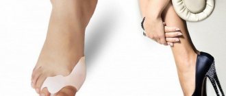
Silicone interdigital roller
Today, many people suffering from this disease know how to treat a chicken spur on the finger of the lower or upper limb. Although this problem is more often encountered by older people, a third of whom know firsthand what chicken spur is. An orthopedist can give the following recommendations on how to get rid of the disease in question:
- corrective bandage;
- inserts with orthopedic properties;
- silicone rollers (photo on the right);
- special shoes with orthopedic insoles.
If the growths appear due to inflammation of the ligaments, then you can try to get rid of them with the help of therapeutic massage, physiotherapy or herbal baths.
Surgical intervention involves the following steps:
- correction of the transverse arch of the foot;
- complete removal of the spur;
- removal of a small part of bone tissue and subsequent editing of the remaining;
- replacing problematic bone with an implant.
For each of these operations, which last 60 minutes, general anesthesia is provided. Today, surgery is radically different from the operations that were performed 20 years ago. Specialists replace damaged phalanges and bone fragments with implants made using innovative technologies. Their main advantage is that they all take root and there are no rejections. The sutures are removed a few days after the operation, and the patient gets back on his feet within a week. Surgeries under general anesthesia are contraindicated for people suffering from heart disease.
Positive results and treatment methods depend on the stage of development of the disease. If a person consults a doctor as soon as he feels the first symptoms, then surgical intervention will not be required.
What complications does it cause?
The very first and most unpleasant complication of a callus is acute pain when stepping on the foot and walking. Painful sensations interfere with normal movement, exercise, walking for a long time, leading an active lifestyle, and reduce performance. It is especially difficult for people whose work involves constant standing.
Due to pain, a person’s gait is disrupted, lameness appears, and the bones and joints of the foot receive additional stress. As a result, due to gait disturbances and an instinctive decrease in support on the sore spot, body weight is distributed unevenly across the foot. Subsequently, this can lead to deformation, overload and inflammatory processes in individual structures of the foot, and curvature of posture.
If the callus is not removed, the rod will continue to grow deeper, compressing blood vessels and nerve endings. This will lead to a deterioration in the supply of nutrients to the location of the callus, and will create favorable conditions for its further growth.
If an infection occurs, which often happens when trying to remove it at home, severe purulent inflammation in the form of an abscess or phlegmon can develop underneath it. Such a complication will require surgical intervention to open the purulent focus and the use of antibiotics.
The keratinized areas of skin on the feet, whether they are corns, a regular dry callus or a callus with a core, cannot be ignored. If left untreated for a long time, they will negatively affect the health of your feet.
