Children have fairly flexible ligaments and joints, but sometimes they have problems while playing. A dislocated arm in a child is a definite dysfunction of the joints. If the movement is awkward, if you pull the handle too hard, the bone will pop out of the elbow or shoulder joint. Unlike a sprain, children experience severe pain and difficulty moving their limbs. What to do in such cases? In such a situation, you should immediately immobilize the injured limb and call an ambulance.
Symptoms of dislocations
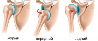
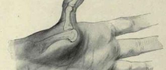
A dislocation of the wrist joint is the emergence of the articular part of the bone from its cavity, in which the articular capsule and ligaments are disrupted.
- When a joint dislocates, it changes shape.
- Motor functions are associated with severe sharp pain, they are limited or completely blocked.
- The area of the joint and proximal hand are swollen.
- Dorsal dislocations are characterized by a bulge on the back of the hand. If the median nerve is compressed, sensory impairment occurs.
Causes of illness and its symptoms
The shoulder can also be dislocated during vigorous play when a child falls. Often this nuisance appears during sports, especially those associated with lifting weights. The number of sprains increases in winter when there is a lot of ice outside.
A bone in a joint may be completely or partially dislocated. In addition to traumatic dislocations, congenital and pathological dislocations are observed. They may appear in the shoulder, elbow, forearm, hand or finger. If there is no skin damage, it is a closed dislocation, and if the skin is damaged, it is an open dislocation.
There are some signs that it is a sprain. After such an injury, the arm takes on an unnatural position, and bruising and swelling may be observed on the damaged part of the arm. Symptoms such as sharp and severe pain in the damaged part of the limb appear, which intensifies when palpating the arm or trying to move it. Your heart rate may also change. Blood circulation may be impaired and the lower arm may become numb.
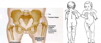
Injuries sustained during childbirth or as a result of careless treatment by parents or doctors are quite serious. Symptoms of such a dislocation may not be noticed immediately, because the child cannot independently determine the uncomfortable place for up to a year. If the child is very anxious, there is an unnatural position of the hands, some deformation and swelling, it is necessary to urgently consult a doctor.
A child under 3 years old has a very weak musculoskeletal system and ligaments. Subluxation can occur if a child suddenly raises their arm to avoid falling. Its characteristic symptom is a strong crunch in the hand, then the limb hangs lifelessly, and the child feels a sharp pain.
What should be done?
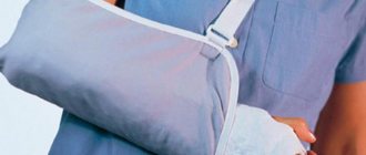
After an injury occurs, an adult should immobilize a limb and seek professional help at an emergency room or the nearest traumatology department.
If signs of dislocation are observed in a child, it is necessary:
- Fix the limb and tape it to the body;
- Apply cold to the site of dislocation;
- If you complain of pain, give painkillers.
It is forbidden to carry out treatment on your own, as there is a serious risk of worsening the situation.
How do hand injuries occur?
Symptoms of hand injuries depend primarily on the degree of damage. Contrary to popular belief, some patients do not feel significant pain even with arm fractures. Therefore, without professional skills and knowledge, the patient cannot visually determine what kind of injury was received.
Main signs of hand injury:
- pain at the site of injury;
- swelling;
- hematoma formation;
- limited mobility;
- tissue redness;
- local temperature increase.
Immediately after the injury, carefully examine the hand: there are no deep abrasions, cuts on the skin, or any visible bone fragments. Even if the integrity of the skin is not compromised, carry out antiseptic treatment before contacting a doctor. Use any antiseptic you have at home - hydrogen peroxide, chlorhexidine, diluted alcohol. Wipe or irrigate the injured area with an antiseptic solution to reduce the risk of secondary infection. Then apply a loose sterile bandage and go to see a traumatologist.
Many patients cannot accurately describe the mechanism of injury. If the pain intensifies even after first aid, the specialist may suspect the development of compartment syndrome, which is characterized by increased pressure in the fascial space. A common cause of the development of compartment syndrome is shrapnel injuries, which often lead to disruption of the nutrition of muscle tissue and the spread of the infectious process.
In most cases, hand injuries are not accompanied by long-term pain and a high risk of complications. The most dangerous are open injuries, which can be complicated by infection and occur with severe disruption of the blood supply (ischemia). In such cases, timely medical assistance is necessary. Only a qualified specialist can assess the degree of damage to the soft and hard tissues of the hand, identify hidden and obvious disturbances in blood flow and injuries to large or small vessels.
Fortunately, up to 70% of hand bruises are mild and do not require hospitalization or serious treatment. Most often, patients injure their fingers during sports training and solving everyday problems. With age, bone density decreases, so even gentle behavior or compression can cause a crack or fracture.
Hand injuries often occur in childhood. They arise as a result of active games during physical education. The problem with early diagnosis of trauma in children is that the child often cannot specifically describe his feelings and evaluate them. Even with cracks and fractures, some children do not complain of severe pain, and sometimes children cry for a long time even from a slight bruise, greatly frightening their parents. It is better to play it safe and still contact a specialist so that the doctor examines the site of the injury and, if necessary, prescribes additional diagnostics.
Treatment
A dislocation of the ulna in the wrist joint is reduced by a traumatologist. The manipulation is performed under anesthesia:
- The arm is bent at a right angle at the elbow joint;
- The traumatologist stretches the joint and presses on the bulging area;
- After restoring the correct position, the hand is bent (angle 40ᵒ);
- The hand is fixed from the elbow to the base of the fingers.
The hand remains in this position for 2 weeks, after which fixation is carried out in a neutral position. For joint instability, Kirschner wires are used. If the patient has an old dislocation, or closed reduction cannot be performed, a distraction device is applied to the hand. If the median nerve is compressed, surgery is required.
If we are talking about transscaphoid-perilunar dislocations, rehabilitation after surgery will require at least 3 months. After other types of wrist joint dislocations, patients recover faster – 1-1.5 months. Physiotherapy will help speed up the process. It is important to perform therapeutic exercises, begin motor movements with your fingers in accordance with the recommendations of the orthopedist
.
First aid for a child with a dislocated arm
If a child is suspected of having a fracture or dislocation, he should be examined by a doctor as quickly as possible, an exception may be “nanny’s elbow,” when parents are able to provide assistance on their own. Subluxations and sprains are usually treated at home. In order for the treatment of all injuries to be successful, rest is needed, since under load the damaged joint will heal slowly.
- When treating sprains and subluxations at home, the first step is to prevent swelling. It is advisable not to disturb the damaged joint. Ice packs should be applied for half an hour (ice is placed in a plastic bag, which is wrapped in a towel, or is in a special bubble). If the swelling does not decrease, then the interval between half-hour ice applications must be increased to fifteen minutes until the swelling begins to subside.
- Pain medication given to the affected person during this period may be helpful. Specific recommendations should be obtained from your doctor or by choosing over-the-counter medications. For children, Nurofen syrup or suppositories can be used as an analgesic and anti-inflammatory agent. This drug, in addition to the antipyretic effect, has a fairly pronounced analgesic effect and can be used for various pain syndromes. According to the official instructions, Nurofen syrup can be used from 3 months.
- After swelling has subsided, the subluxated or sprained hand is wrapped with an elastic bandage or a splint is applied to provide support and reduce pain. Before applying a bandage, it would be advisable to consult a doctor or qualified first aid specialist regarding bandaging techniques. The bandage should not be too tight so as not to disrupt proper blood circulation. The damaged hand can be secured in a sling. If you have a sprain, it is not recommended to wrap your shoulder with an elastic bandage; however, it should be immobilized using a sling.
- When treated at home, the pain caused by sprains and moderate subluxations decreases significantly within one or two days. After the pain goes away, the child will be able to slowly rotate the joint to increase its mobility several times a day.
- Warm baths are used to relax ligaments, muscles and facilitate movement. If there is pain during movements, the child should not move his arm. In this case, a doctor's consultation is required.
Experience in the treatment of congenital malformations of the hand
Congenital malformations and deformations of the hand are among the most complex and diverse. According to literature data, they range from 0.1 to 1.94 per 1000 newborns. It has now been established that congenital deformities of the limbs can be either hereditarily determined or arise as a result of the pathological action of exogenous factors on the developing embryo (embryopathies) or fetus (fetopathies). Therefore, it is possible for malformations of the hand to manifest both as an independent nosological form and in combination with other diseases (formation of syndromes). Often, developmental defects on the same hand are combined with each other, so surgical treatment of this pathology is very difficult.
The most common congenital anomalies of the hand that require surgical treatment can be grouped into the following clinical groups:
- Hypoplasia - includes all anomalies accompanied by varying degrees of underdevelopment of the anatomical structures of the hand - skeleton, muscles, blood vessels, tendons, articular-ligamentous apparatus: syndactyly, brachydactyly, camptodactyly, amniotic deformities, ectrodactyly, oligodactyly, hypoplasia of the thumb.
- Hyperplasia - combines all congenital anomalies accompanied by the presence of additional anatomical structures or their hypertrophy: polydactyly, hyperphalanxia, macrodactyly, gigantism, elephantiasis.
- Syndromes - all congenital anomalies of the hand of the hypo- and hyperplastic type are identified in combination with a complex of typical congenital defects in the development of other localizations of the body: arthromyodysplasia, acrocephalosyndactyly, oculodentodigital, orofacial digital, osteoonychodysplasia, as well as syndromes: Robin, Poland, Marfan, Charcot-Marie-Tooth, Maffucci et al.
Syndactyly is a congenital fusion of fingers with cutaneous or osteocutaneous tissue. Accounts for more than half of all hand development anomalies. Syndactyly occurs 2 times more often in boys. The III – IY fingers are most often connected. In 41% of cases there is a bilateral symmetrical lesion, in 35% - on the right, in 24% - on the left. It can be combined with any congenital anomaly of the hand, foot, or other organs.
Methods of surgery for syndactyly can be divided into 3 groups - phalangization with replacement of the skin defect:
- Local tissues;
- Local tissues and free skin autograft;
- Free skin autograft.
After surgery, after removing the corrective bandage, restorative treatment is recommended - massage, therapeutic exercises. As the child grows, in some cases the need for corrective operations arises.
Brachydactyly is a congenital underdevelopment of the hand, shortening or absence of one or more phalanges (brachyphalange), isolated or multiple shortening of the metacarpal bones (brachymetacarpy). Often accompanied by cutaneous or bone forms of syndactyly. Most often, fingers II – IY are subject to deformation. Often with brachydactyly, synphalangia and synostosis of the metacarpal bones are observed. In 40-50% of children, brachydactyly is accompanied by a similar deformation on the feet.
Treatment of brachydactyly is mainly surgical. In each case, an individual surgical plan is chosen. In order to prevent inhibition of hand growth, elimination of finger fusion is recommended at an early age. At the age of 8-10 years, lengthening of the shortest metacarpal bones and phalanges of formed fingers can be undertaken using external fixation devices.
Camptodactyly is a congenital flexion contracture of the finger. It occurs relatively rarely, with equal frequency in boys and girls. There is a flexion contracture at the level of the proximal interphalangeal joints of the Y finger, less often IY-Y and very rarely II-Y fingers. Typically a bilateral symmetrical lesion with a more pronounced pattern on the right or left. The anomaly is accompanied by underdevelopment of all structures: shortening of the skin with the absence or smoothing of skin folds, dysplasia of the tendon-ligamentous apparatus and blood vessels of the hand, underdevelopment of the head of the proximal phalanx. There is a noticeable limitation in the extension of the middle phalanges of the fingers; there is no differentiated flexion of the distal phalanges.
Treatment of camptodactyly is predominantly conservative. From the moment the disease is diagnosed, thermal procedures, massage with skin stretching and joint extension are recommended. In general, hand function does not suffer. In severe cases, surgical treatment is used: Z-shaped skin dissection, capsulotomy of the proximal joint, cutting off the superficial flexor legs, dosed distraction using an external fixation device. The study of long-term results indicates recurrence of the disease.
Clinodactyly is a congenital lateral deviation of the fingers. It is rare, but if the deviation is less than 10° from the normal axis, it is usually not fixed. Always bilateral, symmetrical, twice as likely to affect boys, and progresses as the hand grows.
According to the degree of lateral deviation of the finger, they are distinguished: mild - up to 10°, moderate - up to 20°, severe - more than 20°. In mild cases, hand function does not suffer. With average, there is a limitation in subtle professional activities. With severe deviation of the fingers in ulnar deviation, tip and pinch grips of small objects are sharply limited, and the strength and shape of other types of grip are reduced. This pathology is sometimes accompanied by similar developmental anomalies on the feet, as well as syndactyly and brachydactyly. The deformation increases with the beginning of active growth.
Conservative treatment includes thermal procedures, massage with skin redressing and stretching. Corrective bandages are used as a preparatory stage for surgery.
Surgical correction begins at 7-8 years of age. The choice of surgical method depends on the nature of the anomaly. As the hand grows, additional correction of the deformity is sometimes required.
Ectrodactyly is a congenital cleft hand. It can be unilateral, symmetrical bilateral, in combination with a similar deformation on the feet, as well as with other anomalies. In appearance, the brush resembles a crayfish claw or a double brush. As children grow, they actively adapt the shape of the hand to its function. For wide clefts that limit hand function, surgical treatment is indicated. Considering the degree of impairment of blood flow to the hand, the reconstruction technique is individual in each case. In the postoperative period, special attention is paid to the biomechanical restructuring of hand function.
Oligodactyly is a congenital defect of one or more fingers. Depending on the number of preserved fingers, they are called tetradactyly or tridactyly. It can be combined with hypoplasia of the first finger, underdevelopment or absence of metacarpal bones, syndactyly and other developmental anomalies. It is relatively rare. If the function of the remaining fingers is compensated and the patient is satisfied, surgical treatment is not performed. Surgical treatment is possible to improve grasping function or eliminate associated abnormalities.
Hemimelia is a congenital aplasia of parts of the hand. With radial aplasia, the radial half of the hand is absent, and with ulnar aplasia, the ulnar half is absent. Often combined with syndactyly of the remaining fingers and other birth defects. Treatment is carried out according to an individual plan: syndactyly is eliminated, the function of opposition is restored using muscle-tendon transpositions.
Ectodactyly is complete or partial aplasia of the bones of the wrist and metacarpus; hypoplastic shortened fingers extend directly from the end of the forearm. It is extremely rare. Surgical treatment is not performed until the growth and formation of the skeleton is complete. In the future, an individual approach to surgical treatment or prosthetics and social rehabilitation is possible.
Hypoplasia of the first finger is the most common congenital anomaly. Among all congenital anomalies of the hand, the first finger accounts for more than 16%. Often combined with other hand developmental anomalies and congenital syndromes. Almost all congenital deformities of the first finger are characterized by varying degrees of underdevelopment of all finger structures: skeleton, thenar muscles, tendon-ligamentous apparatus, vascular system and innervation.
Aplasia of the first finger is characterized by its complete defect. The brush has a four-fingered shape. The grip is carried out with the lateral surface of the fingers. In adults, during the process of function, a semblance of opposition of the second to third finger is formed.
Microdactyly of the first finger is underdevelopment of the first finger. May vary in degree of underdevelopment. In some cases, the elements of the first finger can be well developed, the flexor tendons are present, and the first metacarpal bone and phalanges of the finger are preserved. However, the thenar muscles are completely absent. Such a finger is unstable and incapable of grasping function. There are methods for surgical correction of this pathology. Operations are most often carried out in several stages. Our clinic uses the Blaut operation as modified by Godunova. Stage 1, starting from 3-5 months of age, plastic surgery is performed with local tissues of the base of the first finger, which further helps to improve blood circulation and the development of the rudiment. Stage 2 involves reconstructive surgery. From an incision along the dorsal surface of the hand along the radial edge of the second metacarpal bone in the sagittal plane, a longitudinal detachment of a fragment from the second metacarpal bone is performed, maintaining its connection at the base. The distal end of the fragment is connected to the rudiment.
Amniotic deformities of the hand - occur as a result of intrauterine constrictions at the level of various segments of the upper limb and are observed in 1:2000-2500 newborns. According to the severity of the hand lesion, amniotic constrictions are divided into complete ones, when as a result of the constriction, intrauterine spontaneous amputation of various segments of the hand, fingers, metacarpal bones, and wrist occurs. Incomplete, when circular, palmar or dorsal strangulations of varying depth remain - superficial or deep. With complete amniotic constrictions, the remaining segments of the hand are rounded stumps covered with thin skin. Superficial strangulations are characterized by atrophy of fatty tissue at the site of the constriction, without circulatory impairment, with preservation of sensitivity distal to the strangulation. Deep strangulations are characterized by adhesion of the skin to the bone along the entire circumference or part thereof. This disrupts lymph and blood circulation. As the hand grows, trophic disturbances intensify, ulceration and necrosis of the amniotic stump occur.
Treatment of amniotic bands is surgical.
Ectrophalangia is a congenital splitting of the first finger at the level of the proximal or distal phalanges. It is often combined with syndactyly and triphalangy. For this defect, the Bilhout operation is performed, which consists of resection of the inner halves of the phalanges facing each other and joining the two outer halves into one finger, followed by fixation. When combined with triphalangia of the first finger, it is subsequently politicized.
Polydactyly is a congenital increase in the number of fingers from one extra finger to multiple fingers. It can be unilateral, bilateral, and also in combination with polydactyly of the feet, syndactyly and other anomalies. An increase in the number of individual phalanges is called polyphalanx. The most common types of polydactyly are radial polydactyly—extra I-II fingers and ulnar polydactyly—additional to the Y finger. They may have normal development of all structures or their underdevelopment in the form of a rudiment hanging on a skin stalk. The frequency of occurrence is from 1:3300 to 1:630. Treatment is surgical. When choosing tactics and method of surgery, it is necessary to take radiography to establish the nature of bone changes, various options for connection with the main finger, and examine the functions of the accessory and main fingers.
Macrodactyly is congenital partial gigantism. There are 2 forms: progressive and stable. In one case, only soft tissues are affected, in the other, both bone and soft tissues are affected. Most often, fingers I-II and II-III are affected, less often the forearm and ulnar half of the hand are involved in the process. The most common method used is amputation of hypertrophied fingers.
Triphalangy of the first finger - characterized by the fact that the hand contains 5 triphalangeal fingers lying in the same plane, with the swimming membrane located at the same level and the absence of thenar muscles. The basic functions of the first finger and, above all, the opposition are completely impaired. Surgical treatment is carried out from 2-3 years of age
From 1998 to 2004, 146 children with hand defects were operated on in the traumatology and orthopedic department. 200 surgical interventions were performed. The choice of surgical tactics depended on a detailed clarification of the isolated or combined nature of the anomalies.
The clinic performed 65 surgical interventions for syndactyly. Elimination of this defect was carried out in stages, from the age of 1 to 5 years. During phalangization, wavy incisions were used to cross the dorsal transverse ligament between the heads of the metacarpal bones. Plastic surgery of the interdigital space was performed using split skin autograft, local tissues, and a combination thereof. In the postoperative period, the hand was fixed with a simulated plaster splint for 4 weeks. This made it possible to avoid scar contractures of the fingers. Scar contractures developed in 3 patients due to non-compliance with recommendations for performing rehabilitation measures at the outpatient stage of treatment.
For congenital clefting of the first finger of the hand at the level of the proximal and distal phalanges (ectrophalanges) in children aged 7 months to 4 years, 8 Bilhout operations were performed.
The next group is represented by 67 surgical interventions for polydactyly, polyphalanx, when one of the fingers or one of the phalanges is hypoplastic, as well as in the presence of an additional ray. The scope and stages of correction included amputation of accessory fingers, isolation of accessory rays, including 9 with tendon transposition. Most children are operated on before the age of 1 year.
Removal of amniotic constrictions was performed in 13 children aged 10-12 months by excision of the constriction itself, followed by skin grafting according to Limberg.
For microdactyly of the first finger and aplasia of the first metacarpal bone, we consider the stabilization of the first finger according to Blaut-Godunova to be the operation of choice, which was performed in 3 patients. It was possible to create a strong bone block in all children.
Patients with camptodactyly underwent application of an external fixation device, followed by distraction and elimination of deformity in 5 cases. Children were operated on in adolescence. The immediate and long-term results in all patients are satisfactory.
For stenosing ligamentitis, 27 operations were performed - ligamentotomy of the annular ligament. The function of 1 finger was completely restored in all patients.
Thus, complex staged reconstructive and plastic surgeries for malformations of the hand allow, first of all, to restore the functional state of the underdeveloped hand, and secondly, to achieve a cosmetic result.
“It can be said without exaggerating that the projectiles, remarkable in their mechanism, invented by the human mind, are only imitations of the wonderful mechanism of the hand, far removed from it in convenience, simplicity and elegance of form.” N.I. Pirogov.
Diagnostics
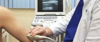
During a clinical examination, the doctor makes a preliminary diagnosis. It evaluates the appearance of the limb and the function of the affected arm compared to the healthy arm. If necessary, additional examinations may be prescribed.
In case of severe injuries, impaired joint stability, or unnatural position of the limb, the doctor prescribes an additional examination - radiography. It can be used to determine bone displacement, fragment separations, and fractures. To clarify the nature of the injury, x-rays are taken in several projections. When a child has a dislocated shoulder joint, three projections are needed.
What to do after the doctor sets the joint
Many parents wonder what the symptoms and treatment of a dislocation in a child are. Unfortunately, setting the bone back does not mean that the baby is completely free of the problem. When a dislocation occurs, there is a rupture and severe damage to the joint capsule. This suggests that both time and conditions will be needed for complete restoration of the limb and tissue fusion.
In the first 2-3 days after the specialist sets the joint, cold must be applied to the damaged area from time to time. The procedures are carried out for 15 minutes up to 5 times a day. You will also need to remain immobile.
After eliminating the swelling a couple of days later, the sore spot no longer needs to be cooled, but, on the contrary, it needs to be warmed. This will increase blood flow in the tissues and speed up healing. To warm up the joint, simply apply a cloth ironed or heated in a microwave oven.
For about 2-3 weeks after the injury, the child must be careful in his movements. This does not mean that bed rest is necessary. Don't just play too active games.
Medical treatment
If a child has been diagnosed with a dislocated arm, a traumatologist will tell you what to do. First, he prescribes desalting drugs, and then realigns the joint. In this case, anesthesia is used to completely relax the arm muscles. After the procedure, the arm is fixed in a cast, which the child will wear for about two weeks. After removing the cast, the doctor prescribes a rehabilitation course to restore hand mobility. For this, exercise therapy and physiotherapy are used to normalize blood circulation and strengthen joints.
It is impossible to self-medicate when a limb is dislocated, as it can provoke a fracture and other negative consequences.
If you do not go to the hospital immediately after your child is injured, further treatment will be longer and more painful.
If the child was taken to a medical facility several weeks after the injury, treatment is carried out according to the following scheme:
- Sprain;
- Reduction of the joint by surgical method;
- Fixation of the joint with knitting needles;
- Plaster application;
- Long-term physiotherapeutic procedures after removal of the cast.
Rehabilitation
After considering what to do if your child has a sprained arm, it is important to begin recovery. The rehabilitation period after injuries is very important in childhood. This is due to the fact that prolonged immobilization provokes the rapid formation of contractures and joint stiffness. Returning to sports too early aggravates the severity of the injury and provokes severe extensibility of the ligamentous apparatus.
During recovery, the following methods are used:
- physical therapy;
- physiotherapy (electrophoresis, magnetotherapy, heat treatment);
- massage;
- aquatherapy
The rehabilitation period can last from 1 to 6 months. Active movements of the healthy limb begin during immobilization. This prevents the formation of contractures and improves the muscle corset. Massage plays an important role in babies. Initially, the first sessions are conducted by a specialist who teaches the parents the techniques. Afterwards, parents massage the sore limb themselves. The most commonly used techniques are stroking and rubbing.
First aid for bruises and other injuries to the hand
Immediately after the injury, the patient must make sure that he received a minor bruise and that no bone remains are visible at the site of the injury. The wound is washed with warm water and soap, gently dried and treated with antiseptic. Then you need to apply dry ice for 5-10 minutes. After this time, the hand must be examined again, finger activity and range of motion checked.
In case of damage to the hand with a violation of the integrity of the skin, it is necessary to apply a bandage of a sterile bandage. When applied correctly, the bandage completely covers the damaged tissue, does not hinder movement and does not cause any pain. Make sure that the bandage does not squeeze the skin. If the tissues begin to turn blue and sensitivity decreases, this indicates that the bandage urgently needs to be loosened or replaced.
Dry ice must be applied every hour for 5-10 minutes. Usually this is enough to reduce pain and prevent the appearance of a hematoma. Intermittent cold therapy is an effective treatment for minor bruises and injuries. More serious injuries require specialist consultation and a comprehensive examination.
The main tasks of first aid for hand injuries:
- immobilization of the limb to prevent the development of complications;
- stopping bleeding from a wound;
- antiseptic treatment for prophylactic purposes;
- reduction of swelling, pain, signs of an inflammatory reaction.
Patients are not always able to provide first aid for hand injuries, especially if the wound is bleeding and there is severe pain. If you cannot adequately assess the complexity of the situation and your condition, it is recommended to immediately contact medical professionals. They will carry out antiseptic treatment themselves, relieve pain and, if necessary, use immobilization.
Prevention
In order not to wonder what the symptoms and treatment of sprains in children are, it is important to adhere to preventive measures. The main measure is to avoid injury. Parents should listen to all the pediatrician's recommendations for caring for their babies. Do not grab it sharply or pull it. It is forbidden to lift the baby by the fingers, wrists and hands without fixing the forearm. Also, do not wear clothes with narrow sleeves or quickly dress and undress.
When older children play sports, sore areas are taped. This will help avoid re-dislocation. It is forbidden to neglect the recovery period - this is the best prevention of recurrent dislocation.
Every parent knows that babies are characterized by incredible mobility, hyperactivity, and an amazing ability to jump. Very often such activity results in injuries. Therefore, it is very important to adhere to preventive measures and monitor children. If a dislocation occurs, then competent first aid is important. Reduction is carried out only by the attending physician.