- Home /
- Branches /
- X-ray /
- X-ray of the calcaneus
X-ray of the heel bone is an indispensable and accessible research method and is widely used by traumatologists and orthopedists to diagnose heel bone pathology.
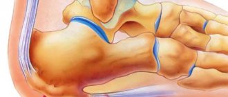
The heel bone is the largest bone of the foot and, due to its anatomical features, it bears most of the load, which provokes injury. An X-ray of the heel bone is indispensable in diagnosing and assessing the effectiveness of treatment of heel spurs (plantar fasciitis); in addition, the study is carried out to determine neoplasms and injuries of the heel bone. Advantages:
- Non-invasive and accurate diagnostics
- Minimum radiation dose with high image quality, which is ensured by our X-ray system
- Possibility of recording images onto the patient’s electronic media (digital equipment)
X-ray of the calcaneus
Posted at 14:47h in Services by doctor
X-ray of the calcaneus
is the most accessible, informative and relatively safe way to study the bone and soft tissues of the foot. Such a study is often used in traumatology, surgery, orthopedics and helps to identify both mechanical damage to the heel and various congenital or acquired pathologies in this area.
- non-invasive and accurate;
- minimum radiation dose with high image quality.
When using digital X-ray equipment, images can be enlarged for a more detailed examination, and also recorded on electronic media.
Treatment
The nature of treatment depends on the stage of the disease being diagnosed. If the disease is in the first or second stage, then the question arises of surgery, which can help in the fight against cancer. Radiation therapy and chemotherapy will be auxiliary at this stage.
If the disease has progressed to the third or fourth stage, then the operation will no longer be effective. The main thing here will be to prevent the spread of metastases to other organs and slow down the growth of the malignant tumor. Therefore, the main treatment options will be radiation therapy and chemotherapy. The patient is also given strong painkillers to reduce pain.
What is a calcaneal x-ray?
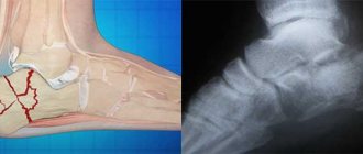
Of all the bones of the foot, the heel is the largest. When walking, running, jumping, a large load is placed on it, which is associated with increased injuries. An X-ray examination allows you to understand how serious the heel injury is, to see whether there is a fracture and what type it is (with or without displacement).
Another common foot problem is heel spurs. It occurs due to wearing the wrong shoes, the development of arthritis or arthrosis, but most often due to formed flat feet. If this disease is not treated, it can cause complications such as heel bursitis (inflammation of the mucous bursae) and fasciitis (inflammation of the muscle tissue of the foot). The main method for diagnosing the listed pathologies is x-ray, which today can be done in almost every public or private clinic.
Development of the disease
With further development of the disease, the following picture is observed:
- Loss of functionality of nearby muscles;
- Loss of joint mobility;
- Inability to bend and straighten the leg to the end;
- Limiting movements and staying in one position to reduce pain;
- The appearance of weakness, loss of appetite, fever and weight loss;
- Further metastasis throughout the body through the bloodstream and lymph, the development of new cancer foci.
Leg cancer
Indications for radiography of the heel
- Foot injury in the heel area. The causes are varied - work injury, sports injury, fracture due to an accident, and so on.
- Severe pain in the heel area. It appears mainly in the morning or after a nap, when a person, after some time of rest, gets up and rests on his heel.
- Pain in the heel area when walking.
- Changed gait (shift of the center of gravity to the toes).
- Suspicion of the development of joint diseases - arthritis, arthrosis.
- Suspicion of the presence of inflammatory diseases of bone tissue – osteomyelitis, etc.
- Suspicion of malignant and benign osteoarticular neoplasms.
X-rays are prescribed as a primary diagnostic procedure when the task is to identify the correct diagnosis and prescribe treatment. It is also carried out to monitor the condition of the heel bone, which has previously been fractured - doctors look at how correctly and quickly the bone tissue heals. Using X-rays, you can track the dynamics of various diseases of the heel bone and evaluate the effectiveness of the treatment.
What does an x-ray of the heel bone show?
Using x-rays, the integrity of the heel bone is first assessed. Then its shape and location in relation to other bones of the foot are analyzed. When identifying heel fractures, their location and direction are studied. If the fracture is comminuted, it is important to evaluate how the fragments are located and whether there is any displacement.
The image can clearly visualize neoplasms on the heel bone. In this case, their size and location are assessed, after which a conclusion is made about the possible nature of the deviation.
Diagnostics
Inspection
If an oncologist suspects heel cancer, he will examine the limb and find out if there is swelling and redness in the heel area. The doctor palpates the sore area and looks to see if the patient has problems with motor activity.
X-ray
Using radiography, the area of the disease is determined. If it is malignant, then in the picture it will be light with unclear outlines.
Scintigraphy
This modern method is a bone scan. The image will show the condition of the bone tissue.
Biopsy
This is a procedure for collecting cells for analysis, which will show whether the collected tissue contains cancer cells.
MRI and CT
These procedures will allow you to see whether there are metastases in the body, what their location, size, structure are, and what is happening in the tissues adjacent to them.
Histology
This analysis is performed in order to obtain the most accurate information about the development of pathology.
Interpretation of results
Immediately after the shooting, the images are developed, and the radiologist begins decoding. An x-ray allows you to find out the cause of pain and determine the nature of the pathological process. To accurately interpret the result, the doctor can compare images of healthy and diseased heels.
If plantar fasciitis (the so-called heel spur) is detected, the specialist gives a detailed description of the image, where he notes the presence of an osteophyte, indicating its length and location. Typically, osteophytes are visible in the image in the projection of the upper edge of the posterior tuberosity of the talus.
Symptoms of a calcaneal fracture
The first thing a patient complains about when a calcaneal bone is fractured is pain. It can occur during movement or persist at rest. Upon examination, swelling may be detected that extends to the Achilles tendon. Also among the symptoms of a fracture are: flattening and widening of the heel, hematoma in the center of the sole.
When palpating the site of injury, the patient feels sharp pain. While standing, he cannot support his injured foot.
Symptoms
Symptoms of heel cancer can be divided into two groups.
Nonspecific symptoms
Occurs in the initial stages of the disease. They may also be present in other non-cancerous diseases, so diagnosing foot cancer at an early stage is not so easy. Let's list these symptoms:
- Painful – when walking, putting pressure on the heel, a person experiences pain in it, which disappears at rest;
- Tissue swelling near the bone;
- Increase in size of the heel due to tumor growth;
- Fixing the foot and restricting movement does not relieve pain.
Specific symptoms
These are typical symptoms of heel cancer. They are as follows:
- Expansion of swelling due to the spread of the disease to areas close to it;
- Severe pain at night, poor sleep;
- The density and immobility of the neoplasm attached to adjacent tissues when palpated;
- Redness of the affected areas of the heel;
- Increased temperature in the heel area.
Features of the study
X-rays of the heel bone can be performed in two ways: under load and without it. In the first case, the procedure is performed in a standing position, with the patient resting on his heel. In the second case, the patient lies on his back, his legs are bent at the knees, and his feet are straight.
During the study, X-rays, passing precisely and in micro doses through an area of the human body, fall on a highly sensitive film with a special composition. The result is an image from which you can assess the condition of the bone and connective tissue of the foot and ankle joint.
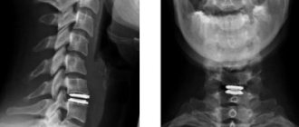
An X-ray examination does not require any preparation and can be scheduled either routinely or urgently. Also, as prescribed by the doctor, an x-ray of the heel bone may be required in two projections - lateral and axial.
Contraindications for the X-ray procedure are:
- pregnancy and breastfeeding;
- circulatory disorders and bleeding;
- a critical condition of the patient, in which even minimal effects on the body can become fatal.
How is the examination carried out?
X-ray photos of the heel bone can be taken in various projections.
Direct radiography
To take a frontal view, the patient needs to lie on his back on the X-ray machine table. Legs bend at the knees and feet rest on the table.
X-ray with stress
The load on the heel bone occurs when a person stands. Accordingly, to perform weight-bearing x-rays, the patient must stand and place weight on the foot.
Using X-rays for heel spurs
A heel spur or plantar fasciitis is a pathological growth of bone tissue in part of the heel bone. This provokes discomfort, pain, difficulty in choosing shoes, trauma to the affected area and, as a consequence, further growth of bone tissue and an even greater aggravation of the problem.
X-ray therapy of the heel bone is one of the methods of treating this disease of the heel bone - under the influence of targeted irradiation, pathologically overgrown tissues are destroyed, the growth decreases. Also, due to the blockade of nerve endings, as a result of X-ray treatment, pain and inflammation are significantly reduced.
Preparing for the study and carrying out the procedure
X-ray of the foot does not require special preparation. The patient should remove clothing (including shoes), jewelry, or any metal objects that may interfere with the image.
A developing fetus is more sensitive to the effects of radiation than an adult and therefore is at greater risk of harm from X-ray examinations, so if the patient is pregnant, the doctor should be informed.
Although the procedure may take about 15 minutes or longer, the actual radiation exposure is usually less than a second.
The patient enters a special room, which will likely contain a table and a large X-ray machine hanging from the ceiling.
Parents can usually come with the child to ensure peace of mind. Accompanying persons will need to wear a special protective apron to protect certain parts of the body.
The x-ray technician positions the patient both on and off the table to achieve the required position. Then he goes behind the wall, or into the next room to control the machine.
The three x-rays are usually taken from the front, side and angle, so the technician will return to reposition the leg for each new x-ray. Sometimes doctors ask for x-rays of the opposite leg for comparison.
Getting Results
The patient will not feel anything during the x-ray. The x-ray room may be cool due to the air conditioning used to keep the equipment operating properly.
The positions required for x-rays may be uncomfortable, but only need to be held for a few seconds.
If the patient is injured and cannot remain in the required position, the technician may find another position that will be easier for him.
A radiologist is a doctor specially trained in interpreting x-ray images. He will be the one to review the patient's x-rays.
Best materials of the month
- Coronaviruses: SARS-CoV-2 (COVID-19)
- Antibiotics for the prevention and treatment of COVID-19: how effective are they?
- The most common "office" diseases
- Does vodka kill coronavirus?
- How to stay alive on our roads?
After evaluating the results, the radiologist will send a report to the attending physician, who will discuss with the patient what the images show and explain what it means.
In emergency situations, X-ray results can be available quickly. And usually the results are ready in 1-2 days. In most cases, results can be given directly to the patient or family during the examination.