This information explains how a lumbar puncture procedure is performed at Memorial Sloan Kettering (MSK).
A lumbar puncture is a procedure performed to obtain a sample of cerebrospinal fluid (CSF). Cerebrospinal fluid surrounds your brain and spinal cord. Lumbar puncture (LP) is also called a lumbar puncture.
Your healthcare provider may need to take a sample of your cerebrospinal fluid to check for the following:
- development of infection, such as meningitis;
- bleeding;
- spread of cancer to tissues surrounding the brain or spinal cord.
A healthcare professional may also perform a lumbar puncture to administer certain types of medications directly into the area around the spinal cord. These may be the following medications:
- anesthesia (medicine that blocks pain);
- anti-cancer drugs (such as chemotherapy drugs);
- antibiotics.
to come back to the beginning
Indications for lumbar puncture of the brain
Changes in the composition of brain fluid are observed in various diseases of the brain and spinal cord. In the following cases, cerebrospinal fluid examination is an important diagnostic method:
- Inflammation of the brain and meninges (encephalitis and meningitis) - in these cases, the causative agents of the disease can be confirmed;
- Multiple sclerosis - In this inflammatory disease, which is caused by an overreaction of the immune system, certain proteins (proteins) and inflammatory cells multiply in the cerebrospinal fluid;
- Cancer of the meninges, such as lymphoma;
- Hemorrhages in areas adjacent to the cerebral fluid, primarily subarachnoid hemorrhages.
Contraindications
Lumbar puncture is contraindicated if there is a threat or signs of axial displacement of the brain in the presence of an intracranial space-occupying process of various etiologies. The absence of congestive processes in the fundus is not a sign that allows a lumbar puncture. In such cases, one should rely on data from magnetic resonance and computed tomography scans.
Another contraindication is the occlusive form of hydrocephalus; pathology of the spinal canal and spinal cord with impaired cerebrospinal fluid circulation; the presence of infection in the lumbar region, including bones, subcutaneous tissues and epidural space, as well as skin; long-term use of anticoagulants, the presence of hemorrhagic diathesis with severe pathology of the blood coagulation system.
You should know that cytostatics also affect the blood coagulation system. In the case of neuroinfection, contraindications lose their force, since determining the pathogen and sensitivity to antibiotics gives a prognosis for the patient’s life.
How is a lumbar spinal puncture performed?
The brain and spinal cord are surrounded by three layers: the outside is the dura mater, the inside is the pia mater, and between them there is a narrow gap, the so-called subarachnoid space, filled with brain fluid, cerebrospinal fluid. It is this fluid that is taken for analysis during the lumbar puncture procedure.
Performing a cerebrospinal fluid puncture involves positioning the patient with an arched back, so that the chin is pulled towards the chest. In this position, the processes of the vertebrae open and access to the intervertebral space becomes possible, through which the material is collected. As a rule, the space between the third and fourth or fourth and fifth lumbar vertebrae is punctured. Very carefully, the doctor inserts a thin puncture needle between the vertebral bodies into the spinal canal, and the brain fluid begins to flow out on its own.
After performing a lumbar puncture of the spinal cord, the first glance at the obtained material gives some results. In a healthy person, cerebrospinal fluid is colorless and transparent. The reddish color of the cerebrospinal fluid indicates fresh bleeding, yellow indicates old bleeding, and with inflammation, the cerebrospinal fluid may be cloudy. In the laboratory of the Nordwest clinic, a sample taken during a diagnostic puncture of lumbar (cerebrospinal) fluid is studied in more detail, and the following is examined:
- quantity;
- color;
- transparency;
- protein;
- Pandey reactions;
- Nonne-Apelt reactions;
- cytosis (number of cells);
- number of neutrophils;
- lymphocytes;
- monocytes;
- eosinophils;
- ependymocytes;
- red blood cells
Depending on the patient’s health condition, the procedure is performed on an outpatient basis or in an inpatient department where a lumbar (spinal) puncture is performed.
Diagnostic lumbar puncture: indications, contraindications, technique
A clinician who is familiar with lumbar puncture techniques can play a critical role over other specialists in diagnosing conditions such as meningitis, subarachnoid hemorrhage, and Julian-Barre syndrome.
Indications for lumbar puncture
- Meningitis : bacterial, viral, fungal or tuberculous infection of the central nervous system Viral meningitis is an inflammation of the pia mater and arachnoid membrane of the brain, which is a clinical manifestation of infection of the central nervous system. Lumbar puncture is the most important procedure in diagnosing viral meningitis. A high cerebrospinal fluid (CSF) white blood cell count (especially neutrophils), high protein levels, and low glucose levels are grounds for suspicion of bacterial meningitis, although some viral pathogens can produce similar findings. CT and MRI of the brain with contrast help identify intracranial pathology, such as intracranial abscess or subdural empyema.
- Typical CSF changes in patients with meningococcal meningitis include: Increased open pressure (above 18 cmH2O)
- White blood cell count between 10 and 10,000 cells/µl, predominantly neutrophils
- Decreased glucose concentration (below 0.45 g/l)
- Increased protein concentration (above 0.45 g/l)
- Multiple sclerosis is a chronic demyelinating disease. The course of the disease is usually characterized by relapses that develop after remission. Multiple sclerosis is difficult to diagnose, so the use of MRI of the brain, lumbar puncture and induced potential testing (visual and sensory) should be considered. After lumbar puncture, CSF is analyzed by electrophoresis to detect oligoclonal IgG bands
- During the active phase of Julian-Barré syndrome, characteristic CSF changes include albuminocytological dissociation, which manifests itself as an increase in CSF protein levels (above 0.55 g/L) without an increase in the number of leukocytes. This increase is thought to reflect the spread of inflammation along the nerve fibers. MRI and CT scan of the spinal cord help rule out other diseases.
Contraindications to lumbar puncture
- Increased intracranial pressure. In patients, this may manifest as: Severe headache
- Blurred vision
- Vomiting
- Decreased level of consciousness
- Papilledema Papilledema develops secondary to increased intracranial pressure. Many symptoms in a patient with papilledema develop secondary to increased intracranial pressure, such as headache, visual disturbances, nausea and vomiting. Performing a lumbar puncture on a patient with increased intracranial pressure may cause herniation (incarceration) of the brain
- There is a high risk of bleeding after lumbar puncture due to coagulopathy. You should minimize this risk by correcting any tendency for any bleeding to occur if possible.
- Cauda equina syndrome can be caused by central disc prolapse, tumor, abscess/tuberculosis, hematoma, or trauma. Symptoms include urinary incontinence or retention (may be painless), fecal incontinence, bilateral leg weakness, and pain. Signs may include bilateral decreased strength and sensation, decreased peripheral (lumbar) sensation, decreased anal tone, and bilateral absence of foot reflexes. The disease is diagnosed by performing an emergency MRI of the spinal cord. Patients with cauda equina syndrome require immediate orthopedic consultation and neurosurgery. The disease is not diagnosed by lumbar puncture.
You should not perform a lumbar puncture on patients with increased intracranial pressure, as this can cause herniation (pinching) of the brain and be fatal. Performing a lumbar puncture in these patients reduces pressure by dumping CSF into the puncture area, which may leak. As CSF pressure decreases, CSF and brain tissue may shift into the area of low pressure (towards the puncture), leading to both transtentorial and uncal herniation (hooking out) with partial acute neurological symptoms. Strangulation can lead to coma and death. Careful selection of patients suitable for lumbar puncture and the use of CT scans of the brain reduce the risk of brain herniation during lumbar puncture. At a minimum, all patients referred for lumbar puncture should undergo fundoscopy to examine the optic disc and rule out papilledema.
Coagulopathy
As with other invasive procedures, the presence of coagulopathy is a contraindication to performing a lumbar puncture. This includes patients receiving heparin, warfarin, or those with coagulation defects such as disseminated intravascular coagulation, thrombocytopenia, or hemophilia. When coagulation abnormalities are corrected, lumbar puncture is not a contraindication.
You can adjust your INR with vitamin K, fresh frozen plasma, or prothrombin complex concentrate. Patients with hemophilia require clotting factor replacement and patients with thrombocytopenia require platelet transfusions to safely perform a lumbar puncture.
Anatomy
You must review the basic anatomy of the spine, spinal canal, and meninges before performing the procedure.
Rice. Figure 1 shows a simplified cross-section of the spine along the midline with the structures that the needle traverses as it moves from the skin (superficial) to the subarachnoid space (deep).
These structures include:
- skin
- Superficial fascia and adipose tissue
- Supraspinal ligament
- Intraspinal ligament
- Ligamentum flavum
- Dura mater
- Arachnoid membrane
- Subarachnoid space (containing CSF).
Lumbar puncture procedure
Consent to the procedure
You should inform the patient and relatives or caregivers about the planned procedure and, if possible, sign an informed consent and place it in the medical record.
You should always:
- Explain why a lumbar puncture is prescribed and ensure that the patient and relatives understand this
- Explain the risks of the procedure and
- Possible complications.
Risks and complications
- Bleeding : Minor bleeding is common, but the formation of an epidural or spinal hematoma can cause neurological deficits. If this occurs, you should immediately seek advice from a neurologist
- Headache : Almost 30% of patients develop a headache after a lumbar puncture. It usually appears between 48 and 72 hours after the procedure and may last for two weeks with subsequent resolution. Patients describe constant, tiresome bilateral pain, predominantly in the frontal rather than occipital region. The most characteristic symptom is its positional orthostatic aggravation, that is, the headache increases in a standing position, while it may not be present in a lying position.
- The pain is thought to be caused by ongoing CSF leakage from the puncture site and intracranial hypotension. You can prevent headaches to a certain extent by using a finer needle than usual (22G) for puncture, placing the bevel of the needle parallel to the fibers of the dura mater
- If headache occurs during a lumbar puncture, you should give the patient simple analgesia and intravenous rehydration, and antiemetics if the headache is accompanied by nausea. Bed rest may delay the onset of headaches, but research does not show that it can prevent headaches from occurring. For severe or prolonged headaches, ask your anesthesiologist for a special patch through which a small amount of your own venous blood is injected into the adjacent epidural space to “clog” the puncture hole. Intravenous caffeine is an option for treating headache after lumbar puncture, but evidence for the effectiveness of this approach is minimal.
Preparation
Prepare all equipment on a sterile cart with the assistance of an assistant.
Equipment includes:
- Sterile gloves, gown and eye protection
- Porous sterile wipe
- Antiseptic solution for skin
- Tampons
- 10 ml syringe
- 25G needle (orange)
- 21G needle (green)
- 1% lidocaine for local anesthesia
- Spinal needle (ideally atraumatic)
- Pressure gauge tube with three-way stopcock
- Three clean sample tubes (or four if you suspect subarachnoid bleeding)
- One blood vial with fluoride oxalate for glucose (gray top)
- Occlusive dressing.
Patient position
Correct positioning of the patient is the key to success. The patient is placed on his left side, with a horizontally bent spine and knees bent and pulled up to the chin. The patient's back should be positioned directly along the edge of the bed.
Risks and side effects
The following are noted as consequences and possible complications after lumbar puncture:
- bleeding and hemorrhage;
- infections and inflammations;
- circulatory disorders and fainting;
- passing numbness and paresis.
In patients suffering from epilepsy or migraines, the procedure may provoke an attack. In addition, the risks of such a study include the so-called liquor hypotension syndrome, in which the patient experiences headaches, neck stiffness, tinnitus, nausea and photosensitivity.
Common side effects are:
- headache;
- back pain;
- nausea, vomiting;
- pain in the injection area.
After the procedure
In the hospital
You may be asked to lie on your back for 30 minutes.
At home
You can remove the bandage the day after the procedure.
Side effects
After a lumbar puncture, you may have a headache. This happens because the hole from the needle insertion may not close immediately. If it remains open, it can leak cerebrospinal fluid, causing headaches.
In most cases, the hole closes on its own and the headache goes away within 1–2 days. For headaches, you can take an over-the-counter pain reliever such as acetaminophen (Tylenol®) or ibuprofen (Advil®). You may also feel better if you lie down.
Caffeine can also relieve headaches. One 8-ounce (240 ml) cup of coffee contains about 150 mg of caffeine. To get rid of headaches, you can try drinking 1-2 cups of coffee a day.
If your headache doesn't go away within 2 to 3 days, call your doctor. You may be given a so-called “blood patch”. To do this, a small amount of blood will be taken from you and injected into the puncture site. The blood will fill the hole and the headache will go away.
to come back to the beginning
Therapeutic lumbar puncture
The procedure is used not only as a diagnosis, but also during therapeutic measures:
- Injecting medications directly into the spinal cord, such as some chemotherapy drugs;
- Surgical pain relief - in this case, lumbar puncture is used as lumbar anesthesia, for example, for caesarean section or hip surgery;
- Therapy for prolonged headaches, for example, with the so-called spontaneous syndrome of liquor hypotension, accompanied by severe headaches (during a liquor puncture, the doctor injects his own blood into the epidural area).
Preparation measures
Diagnostic puncture of the thyroid gland does not require complex preparation. It is enough to follow some recommendations:
- Before the procedure, you do not need to diet or fast, but it is not recommended to eat food immediately before the procedure; it is better to refuse food 2-3 hours before the procedure.
- If you plan to undergo the procedure under general anesthesia, you will need to avoid eating 7-8 hours before the procedure.
- Be sure to tell your doctor about all medications you take on an ongoing basis.
- Choose clothing that can be worn completely while examining the neck (without high collars or collars).
- It is recommended to remove all jewelry before the procedure.
If you have any concomitant diseases, you may first need to consult with specialists.
Mental preparation is also important. Often people worry about the study, believing that it is painful. In fact, this is not true. For a biopsy, the thinnest needles are used and local anesthetics are used. Most patients tolerate this procedure well and do not experience any discomfort.
Cost of lumbar puncture in Germany
Cerebrospinal fluid puncture is prescribed in accordance with the clinical recommendations of your doctor at the Nordwest Clinic. The cost depends on the purpose of the procedure: diagnostic or therapeutic. You can find out the prices for a lumbar (spinal) puncture by calling the clinic at 8 800 551 8099. You can discuss the feasibility of this procedure remotely, without traveling abroad. To do this, you can use services such as a video consultation with a doctor at the Nordwest clinic or a “Second opinion from a specialist from Germany.”
Our advantages
- Punctures are performed in modern outpatient settings;
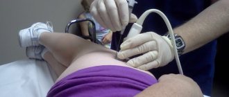
- The use of ultrasound guidance and fluoroscopic control of the manipulation allows one to avoid damage to vital structures and complications inherent in this type of intervention.
- As a rule, does not require special training;
- In most cases, local anesthesia is used, which makes the examination painless.
- Histological, cytological and other necessary studies of biological material are performed by expert class specialists - employees of the National Medical Research Center of Oncology named after. N.N. Petrova.
Ultrasound-guided punctures with drainage, percutaneous nephrostomy, as well as punctures of the female genital organs are carried out only at the KDO in the village. Sand.