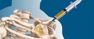
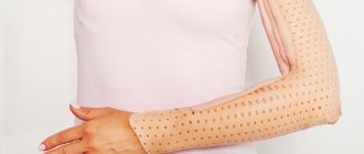
A joint puncture is a diagnostic procedure during which the doctor inserts a thin needle into the joint and removes a small amount of synovial fluid. The technique can provide useful information in the diagnosis of various orthopedic, rheumatic and other joint-related diseases.
In addition to diagnostic purposes, puncture can also be performed for therapy. The technique is relevant for removing excess joint fluid and delivering medications directly to the site of the lesion (this helps relieve swelling, reduce pain, and speed up the regeneration process). The article will discuss puncture for diagnostic purposes.
How the procedure is performed
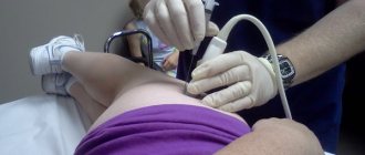
Synovial fluid testing is an invasive procedure that usually takes a few minutes. Depending on the punctured joint, the patient takes the appropriate position - lying or sitting, with the joint straight (for example, knee) or bent (for example, elbow). The doctor chooses the injection site taking into account the minimal risk of damage to blood vessels, nerves and cartilage. The area is treated with disinfectants - maintaining sterility is important.
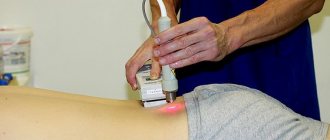
The doctor then uses a thin needle to inject local anesthesia into the area being examined. After the anesthetic has taken effect, he inserts a slightly larger needle into the selected area and draws synovial fluid into the syringe. In some cases, the puncture is performed under ultrasound guidance - this is largely due to the size of the joint.
The minimum volume required for analysis is 3-4 ml, but sometimes much more is taken - removing excess fluid from the joint can be a pain relief in itself. Depending on the suspected disease, the liquid is divided into 2-3 test tubes and sent to the laboratory. Biological material should be examined within 4 hours after collection.
Joint punctures
Posted at 19:37h in Services by doctor
Joint puncture is performed with a 10-20 g syringe, with a needle 5-6 cm long and 1-2 mm thick. Thin needles are used to inject medications into the joint when there is no need to take fluid from the inside, which can significantly reduce trauma. For pumping, 2 mm needles are used, which reduces the risk that they will become clogged with solid particles. The doctor is required to make extremely careful movements. Thus, the needle should not enter the joint capsule more than 1-1.5 cm, and the slightest vibrations of the tip when it passes through the synovial membrane seriously injure it. There is a technique that prevents infection and leakage of joint contents through the hole: the skin must be pulled back, thereby achieving curvature of the puncture cavity.
In order to reduce the risk of pathologies, a tight bandage is applied to the leg after the puncture, or it is immobilized with a splint. To prevent complications, the doctor carefully monitors the progress of healing, although outpatient treatment is also possible.
How to prepare for the procedure
Joint puncture and examination of synovial fluid does not require special preparation. However, the doctor should be informed about the medications taken, especially those that affect blood coagulation processes (for example, acetylsalicylic acid, clopidogrel, acenocoumarol), and about hypersensitivity to anesthetics. After the examination, no special rehabilitation is required, and the patient can return to normal activities. The procedure is painless, and there are no scars or cosmetic defects left on the skin. After the effect of the local anesthesia wears off, a sensation similar to the state of recovery from dental anesthesia may be present at the puncture site for some time.
Synovial fluid testing carries a low risk of complications. If you feel discomfort and slight swelling immediately after the examination, you can apply a cool compress to the area being examined.
However, if the procedure is performed in institutions with a poor reputation, the possibility of complications cannot be 100% excluded: the risk of infection remains. It may appear for several days after the procedure as significant swelling, pain, warmth and redness in the area of the joint being examined. Intra-articular bleeding or damage to articular cartilage may also occur. In a good medical clinic, such side effects are completely excluded: doctors are fluent in puncture technology. The procedure is safe and is also prescribed for children.
Preparation
Puncture of the knee joint is an outpatient procedure for which no special preparation is required. Situations have arisen more than once when the procedure was carried out urgently, because there was simply no time to take tests. If the operation is not urgent, then blood and urine tests may be taken from the patient. The quality of blood clotting is also determined, and the patient is sent for an X-ray, computed tomography or ultrasound.
The next stage of preparation for the operation is that the doctor finds out if the patient has an allergic reaction to anesthetics. There are people who are allergic to standard painkillers, but they can be replaced with other drugs that do not differ in quality from the standard ones.
Indications and contraindications for the diagnostic procedure
Joint pain, swelling and redness, or recent injury can all be reasons to schedule a procedure. Synovial fluid examination is carried out to identify the causes of joint diseases.
Among them:
- acute and chronic inflammation of the joints caused by microorganisms (bacteria, viruses, fungi);
- rheumatoid arthritis;
- systemic lupus erythematosus;
- ankylosing arthritis;
- systemic sclerosis;
- reactive arthritis and other aseptic arthritis;
- metabolic disorders associated with arthritis - gout, pseudogout (chondrocalcinosis);
- osteoarthritis;
- other causes of excessive fluid accumulation in the joints.
Contraindications to joint puncture and examination of synovial fluid are:
- skin infection around the joint (risk of transferring germs into the joint);
- bleeding disorders or taking drugs that reduce blood clotting (anticoagulants): INR > 1.5 or aPTT twice the normal;
- platelet count <50,000/mm3.
If an X-ray examination with contrast (arthrography) is prescribed, a puncture is performed to provide access to the intra-articular cavity. In this case, the list of indications and contraindications expands.
Indications
The following factors are considered indications for surgery for therapeutic purposes:
- bleeding into the joint cavity of the knee as a result of injury;
- the presence of a purulent focus caused by inflammation;
- the presence of inflammatory diseases that require the introduction of an antibacterial drug into the joint cavity (bursitis);
- the need to administer painkillers (during reduction of a dislocation);
- treatment of deforming forms of arthrosis with medications;
- fibrous adhesions, characterized by the need to administer oxygen to restore mobility.
It is impossible to get rid of these pathologies on your own, which is why they use a method of therapy such as puncture of the knee joint.
During puncture, the resorption of the hematoma is accelerated and the spread of inflammation is prevented.
In some situations, puncture is the only opportunity to find out an accurate diagnosis. To perform the operation for diagnostic purposes, the following indications are distinguished:
- the need to detect fluid in joint cavities;
- confirmation of the diagnosis of meniscus injury;
- identifying the cause of the development of the inflammatory process and swelling;
- the need to detect “rice bodies” in the joint.
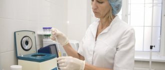
How is biomaterial analyzed?
The examination of synovial fluid is performed under a microscope and consists of assessing a number of parameters. Among them:
- color, transparency and viscosity of the liquid, its pH;
- glucose, protein, uric acid and lactate dehydrogenase (LDH) levels;
- number of leukocytes and other cells;
- assessment of the presence of bacteria, if necessary, a culture is prescribed (microbiological test).
Depending on the results, the doctor decides on further diagnosis of the disease or initiation of therapy.
The nuances of puncture of different joints
The technique of performing the operation is different for different joints due to their structure; the doctor must clearly understand how and where certain tissues, bones and other anatomical formations are located in order to lay out the puncture path between them as accurately as possible, without touching anything unnecessary or resting on bone.
Shoulder joint puncture
It is customary to distinguish three types of punctures of the shoulder joint: anterior, posterior and lateral. There is no fundamental difference in the technique; in all cases, the acromioclavicular joint serves as a reference point, and the needle is inserted respectively in front, behind and to the side of it.
The skin is punctured strictly perpendicular to its surface, after which the direction of advancement changes to the desired one. Injection is carried out until it feels as if the needle has “failed” or until punctate appears in the syringe.
Puncture of the elbow joint
Before starting, the arm is bent at the elbow at an angle of 90°. The needle is passed into the cavity behind, between the outer edge of the olecranon and the lower edge of the outer epicondyle, directly above the head of the radius.
If you need to take a puncture from the posterior inversion of the joint, then the needle is inserted through the triceps tendon, above the apex of the olecranon process, and carefully passed towards the synovium.
Wrist puncture
The puncture is made on the posterior surface, at the distal end of the radius, between the extensor tendons of the thumb and index finger.
Hip puncture
Performed from the front or side. In the first method, the puncture point is in the middle of a line drawn from the upper edge of the greater trochanter to the point between the middle and inner third of the inguinal ligament. The needle is inserted in front, perpendicular to the upper edge of the femoral head, at the edge of the acetabulum.
In the second method, the needle is inserted from the outside, above the apex of the greater trochanter, and advanced in the frontal plane towards the corresponding point on the opposite side.
Knee joint puncture
The puncture is made near the upper edge of the patella, the needle is advanced through the quadriceps tendon towards the bone, making rotational movements. It is possible to perform a puncture on the upper-outer, upper-inner and lower-outer edges of the patella.
Content
- Puncture (piercing) of the knee joint: technique and indications
- Who carries out the treatment?
- Why do they contact us?
- Reviews from our patients
- Sign up for treatment
If the rules are followed, puncture (piercing) of the knee joint takes place without consequences or complications. The procedure is considered very effective and painless. In addition, it takes no more than 15 minutes, and the cost is quite affordable. To verify this, you can look at the photo of the puncture of the knee joint, and also read the reviews left by our patients.
Baker's cyst symptoms
Most often, patients develop a single cyst on one limb, but other variations occur when both legs are affected or multiple formations appear. At first, Baker's cyst practically does not manifest itself at all, but gradually its size increases from two to fifteen millimeters.
As it increases, the following symptoms appear:
- There are difficulties in bending and straightening the knee.
- Gradually a dense formation forms.
- Its palpation can lead to pain.
- Over time, the cyst begins to cause pain, even if it is stationary, and with movement these sensations intensify significantly.
- When the cyst affects the nerve endings, there may be numbness in the limb, goosebumps or a tingling feeling in the foot.
- Sometimes there is aching in the calf muscles.
Baker's cysts in children most often appear against the background of injuries, including knee bruises and various sprains that also affect the meniscus. Typically, pathologies develop between the ages of four and seven years, but few people go to the doctor, since it tends to go away on its own.
Baker's cyst of the knee
The knee joint is one of the most complex in structure in the human body. He probably experiences the greatest physical stress. This is why knee joint diseases are most common. Baker's cyst of the knee joint is one of these pathologies, which is caused by inflammation of the joint.
The disease is characterized by pain in the knee that occurs when the limb is loaded, discomfort in the knee fossa. The advanced form is characterized by swelling of the joint, limited mobility, and partial deformation. That is why diagnosis and treatment of pathology should begin immediately.
Causes of Baker's cyst development
Most often, the disease develops due to metabolic-dystrophic or inflammatory processes that occur in the knee joint. Baker's cyst is caused by pathologies such as osteoarthritis, arthrosis, rheumatoid arthritis, but other options are also possible.
The main causes of Baker's cyst:
- Inflammatory processes in the knee joint.
- Pathological changes in the menisci.
- Mechanical damage to the knee.
- Excessive physical activity.
- Problems with cartilage, their gradual destruction.
Diseases leading to the formation of Baker's cyst:
- Rheumatoid arthritis.
- Psoriatic arthritis.
- Knee injuries.
- Osteoarthritis.
- Osteoarthritis.
- Metabolic disorders with joint damage (chondrocalcinosis, gout, etc.).
- Reactive arthritis, complicating some infectious diseases (tuberculosis, enterocolitis of various etiologies, respiratory tract infections, urogenital infections, Lyme disease, etc.).
Regardless of what exactly caused the pathological process to start, this causes increased production of synovial fluid, which fills the tendon bursa, which is why a cyst is formed.
A Baker's cyst can also occur due to mechanical damage to the knee.