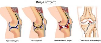
An inflammatory process that affects one or more joints is called arthritis.
It has several varieties, differing in causes, symptoms and treatment. The rheumatoid form of the disease can appear at any age, but most often affects people over 30 years of age. It has been noted that the proportion of women among the sick is over 70%. The disease causes destruction of the synovial membrane of the joint, cartilage, and bone. In its chronic form, the disease causes pain for many years and interferes with leading a full life. Over time, the degree of damage to the knee joint will only increase, so treatment should begin as early as possible.
Classification of the disease by radiological stages
The pathogenetic mechanism for the development of rheumatoid arthritis is an autoimmune cross-reaction. The antigenic structure of hemolytic streptococcus, belonging to group B, is similar in structure to the connective tissue of the human body.
When infected with a microorganism (acute or chronic tonsillitis, pyelonephritis, intestinal pathology), the produced antibodies may damage one's own tissues. The joints are the first to suffer. With each new rheumatic attack, the affected area expands and the changes become irreversible and severe.
In the early stages, at the first signs, changes may not be detected on an x-ray. But the further the process progresses, the more obvious the damage becomes. There are four radiographic stages of pathology.
First stage
The first stage is characterized as the easiest, the changes are reversible and treatable. Clinically, the patient complains of morning stiffness in the movements of the hands, swelling is visually noted in the area of the small joints of the fingers. More often, the symptoms are associated with a recent infectious pathology (sore throat, tonsillitis, urinary tract infection, streptoderma, etc.).
First stage
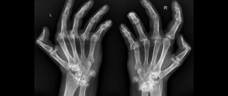
The small joints of the hands and feet are the first to suffer. X-ray signs of the first stage of rheumatoid arthritis:
- predominant inflammation of the soft tissue component, which has the form of compaction of structures above the affected joint;
- slight narrowing of the interarticular space, which is not visualized on an x-ray;
- initial manifestations of osteoporosis in the form of local clearing of bone structures and loosening of only the articular surfaces.
Second stage
Clinically, the second stage manifests itself in the form of severe stiffness of the hands for several hours, mainly in the morning and evening. The changes are more pronounced, manifested in the form of swelling and visible deformation, manipulative movements of the hands are significantly difficult (it is difficult for the patient to thread a button into a buttonhole, a thread into a needle, or tie shoelaces).
Second stage
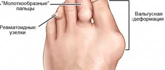
In the area of the joints, a seal is palpated, skin manifestations in the form of rheumatoid nodules are often added, the skin over them is hyperemic. Pain in the legs is acute, walking is difficult, and lameness is observed.
The second radiographic stage of the disease is divided into two subgroups, depending on the degree of damage in the form of erosion of the articular surface:
- What are the stages of rheumatoid arthritis, and how do they differ?
- subgroup A of the second radiological stage is characterized by the absence of erosions on the articular surfaces; the image reveals periarticular osteoporosis in the form of cysts (local clearing of bone structures), areas of bone compaction and a more noticeable narrowing of the gap between the articular surfaces;
- subgroup B of the second radiological stage of the pathology, in addition to the main manifestations, is characterized by the appearance of erosions of the surfaces of the joints, in no more than four places.
Third stage
Clinically characterized by complete immobility of small joints. The pathology extends to large ones: wrist, elbow, knee, ankle, upper shoulder girdle and even intervertebral joints in rare cases. The patient experiences difficulty walking due to severe pain and stiffness of the knee joints.
Third stage
Deformation of the hands occurs in the form of a “walrus fin” (subluxation of the metacarpophalangeal joints) or “swan neck” (formation of persistent flexion contractures of the metacarpophalangeal joints and hyperextension of the interphalangeal joints), inability to abduct the thumb. The patient cannot perform simple manipulative actions with his hands, including difficulty holding a cup, spoon, etc.
Bones become fragile due to a pronounced degenerative process. Cases of subluxations, dislocations, and pathological fractures are becoming more frequent. Healing of damage takes a long time and requires surgical interventions.
Third degree of rheumatoid arthritis on x-ray:
- formation of a single bone block in the area of small joints of the hands;
- more than 5 areas of bone tissue erosion;
- narrowing of the joint spaces of small and large joints;
- the appearance of osteoporotic cysts;
- the formation of soft tissue calcifications (rheumatoid nodules), which in the image appear as rounded areas of darkening, up to 2 cm in diameter, in the area of the soft tissue component around the joints (usually the hands, elbows, knees).
Fourth stage
The terminal stage of the rheumatoid process, which irreversibly disables the patient. Performing simple everyday activities is impossible due to severe pain and persistent joint contractures (immobility). The patient's muscles atrophy due to lack of movement. A person cannot take care of himself (eat, go to the toilet) and requires regular assistance.
Fourth stage
Based on the results of radiography of the joints, the following changes are revealed:
- absence of joint space with compaction in the articulation area and the formation of ankylosis (joint contractures) and subchondral osteosclerosis (due to increased friction of cartilaginous structures in the absence of joint space, they calcify and sclerosis, become dense, lose shock-absorbing capabilities);
- bone growths are formed on the articular surfaces - osteophytes, which have a pointed shape;
- osteoporosis can develop into osteonecrosis.
Rheumatoid arthritis: symptoms, diagnosis and treatment
Joint pain, feeling of “stiffness” in the morning. What is this? Are you overtired at work? “Re-exercise” at the gym? Got a cold?
The manifestations listed above may indicate an illness, the cause of which remains not fully understood to this day.
Rheumatoid arthritis. We are talking about it with Candidate of Medical Sciences, rheumatologist at the Expert Voronezh Clinic, Inna Alekseevna Strelnikova.
— Inna Alekseevna, what is rheumatoid arthritis and for what reasons does this disease occur?
This is an immunoinflammatory rheumatic disease of unknown etiology. It is characterized by chronic erosive arthritis and systemic damage to internal organs.
The causes of rheumatoid arthritis are currently completely unknown to medicine. According to world statistics, it affects on average about 1% of the population.
— Is rheumatoid arthritis coded in ICD-10?
Yes. It is reflected under code M05 (seropositive rheumatoid arthritis) and M06 (other rheumatoid arthritis).
— What happens to the body with rheumatoid arthritis?
The development of this disease is based on inflammation of an immune nature, mainly affecting joint tissue.
In the initial stages, the patient complains of pain and swelling in the joints, and morning stiffness for more than 30 minutes. The small joints of the hands and feet are most often affected.
This pathology is also characterized by extra-articular manifestations. These include vasculitis of the skin and other organs, neuropathies, pleurisy, Sjogren's syndrome, rheumatoid nodules.
With rheumatoid arthritis, so-called constitutional signs are sometimes observed: general weakness, loss of body weight, increased body temperature. But these signs may not exist.
— Does this disease creep up unnoticed or do rheumatoid arthritis have warning signs?
There are not always warning signs. The disease can begin immediately with classic manifestations. However, nonspecific complaints of joint pain can often be noted throughout the year. The pain changes its localization (i.e., now in one or another joint), or is noted simultaneously in several, symmetrically or asymmetrically. There may also be swelling of the joints (optional). At this stage, a diagnosis of “undifferentiated arthritis” is made.
What is arthritis and how is it diagnosed? Deniz Ruslanovich Matsiev, radiologist, tells
"MRI Expert Sochi"
As an independent manifestation, without articular signs, the erythrocyte sedimentation rate (ESR) may increase and the content of C-reactive protein may increase (laboratory signs of inflammation). The constitutional manifestations that I spoke about may also be noted here.
— Which joints are most often affected by rheumatoid arthritis?
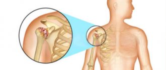
Certain small joints of the hands and feet (metacarpophalangeal and metatarsophalangeal, proximal interphalangeal joints of the hands and feet). Large joints are much less frequently affected: hip, knee, wrist, shoulder.
— What is included in the diagnostic standard for rheumatoid arthritis?
Complaints and a detailed medical history are collected, a thorough examination and an objective examination of the patient are carried out.
Laboratory tests are required. These include a complete blood count, a complete urinalysis, a blood test for C-reactive protein, rheumatoid factor, and antibodies to cyclic citrullinated peptide (ACCP).
X-rays of the joints of the hands and/or feet, as well as any other joints that have obvious manifestations, are also performed.
Are x-rays dangerous? Yulia Aleksandrovna Rutskaya, head of the radiation diagnostics department, tells
"Clinics Expert Kursk"
According to indications, radiography or computed tomography of the chest organs, electrocardiography and other studies may be performed.
— What is the difference between rheumatism, rheumatoid arthritis and polyarthritis?
The concept of “rheumatism” is no longer used in modern medicine. Instead, the designations “acute rheumatic fever” and “rheumatic heart disease” have been adopted.
Acute rheumatic fever occurs more often in young people, often females (from 7 to 25 years). It begins with a sore throat (acute period). After 2-3 weeks, pain in the joints, pain in the heart, and possibly annular erythema and chorea appear.
Acute rheumatic fever can occur without or with the formation of heart disease. In the latter case they talk about rheumatic heart disease. If the defect does not form, rheumatic fever usually goes away; recurrences are extremely rare. Today this pathology is less common.
As for polyarthritis, just by the name we can conclude that we are talking about inflammatory damage to several joints. Polyarthritis can be rheumatoid, psoriatic, etc. Those. This is not an independent diagnosis, but rather a syndrome.
Who gets rheumatoid arthritis more often: men or women?
Among women. The onset of the disease is most often from 40 to 55 years (less often at younger and later ages). The ratio to men in frequency is 3:1.
Why is osteoporosis called a woman's disease? Read more
— Can this diagnosis occur in children?
Yes. It is formulated as "juvenile rheumatoid arthritis."
— Who is at risk for developing rheumatoid arthritis?
These are individuals who, in the complete absence of symptoms of the disease, have an increased level of rheumatoid factor, ACFP, in their blood. This picture can be observed for several years. The disease itself may not develop.
Otherwise, identifying a risk group is problematic. Heredity and predisposition factors have not been fully studied.
Hypothermia or previous infection may be a provoking factor (not a risk factor).
— What specialty do doctors treat patients with rheumatoid arthritis?
Mainly rheumatologists.
Educational program on medical professions. When to contact a rheumatologist? Rheumatologist says
"Clinic Expert Tver" Masyukov Semyon Andreevich
In remote areas where there are no such specialists, patients can be treated by internists and general practitioners, but with periodic consultations with rheumatologists. However, it is still better for such patients to be treated by a rheumatologist.
You can make an appointment with a rheumatologist here
Please note: consultations are not available in all cities
If necessary, orthopedists, neurologists, etc. are involved in treatment.
— Can this disease be cured once and for all, or is this serious diagnosis a death sentence?
Rheumatoid arthritis is not a death sentence. There is an answer to the question whether it is curable or not. With proper treatment, low disease activity or complete remission can be achieved for a certain period of time. Treatment is lifelong.
— Are rheumatoid arthritis and hormonal treatment synonymous or is therapy possible without the use of hormonal drugs?
Treatment without hormonal drugs is possible. Ideally, it is not used at all, or such agents are used locally (in particular, intra-articular injection).
In other cases, such drugs can be prescribed systemically (for example, in the form of tablets) - at the onset of the disease, with pronounced inflammatory activity. They are subsequently cancelled. In some cases, small dosages of hormones are used continuously.
— What are the consequences of rheumatoid arthritis?
In the absence of proper treatment and significant activity of the inflammatory process, early disability, joint deformities, and increased risk of infectious diseases occur. When internal organs are involved in the process (cardiovascular, pulmonary pathology, etc.) - a decrease in life expectancy.
What is tachycardia and for what reasons does it occur? Cardiologist says
LLC "Clinic Expert Kursk" Novikova Elena Viktorovna
— Does rheumatoid arthritis impose restrictions on the patient’s lifestyle? What can and cannot be done with this disease?
When the activity of the process is low or in remission, regular light physical activity, cycling, and swimming are possible. It is recommended to eat rationally and observe a work and rest schedule. In the absence of joint deformation, there are no special restrictions.
Hypothermia and infectious diseases should be avoided. Excessive physical activity is undesirable: rheumatoid arthritis and high-performance sports are, in my opinion, incompatible things. This can lead to worsening of rheumatoid arthritis.
Avoid being overweight and stop smoking. If the process is active in any way, you should not visit saunas and steam baths.
Could an MRI of Michelangelo's joints have helped? Find out here
— Tell us about the prevention of rheumatoid arthritis. How not to provoke an exacerbation of the disease?
Since the cause of the disease is not understood, there is no primary prevention.
Prevention of relapses of rheumatoid arthritis includes constant compliance with the rheumatologist’s treatment recommendations; to give up smoking; avoiding hypothermia, infections, excessive physical exertion; maintaining normal body weight.
For reference:
Strelnikova Inna Alekseevna
Graduate of the Faculty of Medicine of the Voronezh State Medical Academy in 2007.
In 2008, she completed a clinical internship in the specialty “Therapy”.
From 2008 to 2011, she completed full-time postgraduate studies in the specialty “Internal Medicine”. Has an academic degree of Candidate of Medical Sciences.
In 2009, she completed her primary specialization in rheumatology.
From 2015 to the present, he has been working as a rheumatologist at Clinic Expert Voronezh LLC. In Voronezh he receives at the address: Voronezh, st. Pushkinskaya, house No. 11.
Bone erosion in pathology
Erosion of bone structures is divided into three types:
- Marginal or superficial - detected already at the second stage in joints where there is no cartilaginous structure and friction is more severe.
- Compression - occurs due to aggravation of the process and failure of the bone at the site of injury.
- Deforming - formed due to the destruction of the bone plate, visible bone deformation is visually observed, characteristic of stage 4 of pathology.
Single erosions of the bones of the wrist
When bone erosions occur, it is practically impossible to cure the disease. Long-term multicomponent therapy only helps to stop the progressive process.
Treatment and prevention of rheumatoid arthritis
The pathology is treated by a rheumatologist. Treatment of the disease is complex and multicomponent:
- Diet therapy. The diet should be dominated by foods rich in calcium (milk, cottage cheese, fresh vegetables and herbs); if you are overweight, there is a need to lose weight to reduce the load on the joints of the lower extremities. Products that help maintain cartilaginous structures are also useful: jelly, agar-agar, jellied meat, jellied fish.
- Drug treatment.
- Pathogenetic therapy. Non-steroidal anti-inflammatory drugs (Diclofenac, Nimesil) and steroids (Prednisolone, Dexamethasone) are prescribed; they help relieve pain and reduce inflammation. To prevent the autoimmune process, cytostatics (Methotrexate, Cyclosporine) and biological drugs (Rituximab, Enbrel) are prescribed.
- Etiotropic therapy. Antibacterial agents (Bicillin) are used to prevent relapse of streptococcal infection.
- Symptomatic therapy. Includes the use of chondroitin preparations and hyaluronic acid preparations to maintain cartilage structures, calcium with vitamin D3.
- Physiotherapy and therapeutic exercises. Allows, with the help of physical exercises, electrophoresis, magnetic therapy, to improve hemodynamics in the affected area and activate regeneration processes.
- Operative methods. They are used in extreme radiological stages (third and fourth) in the presence of dislocations, contractures and ankylosis, and pathological fractures.
Prevention of rheumatoid arthritis is the timely and correct treatment of infectious diseases and the prevention of the appearance of chronic lesions. In the initial stages (1-2), it is necessary to develop fine motor skills of the hands and arms - this helps to avoid permanent changes due to normal hemodynamics.
Such patients are recommended to start embroidering, knitting, and drawing. It is also necessary to eat properly to prevent degenerative pathologies of cartilage and bone structures.
Rheumatoid arthritis is a serious pathology that, at extreme stages of development, leads to disability of the patient. The disease can develop both in adulthood and in childhood. Progression leads to a deterioration in overall health and the appearance of persistent irreversible changes in the joint area.
- Rheumatoid arthritis: prognosis and life expectancy
Symptoms of rheumatoid arthritis of the knee joint
Characteristic manifestations of the disease can be detected without the use of special diagnostic equipment:
- decreased mobility of the knee, which manifests itself at the initial stage of the disease only after an overnight rest;
- sharp or aching pain that intensifies with movement; may spread to the entire leg;
- inflammation, signs of which are swelling of the knee, local changes in body temperature;
- effusion in the affected area;
- fever;
- increased fatigue.
As the disease progresses, the symptoms of rheumatoid arthrosis of the knee joint will intensify, and its deformation will be visible even to a non-specialist. Start treatment without waiting for the problem to become disabling! At the initial stage, the destruction of the knee can still be stopped.
How effective is X-ray examination for RA?
Rheumatoid arthritis is commonly called an inflammatory disease of the joints, which is autoimmune (the immune system attacks the cells of its own body, mistakenly perceiving them as foreign and pathological). The reasons why this disease develops have not been precisely established. It is believed that RA can be triggered by:
- infectious diseases previously suffered by a person;
- exposure to toxic substances on the body;
- frequent joint injuries;
- constant hypothermia.
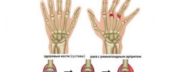
Risk factors include a family history of rheumatoid arthritis. As rheumatoid arthritis progresses, it primarily affects the small joints of the upper and lower extremities symmetrically. The pathological process immediately spreads to the synovial membrane, then to the cartilaginous tissue, eventually forming multiple erosions. The joints become deformed.
An X-ray examination allows the doctor to determine the degree of damage to the joints and determine how deformed the bone and cartilage tissues are. Using the image, the specialist counts the number of erosions that have formed in the bones.
In what cases is x-ray examination indicated and contraindicated?
An X-ray of the joints is prescribed to the patient if he has the following symptoms for a certain period of time:
- pain in the joints;
- limited joint mobility in the morning;
- swelling in the area of the affected joint;
- elevated rheumatoid factor detected in a blood test;
- the presence of rheumatoid nodules (multiple subcutaneous nodular formations near bone structures);
- deformation of the joints noticeable to the naked eye.
Since X-ray radiation is ionizing and can negatively affect the development of the embryo in the womb and small child, X-ray examination is not recommended for women during pregnancy and children. Patients who are in serious condition, suffering from epilepsy, acute and chronic mental illnesses are not examined. But the contraindications listed are relative.
If the attending physician decides that the benefit that radiography will bring to the patient by making a diagnosis will exceed the possible harm, he may, as an exception, prescribe the procedure.
Infection is a trigger for rheumatoid arthritis
Infectious agents—viruses and bacteria—can contribute to the development of rheumatoid arthritis, apparently in many ways. The best known mechanism is so-called molecular mimicry. Some molecules—most often peptides that make up a microbe—may have a structure similar to the structure of the body’s own biomolecules. During an infection, the immune system recognizes such molecules as foreign and effectively fights their carriers, but then attacks its own cells containing similar antigens. In the case of rheumatoid arthritis, this can happen, for example, with infection with the Epstein-Barr virus [15], cytomegalovirus, Escherichia coli and various Proteas.
In addition, during an infectious disease, immune complexes are formed, consisting of antigens and antibodies specific to them. If there are many complexes, then such antibodies themselves can become antigens. Antibodies against antibodies are the same rheumatoid factor that we talked about earlier.
Infectious agents can directly contribute to the development of rheumatoid arthritis. It has recently been shown that oral hygiene may influence the incidence of rheumatoid arthritis [16]. The fact is that the bacterium that causes periodontal disease, Porphyromonas gingivalis, is capable of synthesizing deiminase and, accordingly, taking part in the citrullination of proteins of the host, that is, humans.
Increasing evidence indicates that the composition of the gut microbiota is another important factor influencing the likelihood of developing rheumatoid arthritis [17], [18], [19]. In model systems, it has been possible to convincingly show that certain types of bacteria are associated with rheumatoid arthritis. Perhaps in the near future we will have the development of a special diet that will allow us to keep them under control and thereby reduce the risk of developing autoimmunity.
Stages of RA according to Steinbrocker classification
In modern medical practice, the classification of stages of rheumatoid arthritis developed by Steinbrocker is most often used. He based his research on plain radiographs of the hands and distal feet, performed in a direct projection. With their help, it was possible to identify 4 radiological stages of rheumatoid arthritis, which reflect the nature of the progression of the disease.
Stage 1
This stage is the initial one. It is called periarticular osteoporosis (decreased bone density). The pathological process involves the joints of the hands and metatarsophalangeal joints of the feet. The x-ray visualizes the compaction of the soft tissues surrounding the joint. The bone tissue is thinned, loose, and has increased porosity (these are signs of osteoporosis). There are already cyst-like clearings in its structure. Among the radiological symptoms of the first stage of rheumatoid arthritis may be a narrowing of the joint space.
An appointment with a rheumatologist is necessary for a person who is faced with even a slight restriction of joint mobility in the morning (within approximately 1 hour after waking up). At the first stage, symptoms such as slight swelling of the tissues in the area of the affected joint and short-term pain in the joint during physical activity may also occur.
Periarticular osteoporosis occurs in both adults and children. The disease can progress rapidly, or it can stop in its development for several years and then become more active.
Stage 2
X-ray signs of rheumatoid arthritis at the beginning of the second stage are increased osteoporosis, multiple cysts in the structure of bone tissue, as well as a decrease in the lumen of joint spaces. This stage is called stage 2A, which lasts until the first erosion occurs, that is, bone damage. After this, stage 2B begins, continuing until a maximum of 4 eroded areas appear.
It is customary to classify all bone erosions into 3 categories:
- Marginal superficial (formed in those areas where the articular bones are not covered with cartilaginous tissue).
- Compression (accompanied by the failure of a section of bone due to the formation of a cyst and the progress of periarticular osteoporosis).
- Erosion of the bone tissue of the endplates (appears where the joint connects to the ligaments).
The deformity of the hand or foot is not yet visible on an x-ray.
At the second stage of rheumatoid arthritis, the patient experiences limited joint mobility for several hours a day. During physical activity, pain increases.
Stage 3
This stage of disease development is characterized by the appearance of multiple erosions (from 5 pieces). The muscles around the affected joints atrophy. In addition to periarticular osteoporosis, decreased clearance of joint spaces, and cysts, X-ray signs of rheumatoid arthritis of the hands also include dislocations and subluxations of the joints, and their extended deformation. The most common types of deformities include:
- "button loop";
- "swan neck";
- "walrus fin"
The images show signs of calcification (accumulation of excess calcium salts) in the soft tissues surrounding the affected joint. They are round and dense rheumatoid nodules, the diameter of which is from 2 to 3 cm.
When palpating them, the patient does not experience any painful sensations.
The level of density of calcifications varies, and this is clearly visible on the x-ray. At this stage of the disease, joint pain and immobility are so progressive that many activities require significant effort.
Stage 4
The last stage of RA is manifested by extensive osteoporosis, the spread of erosion not only to the surface of the joints, but also to other areas of bone tissue. The photographs clearly show that osteophytes (bone growths) have formed along the edges of the joints, having different shapes (straight, wavy, with a short or long base).
The fourth stage is also characterized by subchondral osteosclerosis and ankylosis of the joints. A sign of the first is the presence of bone compaction under the articular cartilage, and the second is a fusion of the joint, causing its complete immobility.
The pain in the joints no longer stops. At this stage, the patient may lose the ability to independently care for himself and become disabled.
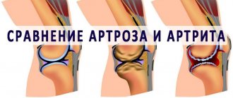
Arthritis stages
Arthritis is diagnosed in at least 2% of the Russian population. Women are more susceptible to the disease - the pathology is detected in the fairer sex 4 times more often than in men. The disease often develops in middle age (after 35 years). Every person approaching the age of 65 is at increased risk.
Types and causes of the disease
Arthritis is an inflammatory disease of the joints, leading to severe pain even at rest. As it progresses, it can cause disability with limited ability to move.
There are mono- and polyarthritis (affecting one or several joints). Depending on the reasons causing the development of the pathology, arthritis is divided into several types. Among which the most common are:
- rheumatoid;
- gouty;
- psoriatic.
Signs of arthritis
Arthritis tends to affect not only the joint elements. There are often cases when the destructive process expands to other organs and systems. The consequences are enlarged lymph nodes, atrophy of skeletal muscles, impaired liver function, damage to the gastrointestinal tract, lungs, heart muscle, and skin. The exact cause of the development of pathology is unknown to modern medicine. Provoking factors include infections, injuries, allergies, weakened immune defenses, excess body weight, unhealthy diet, alcohol abuse, hormonal imbalance, and pathologies of the nervous system. An important role is played by hereditary predisposition to the disease.
Stages of arthritis - initial stage
There are 4 radiological stages of rheumatoid arthritis, differing from each other in the severity of the pathological process. Often the disease begins to affect the joints before the patient manages to notice its clinical symptoms.
The first changes that occur in the early stages of rheumatoid arthritis are the wearing away of the cartilage covering the ends of the bones and the reduction of space between the joints. Age-related phenomena and excessive pressure on the joints lead to gradual wear of the cartilage. The negative effects of the disease are more pronounced in the presence of a genetic predisposition to the disease. The development of the initial stage of rheumatoid arthritis is also indicated by the appearance of the first painful symptoms, which intensify with heavy lifting and physical activity. This condition is accompanied by other typical symptoms such as: swelling; inflammation of the synovial membrane; rapid fatigue.
Diagnosing rheumatoid arthritis in the early stages can become difficult. This is due to the lack of specific laboratory tests aimed at detecting pathology.
Rheumatoid arthritis on x-ray
X-rays in the early stages of arthritis are often negative, but confirm the presence of osteoporosis and swelling in the soft tissues. The only manifestation that is revealed by x-ray is periarticular osteoporosis. The bone in the image becomes less bright than healthy joints. In this case, the patient does not exhibit hyperthermia and the feeling of morning stiffness characteristic of joint diseases.
The greatest danger is seropositive arthritis. Seronegative is easier, but more difficult to treat. The initial stage of arthritis of the fingers is accompanied by mild pain. A typical sign is a change in the shape of the fingers. They resemble “sausages”, and the entire hand loses its former dexterity in handling small objects.
Arthritis of the knee joint occurs in 3 stages. The latter of them has a high risk of developing arthrosis. Gouty arthritis has the same number of x-ray stages. At the initial stage, it is asymptomatic, and x-ray studies are not able to provide the necessary information. Testing (urine, blood, puncture of an inflamed joint), ultrasound, CT, MRI, and scintigraphy help make the correct diagnosis. The same method is used to identify the stages of psoriatic arthritis. If at the first stage of arthritis the patient goes to a medical facility, treatment in most cases is successful and can ensure a complete recovery.
Features of the course of the second stage of the disease
At stage 2 of arthritis, the pain becomes more pronounced. At this stage, no deformities are observed, but the mobility of the affected joint is impaired due to muscle atrophy and changes in the soft tissues surrounding the joints. The process of thinning of cartilage tissue and the formation of bone erosions is activated. Most often they appear in the wrists, ulnas, and phalangeal areas.
Characteristic signs of stage 2 rheumatoid arthritis are:
- feeling of stiffness after waking up in the morning, lasting about 30 minutes;
- slight hyperthermia in the area of the affected joints;
- local increase in temperature;
- exacerbation of weather dependence, aching bones when weather conditions worsen;
- moderate swelling, increasing in the evening and with heavy fluid intake;
- the appearance of single erosions on the cartilaginous surface;
- exudative changes and crunching when moving.
X-rays of this stage of rheumatoid arthritis confirm the presence of periarticular osteoporosis, a decrease in tissue mineral density, and a decrease in the gap between the articulating surfaces of bones due to swelling and the inflammatory process. At stage 2 of gouty arthritis, night attacks of pain are observed. Most patients experiencing such symptoms are forced to immediately visit a medical facility and get advice from a specialist.
Arthritis at this stage of development occurs in a moderately severe form, but is still amenable to conservative treatment and requires the most serious treatment. Otherwise, the risk of developing the third, difficult to treat stage increases.
Third stage of pathology
The third degree of the disease is considered severe. The activity of symptoms at this stage of rheumatoid arthritis increases significantly. In addition to the destructive changes that have already occurred, processes of accumulation of polymorphonuclear leukocytes in synovial fluid and proliferation of synovial cells occur in the body. Cartilage tissue in the affected joints is present in small quantities. Its deficiency is compensated by osteophytes, and the interosseous space becomes narrower.
Arthritis of the hip joint on MRI
Signs of stage 3 rheumatoid arthritis include:
- severe swelling;
- severe pain caused by muscle spasms;
- morning stiffness that does not subside until 12 noon;
- joint deformation;
- moderate hyperthermia and the appearance of exudate;
- forced restriction of physical activity;
- malaise and general weakness.
The advanced stage of rheumatoid arthritis is characterized by symmetrical joint damage. If the hip joints are affected, atrophy of the adjacent muscles (femoral and gluteal) is observed.
The main radiological signs of stage 3 rheumatoid arthritis are joint subluxations in small joints. Some patients exhibit extra-articular symptoms in the form of rheumatoid nodules under the skin, inflammation of blood vessels on the terminal phalanges of the fingers or nail plates. Often, pleurisy, pericarditis, inflammatory processes in the organs of vision, and neuropathy are simultaneously diagnosed.
Other radiological signs of stage 3 rheumatoid arthritis are loss of cartilage and soft tissue swelling. Nodes characteristic of this form of the disease are found on the fingers of the upper and lower extremities.
The victim's physical activity is significantly limited, which requires outside support during movement or the use of assistive devices - crutches, canes, walkers. However, at this stage the patient is able to cope with self-care. At the advanced stage of seropositive rheumatoid arthritis, deformities become visible even to a non-specialist. This stage occurs with paralysis of motor skills, spreading ankylosis to all small and large types of joints. An ECG may demonstrate heart failure and arrhythmia. The hands often deviate to the side, walking becomes deformed, and bilateral flat feet develop.
The advanced stage of seronegative arthritis is somewhat easier. It is characterized by the appearance of erosive formations on the bones. With the development of grade 3 arthritis, a less favorable situation arises for starting treatment. At this stage, you will need to do everything possible to prevent the disease from progressing to the 4th and final stage.
Arthritis at the fourth stage
The fourth stage of rheumatoid arthritis is the last stage in the development of the disease. As a rule, it develops 10–20 years after the appearance of the first signs of pathology. Depending on the effectiveness of the treatment, compliance with the doctor’s basic recommendations and the presence of concomitant diseases, this period may vary. As a result of the steady progression of the pathology, all bone structures of the body are affected. There is a complete loss of lubricating fluid in the joints. Instead, an inflammatory substance accumulates in the affected bones, causing severe swelling, stiffness and acute pain. At this stage, patients constantly take painkillers.
Among the symptoms and signs of the last stage of rheumatoid arthritis are:
- complete loss of performance;
- feeling of stiffness throughout the day;
- the appearance of cystic and erosive formations;
- muscle tissue atrophy;
- severe hyperthermia in the area of the affected joint;
- incessant pain.
Swelling is replaced by persistent deformation, leading to significant limitation of mobility. The development of ankylosis, in which the articular surfaces of the bones fuse together, becomes characteristic. This pathological process makes physical activity completely impossible.
The final radiographic stage of rheumatoid arthritis is characterized by the presence of areas of unequal density. In contrast to grade 3, there is active growth of osteophytes and expansion of chronic inflammation. The consequence is a narrowing of the gap between the bones and its complete disappearance in the future.
The last stage of arthritis leads to irreversible changes in bone and cartilage tissue. A person is deprived of the ability to perform various types of activities, which becomes the basis for disability.
Treatment of stages I and II of arthritis
The main principles of treating arthritis at the initial stage are: - reducing the load on damaged joints; - fight against excess weight; — daily therapeutic exercises; - for injuries of the ankle and knee joint - wearing orthoses and bandages.
Rheumatoid seropositive arthritis at an early stage responds well to treatment, which avoids the risk of developing disability. Medications are prescribed to relieve pain. Non-steroidal anti-inflammatory drugs are used for this. There is often no need for basic therapy. To prevent further progression of stage 1 arthritis, the patient is recommended to change his lifestyle. Regular walks, swimming, and Nordic walking will benefit weakened joints. Regular visits to the attending physician and control X-ray examinations become mandatory.
At stage 2 arthritis, the diagnosis becomes obvious, so the treatment approach is significantly different. The tasks of specialists are: - preventing the transition of pathology to the next level; - reduction in the intensity of symptoms; - prevention of frequent exacerbations. Basic treatment is mandatory. When diagnosing rheumatoid arthritis, the leading drug is Methotrexate tablets.
The product allows you to achieve improvement within 5-6 weeks. A lasting effect is observed after a 6-month course. Often this medicine is taken for several years or even a lifetime.
Approximate treatment regimen with Methotrexate: First 2-3 weeks 6.5 mg per day Then 20 mg for 24 hours After obtaining a pronounced clinical effect, 1 time per week in an individual dosage
The dose can be divided into 2-3 doses, the interval between which should be at least 12 hours. During the treatment period, it is necessary to take folic acid and undergo blood tests once every 3-6 months. If side effects occur, switch to injection of Methotrexate.
To relieve acute symptoms, the patient is still prescribed non-steroidal anti-inflammatory drugs. Orthopedic correction using various types of bandages and physical activity remain relevant. Physical procedures, mud therapy, professional massage, and stay in a medical sanatorium are often recommended. If all medical prescriptions are followed at both stages, it is possible to achieve a pronounced effect from the treatment. After several courses, many patients will need to undergo maintenance therapy, which allows them to live for a long time with minimal manifestations of the disease.
Fighting arthritis at stage III
In the treatment of the last stages of rheumatoid arthritis, the specialist relies on the following principles:
- use of medications;
- use of orthoses;
- physical therapy classes;
- conducting physical therapy;
- compliance with dietary standards.
Treatment of rheumatoid arthritis in the acute stage can be carried out on an outpatient basis or in an inpatient setting. In addition to Methotrexate, the following first-line drugs may be prescribed:
| Name of medicine | Dosage | Duration of admission |
| Tauredon | 10-20 mg once every 7 days with a gradual increase in dose to 100 mg. | Up to several months. |
| Sulfasalazine | Orally 1 g daily with an increase of 0.5 g every day. | 8-12 weeks |
| Auropan | Orally 6-9 mg per day, divided into 2 doses. | From 4-6 months or more. |
| Crinazole | 17-51 mg intramuscularly once a week. | 1.5-2 years |
Before use, be sure to consult your doctor!
It is mandatory to use NSAIDs - anti-inflammatory drugs produced in ampoules, tablets, ointments, and gels. The list of such drugs includes Diclofenac, Indomethacin, Piroxicam, Ketoprofen. The basic drugs of the second and third line include Kenalog, Aristocort, Depo-Medrol, Azathioprine, Chlorobutin. These drugs are used if there is no desired effect from basic therapy. Treatment with genetically engineered biological drugs - Infliximab, Abatacept, Rituximab - is becoming increasingly recognized. These funds are expensive and are often paid for by the government. The formation of persistent deformities in a patient with third degree arthritis is considered an indication for surgical treatment.
At the stage of remission in rheumatoid arthritis, indicators of improvement are the subsidence of the inflammatory process, a decrease in pain and a favorable blood picture. However, there remains a need for regular monitoring of the patient’s condition and supervision by a rheumatologist.
If possible, you should undergo maintenance treatment in a sanatorium (at least once a year).
Rheumatoid arthritis of the temporomandibular joints on CT
The last stage of the disease - how can you help the patient?
Treatment of the fourth stage of arthritis is extremely difficult. At this stage, the prescription of basic therapy continues, but the dosages of medications increase significantly.
The use of necessary medications is most often carried out in a hospital.
The only way to relieve the main symptoms is often the intra-articular administration of glucocorticosteroids. A high degree of pain requires the prescription of narcotic painkillers. Physiotherapy and exercise therapy are indicated if there is a symptomatic effect. Genetic engineering biological therapy, prescribed to patients in the absence of contraindications, is becoming relevant. Surgery becomes the most effective way to help the patient. If the patient’s condition allows, specialists perform joint plastic surgery, and the affected large joints are replaced with prostheses.
Folk remedies and prevention of arthritis
The use of alternative medicine recipes is effective in the initial stages of the disease and can be a good addition to the main treatment regimen. The following will help alleviate the condition: - apple cider vinegar; - garlic; - horse chestnut inflorescences; - bay leaves and juniper.
Apple cider vinegar is used internally. To do this, it is diluted with cool boiled water (a teaspoon of pure product per glass of liquid). The solution is drunk during the day before meals, divided into several servings.
Several peeled garlic heads (4-5 pieces) are poured into 0.5 liters of vodka and kept at room temperature in a dark place for at least 10 days. The finished product is taken daily after meals in the amount of 1 tablespoon. Another preparation option is to mix 3-4 chopped garlic heads, fresh juice of 10 lemons, a teaspoon of chopped horseradish. The product must be infused in a warm place for 3 weeks. To prepare horse chestnut tincture, fill a liter jar 2/3 full with its inflorescences and fill it with alcohol or vodka and leave it for 20 days in a place inaccessible to light. The finished product is used externally to rub the affected areas.
To prepare the ointment, combine a tablespoon of butter, 6 tablespoons of chopped laurel and 1 teaspoon of juniper. After thorough mixing, the product is ready for application. An ointment made from butter (1 tbsp), crushed bay leaves (6 tbsp) and chopped juniper (1 tsp) has an analgesic effect. All ingredients are mixed and applied to the inflamed joint. Primary prevention measures for arthritis include: - timely diagnosis and treatment of infectious diseases; — elimination of foci of chronic inflammation (tonsillitis, caries); - hardening, moderate physical activity; - taking multivitamins; - rejection of bad habits; — minimization of stressful conditions.
Secondary prevention is carried out according to doctor's indications. It consists of a well-chosen course of medications, physical therapy, diet, and a course of Omega-3 fatty acids. Timely preventive measures can help avoid joint destruction, maintain performance and improve quality of life.
Are the radiological signs of RA different from other types of arthritis?
Using an x-ray, the doctor is able to make a differential diagnosis between different types of inflammatory joint disease. For example, with gouty arthritis, urate crystals (accumulations of uric acid) are visualized on x-rays.
The places where crystals are localized are periarticular tissues and joint space.
At the end parts of the epiphyses (edge of the tubular bone), one can notice the presence of a sclerotic border, and in the cortical layers - cystic defects. The soft tissues along the affected joints are enlarged, and the joint space is widened.
Radiological signs of psoriatic arthritis are different. Osteoporosis, which is an integral companion of RA, in this case occurs only in mutilating forms of the disease. Psoriatic arthritis is accompanied by the appearance of erosions in the distal interphalangeal joints. Such lesions form in the marginal zones of the joints, and then spread to the center.
The tops of the terminal and middle phalanges are ground off, the articular surface is concave, resulting in the visual effect of a “cup and saucer” or “pencils in a glass.”
X-ray signs of glenohumeral periarthritis (inflammation of the soft tissues around the shoulder joint) are the presence of deposits of small calcium crystals (calculous bursitis), osteoporosis of the head of the humerus (this symptom is especially pronounced in the ankylosing form of the disease associated with fusion of the joint and, as a result, its complete immobility).
Alternative methods for diagnosing rheumatoid arthritis
If a classic radiograph did not allow the specialist to make a diagnosis and create a treatment program, computed tomography (CT) may be prescribed for clearer visualization of bone tissue. It is also based on the use of X-rays, but the images obtained during this procedure are not flat, but three-dimensional, layer-by-layer and more accurate. Therefore, even minimal changes in tissues are visible on them. However, CT is associated with greater radiation exposure to the patient's body.
To assess the degree of involvement of the soft tissues surrounding the affected joint in the pathological process, magnetic resonance imaging (MRI) is prescribed. It also allows, like CT, to obtain volumetric images, but involves exposing the patient to a magnetic field and radiofrequency pulses. It is known that X-rays tend to accumulate well in bone tissue and pass through soft tissue without hindrance. Therefore, if the nature of the changes in the latter is poorly visible on x-rays, then MRI displays them in the most complete volume.
Ultrasound examination (US) is more sensitive to detecting erosions in joints, compared to radiography. It is also prescribed to those patients who cannot undergo X-rays, CT, or MRI, since ultrasound has no contraindications. Thus, it is impossible to say unequivocally which examination method for rheumatoid arthritis is the best. The choice of diagnostic method is made by the attending physician based on the symptoms with which the patient came to him.
Proper nutrition for rheumatoid arthritis of the knee joint
The diet for rheumatoid arthritis is based on several principles:
- you need to eat in small portions, at least 4 times a day;
- You should avoid cold dishes and limit the consumption of highly heated foods;
- it is necessary to reduce salt intake to the amount indicated by the doctor;
- drink about 2 liters of liquid per day.
Healthy foods that should form the basis of your diet:
- fruits and vegetables;
- bread - rye or bran;
- cereals;
- fresh greens are a source of vitamin C, iron, calcium;
- lean meat;
- sea fish;
- dairy products;
- green tea, natural juices.
You should limit yourself to sour, salty, spicy foods, coffee and alcohol, and avoid fatty foods.
Conclusion
X-ray examination is a fast, inexpensive and very informative diagnostic method that allows not only to identify rheumatoid arthritis, but also to accurately determine its stage. Most often, x-rays are used as an initial examination of patients with symptoms of RA. If necessary, for the purpose of in-depth diagnosis, the attending physician may decide to prescribe a different instrumental research method to the patient.
An x-ray will help you understand how far the disease has progressed. The stages of rheumatoid arthritis are visible on it, but only a specialist can accurately determine the problem. In the later stages, almost anyone can determine the presence of a problem by appearance - deformation of organs occurs. However, there are changes that are not visible to the naked eye.
This:
- Reducing joint spaces;
- Erosion of bones (that is, their thinning, the appearance of “dips”);
- Ankylosis (bones fusion with each other).
But since there are so many diseases of bones and joints, one x-ray is not enough to diagnose and determine the stage of rheumatoid arthritis. A good doctor will supplement this with a blood test.
Benefits of MRI for Arthritis
- Complexity, that is, the ability to analyze the condition of both soft and bone tissues using magnetic waves during one examination.
- Determines the pathological process at its early stage, which automatically increases the effectiveness of subsequent treatment measures and avoids disability as a result of uncontrolled progression of arthritis.
- A detailed, detailed picture of the affected joint, including an image of all articular surfaces: the joint capsule, cartilage and parts of the ligamentous apparatus.
- The ability to enlarge images several times, as well as view the resulting images from different angles in 3D format.
- Control over the treatment process. Since arthritis can be examined on MRI an infinite number of times without any threat to health, using this method it is easy to monitor the effectiveness of treatment by obtaining comparative MRI images of the same joint before the start of the treatment process and throughout its entire duration.
- Health safety and no radiation exposure.
- Possibility of diagnostics in the presence of sutures and surgical staples, patients after heart surgery, with vena cava filters implanted in the inferior vena cava, as well as patients with artificial joints, disconnected medical pumps and shunts for hydrocephalus.
Previous Next
Tests in addition to X-rays
To diagnose rheumatoid arthritis, tests are done for markers of inflammation (that is, markers that show that the body is fighting it) and rheumatoid factor. However, it is important not only to do the analysis, but also to interpret it correctly.
Analysis for inflammatory markers. If the result is positive, this may be an indication of some other disease, not necessarily arthritis. Which one exactly will be revealed by further research.
Analysis for markers of rheumatoid arthritis. It is worth diagnosing with great caution, because it occurs in 5% of healthy people (and indicates a predisposition to the disease, but not the disease itself), and it does not occur in approximately 50% of patients. That is, it is only indirect evidence of the presence of rheumatoid arthritis.
Therefore, tests for other diseases are also necessary. If, as a result of the examination, no other pathogens are found, and the x-ray indicates pathological changes, the doctor diagnoses “rheumatoid arthritis”.
What does the lack of timely diagnosis using X-rays lead to?
Some people believe that due to their young age they cannot get rheumatoid arthritis, especially in the advanced stages. And this is a big mistake, because the disease is rapidly growing younger and occurs in those who are only 30 or 20 years old. The reasons are trivial - environmental degradation, unhealthy diet, sedentary lifestyle.
Heredity plays a big role. If there are people in your family who have had or are suffering from rheumatoid arthritis, the appearance of its symptoms is a very alarming symptom.
What signs of rheumatoid arthritis are visible on x-ray?
On an x-ray, the doctor can see various signs of the disease that are not noticeable at first glance and examination. An x-ray is taken of the part of the body in which the patient complains of pain. Rheumatoid arthritis can be concentrated in one part of the body or affect them all at once (hands, toes, knees, shoulders, elbows). Most often the problem begins in the extremities.
X-rays show erosion and inflammation. How strongly they will be expressed depends on the stage of the disease.
Causes of rheumatoid arthritis of the knee joint
Absolutely anyone can get sick, regardless of gender, age, or lifestyle. Rheumatoid arthritis is caused by a combination of several factors:
- hormonal disorders - this is why women of puberty usually become ill;
- injuries, as well as unsuccessful operations or incorrect rehabilitation after them;
- hypothermia of the body;
- smoking;
- infectious diseases;
- allergy.
The bulk of officially registered patients are men and women aged 25 to 55 years.
Determining the stages of rheumatoid arthritis using x-rays
Doctors have their own requirements for how to make a diagnosis, which they follow. For example, the stage of rheumatoid arthritis is determined by the joint that suffered the most, judging by the x-ray.
That is, if damage to the joints of the legs corresponds to stage 2, and to the joints of the arms to stage 4, then the patient will receive a diagnosis of stage 4. In itself, it is very difficult, but since this concerns not the whole body, but its individual parts, the situation is not as hopeless as it might seem at first glance.
Of course, all this cannot be done by a non-specialist, but with appropriate training a person can roughly understand the condition of the joints. However, to make an accurate diagnosis and determine the stage, a doctor is needed. If there are doubts about the doctor’s qualifications (he is not interested in treatment, he prescribes only ointments and/or tablets), you can show the same x-ray to another specialist.
There is no need to take a new image (unless another doctor insists on this due to the poor quality of the old image or the large amount of time that has passed).
Still, every X-ray is radiation, albeit a small one.
To make a judgment regarding the stage of rheumatoid arthritis, rheumatologists analyze:
- The degree of destruction of joints, bones, cartilage on x-ray;
- Number of erosions;
- Signs of inflammation in the picture;
- Presence of cysts.
But in very early stages or in cases of small erosions, it is difficult to see all the details of rheumatoid arthritis on x-rays. Then MRI is used. The resolution of this equipment is higher and more details can be obtained.
Treatment of rheumatoid arthritis of the knee joint
The methods used to combat the disease are divided into non-drug and drug-based. Surgery (surgery) is used in extreme cases.
Non-drug treatments for rheumatoid arthritis of the knee:
- Pharmacopuncture. Designed to relieve inflammation and provide nutrition to the damaged joint.
- Shock wave therapy, which serves to restore normal blood supply. Returns the ability to bend the leg at the knee.
- Vacuum therapy is a type of massage based on the organization of low pressure zones in a certain area.
- Magnetotherapy. It has an analgesic and anti-inflammatory effect on affected joints.
- Laser therapy. Increases local immunity, has anti-inflammatory and sedative effects, improves blood circulation.
In contrast, some medications, in addition to the obvious effect (relieving pain, inflammation, etc.), may also have side effects:
- non-steroidal anti-inflammatory drugs;
- basic medications, the use of which begins immediately after diagnosis and can continue throughout life;
- hormonal agents.
How to treat rheumatoid arthritis of the knee joint and in what ratio to use Eastern and Western methods, the doctor must determine individually.
What do the stages of rheumatoid arthritis look like on an x-ray?
The first stage is the easiest, but even it can be seen in the picture. As already mentioned, rheumatoid arthritis most often begins in the extremities, so photographs are taken of the hands where changes occur. Signs of the first stage of arthritis:
- The soft tissue around the affected joints is thickened;
- The bones themselves are more porous, even, one might say, loose;
- The joint space is narrowed, but still weakly.
At this stage, a person feels that it is more difficult for him to move his limbs in the morning. There is slight swelling. These two symptoms are enough to start sounding the alarm and taking an x-ray. Moreover, symptoms can appear in both adults and children. Pain may be added to limited mobility in the morning. If rheumatoid arthritis is diagnosed early at a young age, you can avoid its rapid development in adulthood and old age.
Second stage. Cysts are added to existing symptoms. These are growths on bone tissue that deform the arm and further limit its mobility. The joint gap becomes even smaller. Osteoporosis (fragility and looseness of bones) is on the rise. This is what is seen on an x-ray. But in real life, mobility decreases and pain manifests itself not only in the morning, but also during the day.
It is important to note that the second stage is divided into 2 types - 2A and 2B. At stage 2A of rheumatoid arthritis, there is still no erosion in the image - pronounced bone damage, but at stage 2B they already appear. The most common type of erosion is marginal, that is, along the border of the joint.
The most vulnerable areas are those where the cartilage does not cover it. Another type is the failure of a section of bone, this is called compression erosion. The third type is destruction of the endplate (it connects the ligament to the joint). And if only a very good specialist can determine stage 1 rheumatoid arthritis by x-ray, then even an average doctor should be able to handle stage 2. In real life, the patient suffers from pain during exercise.
The third stage of rheumatoid arthritis on x-ray is characterized by a large number of erosions, namely more than 5. The doctor also notes that the muscles atrophy due to rheumatoid arthritis, as a result of tissue damage and limited mobility of the organ. The interarticular spaces are still narrower. Subluxations and dislocations of the joints appear, they are already visually deformed.
In the soft tissues near the diseased joints, in the photographs you can see nodules that form from calcium salts. This is called calcification. The nodules not only contribute to the progression of the disease, but also cause pain when touched. This is about x-rays. But in real life, a person already has to make significant efforts to perform the simplest everyday operations.
This stage is especially painful for those who engage in sports or physical labor, as it deprives them of the opportunity to have a full load.
All the signs that were mentioned in the description of the previous stages are present in X-ray 4, but to a more pronounced degree. New ones are also appearing.
Erosion. At stage 4 of rheumatoid arthritis, on x-rays they are visible not only on the joints, but also on the bones. The joint spaces narrow so much that bone fusion occurs (ankylosis). Osteophytes appear. Roughly speaking, osteophytes are growths along the edges of joints. They have different shapes. At first they are small, and then they grow larger and larger.
Osteophytes even change the appearance of the limb, visible to the naked eye. A bone compaction is visible under the cartilage of the joint. It is called subchondral osteosclerosis. In fact, this compaction is an attempt by the body to protect itself from destruction, as the cartilage breaks down and the bones begin to rub against each other.
And to prevent their complete destruction, the body provokes the appearance of such compactions that are visible on x-rays. In real life, it becomes even more difficult for a person to perform the simplest household operations. The pain is very severe and difficult to endure without taking painkillers.
MRI or ultrasound to diagnose arthritis?
Radiography
The advantages of a diagnostic examination for arthritis using x-rays include low cost and accessibility. It is usually carried out in two projections: direct and lateral, however, if a detailed study of the condition of the bone tissue is necessary, radiography in an oblique projection is additionally prescribed. On radiographs, arthritis appears as an uneven narrowing of the interarticular space, the presence of cysts and osteophytes. Among the disadvantages of this type of diagnosis is the inability to see the degree of damage to soft tissues and the layered structure of joints, as well as to determine the disease at an early stage of development. With x-rays, there is radiation exposure to the body. Radiation doses do not allow the use of radiography as a diagnostic method for pregnant women.
Arthritis on ultrasound
An ultrasound of a joint with arthritis is carried out to study the soft tissues and allows you to determine the presence of an inflammatory process, swelling, thickening of the synovial bursa, protrusion and deformity disorders. Among the main advantages of ultrasound, doctors highlight affordability and the absence of radiation exposure. The biggest disadvantage of ultrasound is that it is impossible to determine the degree of destruction of bone tissue in arthritis, and also that the accuracy of diagnosis depends on the experience and level of qualifications of the doctor who performed the examination.
| Ultrasound services of joints | price, rub. |
| Ultrasound of the shoulder joint | 1500 rub. |
| Ultrasound of the elbow joint | 1500 rub. |
| Ultrasound of the hip joint | 1500 rub. |
| Ultrasound of the knee joint | 1500 rub. |
| Ultrasound of the ankle joint | 1500 rub. |
| Ultrasound of one small joint (hand, foot) | 1000 rub. |
| Ultrasound of two small joints | 2000 rub. |
| Ultrasound of two large joints | 2500 rub. |
CT scan
CT of the joints is one of the most accurate diagnostic methods, since it makes millimeter scans of each layer of the affected joint, but it has one significant drawback - a high dose of radiation. It is 2-3 mSv per scan of one area.
MRI for arthritis
In order to diagnose arthritis, MRI is today the most effective method of instrumental examination. The reason for the popularity of this diagnostic method is the ability to see both bone and soft tissue and the absence of any radiation exposure. MRI images of joints will clearly show the following signs of arthritis:
- increased accumulation of fluid in the interarticular space;
- the joint space is narrowed;
- signs of subchondral osteosclerosis;
- single usuration of articular surfaces;
- edge sharpenings.
| Service | Price according to Price | Discount Price at Night | Discount Price During the Day |
| from 23.00 to 8.00 | from 8.00 to 23.00 | ||
| Shoulder MRI | 4000 rub. | 3190 rub. | 3690 rub. |
| MRI of the elbow joint | 4000 rub. | 3190 rub. | 3690 rub. |
| MRI of the knee joint | 4000 rub. | 3190 rub. | 3690 rub. |
| MRI of the ankle | 4000 rub. | 3190 rub. | 3690 rub. |
| MRI of the hip joints | 5000 rub. | 3190 rub. | 3690 rub. |
| Appointment with an orthopedist | 1800 rub. | free after MRI | free after MRI |
| First aid program for joints (8 studies + appointment with an orthopedist + MRI of the joint) | 13000 rub. | 7500 rub. | 7500 rub. |
How to recognize rheumatoid arthritis on x-ray
To understand that the X-ray shows exactly the signs of rheumatoid arthritis, you need to know how to interpret the image. Let's look at what certain signs look like on an x-ray.
Tissue compaction around the affected joint. This symptom is visible even in photographs of stages 1 and 2 of rheumatoid arthritis. The tissue around the diseased area appears smoother than that around the healthy area. They are also more transparent.
Calcifications. These are the nodules discussed above. On x-rays they simply appear as shadows or round formations. The shape does not need to be round. In rheumatoid arthritis, they can be in the form of dashes or small formations on the edge of the joint. Moreover, their density can also be different.
Some are just forming, while others are bone-like in density. Calcifications are more clearly visible on an MRI image.
Tendinitis and tenosynovitis. This is a hardening of a tendon or ligament where it attaches to the bones. This process is also visible on x-rays. An additional sign is calcifications around such compactions.
Osteoporosis. The actual destruction of the bone, increasing its fragility and weakness. Osteoporotic areas of the bone/joint appear to appear more transparent. The endplates appear thinner.
Narrowed joint space. It is possible to determine that it is narrowed due to rheumatoid arthritis only if there is a similar photograph of the healthy arm. The doctor can do this without such an image, since he knows exactly what the ratio should be. However, in later stages, even a non-specialist can see that the gap on the x-ray is significantly narrowed.
Unevenness of the articular surface. These can be both failures and growths. In the first case we are talking about erosion, in the second - about osteophyte. Osteophytes have different shapes on x-rays in rheumatoid arthritis. But it is important that they change the normal shape of the joint at a later stage. Possible crooked fingers. This is often visible without an x-ray, but the image confirms the suspicions.
Osteolysis. Rarely seen. This is a process in which bone is essentially resorbed. Accordingly, it is absent on x-rays in some areas. This sign indicates a big problem and the need for serious treatment of rheumatoid arthritis.
If tissue necrosis occurs (its death due to insufficient blood supply), this area will appear darker on the x-ray, looking like a shadow. In this case, this shadow is surrounded by a contour lighter than the color of the rest of the bone.
An indirect sign of necrosis in arthritis is the proliferation of osteophytes.
Osteophytes. Growths on the edges of joints in rheumatoid arthritis, which have already been discussed above. The more severe the stage, the larger the osteophyte. Thus, the answer to the question of whether arthritis is visible on an x-ray is yes.
It cannot be cured, but it can be alleviated. Current treatment strategies for rheumatoid arthritis
Since a clear target for the autoimmune process in rheumatoid arthritis cannot be found, it is not yet possible to develop a specific treatment. You can only reliably stop the inflammation and try to maintain the patient’s state of remission for as long as possible. The main modern treatment strategies are aimed at selecting the most effective anti-inflammatory drugs [20]. Below we will talk about their various options.
Nonsteroidal anti-inflammatory drugs (NSAIDs), such as ibuprofen and naproxen , reduce inflammation and reduce pain. They inhibit the activity of cyclooxygenase enzymes, which promote the production of prostaglandins - substances that increase the sensitivity of receptors to pain mediators. Unfortunately, NSAIDs, like any medicine, have side effects. As a result of long-term use of these drugs (especially if they are not selective - Ed.), damage to the mucous membrane of the stomach and duodenum may develop, which will lead to the formation of ulcers and bleeding. Diarrhea and nausea may also sometimes accompany treatment. In addition, suppression of prostaglandin production negatively affects the functioning of the vascular epithelium, which can lead to myocardial infarction, thrombosis and, due to decreased renal blood flow, acute renal failure.
Corticosteroids are another class of drugs that reduce inflammation and pain. These include, for example, prednisolone . These drugs are stronger than NSAIDs and therefore have more serious and frequent side effects, such as type 1 diabetes [21], obesity, and brittle bones caused by their loss of calcium. Because of this, corticosteroids are prescribed only when the severity of the symptoms of rheumatoid arthritis increases, and old methods of pain relief are no longer effective.
The so-called basic anti-inflammatory drugs (DMARDs) are usually considered as a separate therapeutic group. It is very important for the best long-term results that the patient begins taking these drugs as early as possible. The fact is that these drugs have immunosuppressive properties, that is, the ability to suppress the activity and division of immune cells, as well as joint cells. Accordingly, the autoimmune process, which literally eats up the joint affected by rheumatoid arthritis, can be limited at an early stage. The most common and effective DMARDs are methotrexate , leflunomide and sulfasalazine .
The effect of taking DMARDs is observed in 60% of patients with rheumatoid arthritis, but it develops very slowly, so the course of taking the drug lasts at least six months. If during therapy the patient does not feel any improvement, then, as a rule, he is prescribed combination treatment with several DMARDs. However, you need to be careful with this tactic, as it significantly increases the likelihood of side effects.
Side effects of DMARDs typically affect the gastrointestinal tract and hematopoietic system. The fact is that DMARDs suppress the proliferation of the most actively dividing cells in our body, that is, immune cells (which is what their therapeutic effect is based on), but also other blood cells (for example, megakaryocytes, from which platelets are formed, and red blood cells) and cells intestinal epithelium, suffering completely undeservedly.
Biologics are a relatively new treatment for rheumatoid arthritis. They are antibodies or soluble receptors and can therefore specifically block certain molecules involved in the development of rheumatoid arthritis. Such “target molecules” can serve as pro-inflammatory cytokines and receptors on the surface of immune cells (Fig. 6).
Figure 6. Pattern of the immune response and the main biological drugs that can be used in the treatment of rheumatoid arthritis. To see the picture in full size, click on it.
website
The most common drugs that block proinflammatory cytokines are antibodies to tumor necrosis factor (TNF), interleukin-6 and interleukin-1. The former include, for example, infliximab and adalimumab , monoclonal antibodies that bind TNF. To block TNF, etanercept , a drug consisting of a soluble TNF receptor and the Fc fragment of an antibody (IgG1). The therapeutic antibody tocilizumab blocks the interleukin-6 receptor, which also helps reduce inflammation. Anakinra is a genetically engineered version of the IL1-RA protein, a natural interleukin-1 receptor antagonist. By attaching to the receptor, anakinra blocks the transmission of a signal through it and, therefore, the launch of an inflammatory response.
As readers might have already guessed, in the treatment of rheumatoid arthritis it was simply impossible not to use agents that block the costimulatory signal to the T cell from antigen-presenting cells. This is the basis for the action of the previously mentioned abatacept , consisting of the CTLA-4 molecule (a blocker of costimulatory molecules on the surface of antigen-presenting cells) and the Fc fragment of IgG1.
Since B cells play an important role in the pathogenesis of rheumatoid arthritis, suppression of their activity can also be a fairly effective treatment strategy. The use of rituximab , an anti-CD20 monoclonal antibody drug, results in the selective removal of mature B cells (antibody-producing plasma cells are not affected) and a decrease in inflammation.
The use of biological drugs in combination with DMARDs mutually enhances their therapeutic effect, so these two groups of drugs have become the most significant in the treatment of rheumatoid arthritis. Alas, this is not the case in all countries. The high price of biological drugs greatly hinders their widespread use. In Russia, the cost, for example, of infliximab is about 50 thousand rubles per 100 mg. This is not sufficient even as a starting dose in a treatment that requires the patient to receive the drug at least every two months for a year or more [20].
Unfortunately, biological drugs are also not without serious side effects, primarily associated with a lack of selectivity in blocking molecules important for an effective immune response. The use of such drugs may weaken the adequate immune response to infection. Almost all biological drugs contain antibody fragments, and this can lead to the development of an autoimmune response in which they play the role of antigens. Targeting the action and reducing the immunogenicity of the drugs themselves are among the most important problems that can be solved using new approaches to the development of biological drugs.
Constant monitoring of a patient with rheumatoid arthritis and timely change of treatment strategy in case of ineffectiveness of a particular drug can maintain a state of remission for years and even decades. Without proper treatment, inflammation and associated symptoms develop quickly enough that it can lead to surgical removal of the affected joints.
X-ray or MRI for rheumatoid arthritis?
Those who suspect they have rheumatoid arthritis are faced with a choice: X-ray or MRI? It is ideal to use both methods in combination, supplementing them with analyses. At the first stage of the disease, x-rays are practically useless, since pathological disorders are poorly visible. If you are worried about immobility and pain in the morning, it is better to consult an MRI.
At the same time, MRI is a more expensive procedure. Magnetic resonance imaging also has another drawback - the image provides information about the existence of a problem, but it is difficult to determine from it what kind of joint disease the patient is affected by. To classify and confirm that it is rheumatoid arthritis, an x-ray will also be required.
Bibliography:
- https://www.rmj.ru/articles/revmatologiya/stadii_rentgenologicheskih_izmeneniy_v_sustavah_pri_revmatoidnom_artrite/
- https://osnimke.ru/kosti-i-sustavy/revmatoidnyj-artrit-rentgen.html
- https://prorentgen.ru/kosti/kak-opredelit-stadiyu-revmatoidnogo-artrita.html
- https://mysustavy.ru/priznaki-i-stadii-revmatoidnogo-artrita-na-rentgene-i-mrt/