X-ray method
The easiest and most accurate way to diagnose arthrosis is with an X-ray. It will show signs characteristic of one or another stage of the disease:
- Stage 1. The image does not yet show bone growths, but the surface of the joint is uneven, sometimes with slightly ossified areas. The joint space is slightly narrowed, so external symptoms are not yet pronounced.
- Stage 2. Bone growths are already clearly visible, the gap is approximately 2-3 times narrower than normal. The specialist notes subchondral sclerosis - one of the signs of arthrosis, hardening and proliferation of bone tissue.
- Stage 3. The picture does not show the joint space at all. Most of the articular surface has ossified and increased due to marginal growths. One of the signs is a “joint mouse,” the movement of a fragment of the meniscus, cartilage or bone, which radically limits motor activity.
Only x-rays can accurately determine the degree of arthrosis
Stages of development of arthrosis
Arthrosis is a pathological chronic condition of the joints. There are several stages of this disease:
- At the initial stage, a barely noticeable narrowing of the joint space is observed. In this form of the disease, only the cartilage of the joints is affected.
- At the next, second, stage, pathological changes are already significantly pronounced. The narrowing of the joint gap is clearly visualized and has uneven contours. Cartilage begins to break down at the point of greatest stress. The images reveal subchondral osteosclerosis.
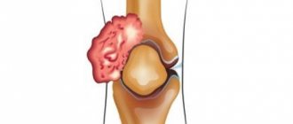
- At the third stage, clearly defined irreversible changes are visible. The cartilage becomes loose and almost completely destroyed. There is obvious dysfunction of the diseased joint. This stage is shown in the photo.
An X-ray of joints with arthrosis will help to diagnose the disease earlier, as a result of which it will be easier to relieve painful symptoms. The degrees of arthrosis on an X-ray are clearly visualized, and can be easily deciphered and described by an experienced radiologist.
- Is arthritis visible on an x-ray, and how to interpret the image?
Inflamed knee
Blood test for arthrosis
Since arthrosis has similar symptoms to other joint diseases, to distinguish it, for example, from infectious or rheumatoid arthritis, the following is prescribed:
- Clinical blood test. As a rule, arthrosis does not cause serious changes in blood counts, with the exception of a slight increase in ESR - up to a maximum of 25. With arthritis, ESR increases much more intensely - up to 40-80 units.
- Blood chemistry. The material is taken strictly on an empty stomach from a vein. With arthrosis, the indicators remain normal, but with arthritis, specific markers of inflammation appear - C-reactive protein, certain immunoglobulins, etc.
A blood test is needed to differentiate arthrosis from arthritis
In what cases are MRI and CT prescribed?
Sometimes, especially at an early stage of the disease, x-ray examination is not informative. In this case, magnetic resonance imaging is prescribed - an expensive but highly accurate method. It is based on the use of magnetic waves. With their help, it is possible to recognize even minor changes in cartilage, including at the initial stage. MRI is contraindicated in:
- an installed pacemaker, since changes in the magnetic field may affect the heart rhythm;
- the presence of electronic or ferromagnetic middle ear implants;
- large metal implants;
- pregnancy up to 12 weeks.
The study is not carried out in case of some relative contraindications, for example, in case of decompensated heart failure, claustrophobia, or other severe conditions of the patient.
If MRI is contraindicated, they resort to computed tomography, a modern analogue of X-ray. This method, which provides an image of all layers of the joint, is more accurate than radiography, but inferior to MRI.
MRI is a method that guarantees maximum accuracy and reliability of three-dimensional images. Unlike CT, MRI does not use radiation. A radiologist talks about these and other features of magnetic resonance therapy of joints:
X-ray diagnosis of primary idiopathic osteoarthritis
ABOUT
Steoarthrosis (OA) is a chronic progressive disease of the joints, characterized by degeneration of articular cartilage, changes in the subchondral part of the epiphyses of bones and in periarticular soft tissues.
The condition of articular cartilage is important not only for diagnosing OA, but also for assessing disease progression and treatment. The total thickness of the articular cartilage on radiographs is determined by measuring the width of the X-ray joint space between the articular surfaces of the epiphyses of the bones. The width of the joint space is still used as the main indicator in the X-ray diagnosis of OA.
, and standard AP and lateral X-rays of the knee joints are recommended by WHO and ILAR as the method of choice for assessing the dynamics of changes in articular cartilage during clinical drug trials.
Radiography remains the simplest and most widely available method for examining joints to assess anatomical changes in bone structure in OA. From the point of view of diagnostic efficiency, simplicity and ease of use, mobile X-ray diagnostic devices with a multi-position C-arm type stand, which are widely used in world practice, are especially interesting. Devices of this class make it possible to conduct examinations of the patient in any projection without changing the patient’s position. In Russia, the only manufacturer of such equipment is the St. Petersburg Research and Production Company “Electron”. The characteristic radiological manifestations of OA are usually easily determined on standard radiographs of the joints, while the narrowing of the X-ray joint space corresponds to a volumetric decrease in articular cartilage, and subchondral osteosclerosis and osteophytes at the edges of the articular surfaces are a response of bone tissue to an increase in mechanical load in the joint, which, in turn, , is the result of degenerative changes and a decrease in the volume of articular cartilage. These radiological symptoms are considered as specific for OA, are used to diagnose OA and are included in the radiological criteria (in combination with clinical ones) for diagnosing OA.
Radiological symptoms required for diagnosis of osteoarthritis
Narrowing of the x-ray joint space is one of the most important x-ray symptoms, which has a direct correlation with pathological changes occurring in the articular cartilage. It is known that the decrease in the volume of articular cartilage is distributed unevenly in different parts of the articular surface, and therefore the X-ray joint space in different parts of the joint has different widths. According to WHO/ILAR recommendations, the x-ray joint space width should be measured in the most narrowed area. It is believed that in a pathologically altered joint it is this area that experiences the greatest mechanical loads (for the knee joint these are the medial sections, for the hip joint – the superomedial sections, less often – the superolateral sections). Anatomical landmarks used to measure joint space on radiographs of large joints are:
a) for convex surfaces (head and condyles of the femur) - the cortical layer of the endplate of the articular surface of the bone;
b) for concave surfaces (edge of the acetabulum, proximal condyles of the tibia) - the edge of the articular surface at the base of the articular cavity.
Osteophytes
– bone growths on the edges of the articular surfaces of bones of various shapes and sizes are an extremely characteristic x-ray symptom of OA. Osteophytes in the initial stages of development of OA joints appear in the form of pointed or small-sized (1–2 mm) bone formations at the edges of the articular surfaces and at the places of attachment of the joint ligaments (in the knee joints, these are the edges of the intercondylar eminences of the tibia, at the place of attachment of the cruciate ligaments; in the hip joints - the edges of the fossa of the femoral head, on its medial surface, at the place of attachment of the own ligament of the femoral head). As the severity of OA in the joints increases and the progressive narrowing of the joint space, osteophytes increase in size, taking on various forms in the form of “lips” or “ridges”, straight or “lush” bone growths on a wide or narrow base. In this case, the articular head and socket can significantly increase in diameter, become more massive and flattened. The number of osteophytes can be counted separately or cumulatively in both joints, and their sizes are measured by width at the base and length. Changes in the number of osteophytes and their size are sensitive indicators of disease progression, and the absence of these changes may indicate success in the treatment of OA.
Subchondral osteosclerosis
– compaction of the bone tissue directly located under the articular cartilage. Typically, this radiological symptom is detected in the later stages of OA, when the joint space is already sharply narrowed and is a consequence of friction of the exposed articulating bone surfaces against each other. The articular bone surfaces become uneven. This indicates a deep degenerative process in the integumentary cartilage or its disappearance. Changes in the integrity of articular cartilage that precede its quantitative reduction may result from compaction of the cortical and trabecular bone tissue immediately underlying the cartilage. The compaction of subchondral bone tissue in the area of the articular surfaces of the bones is measured at 3 equally spaced points along the articular edge; The measurement results can be averaged.
Radiological symptoms not necessary for diagnosis of primary osteoarthritis
Periarticular marginal bone defect. Although this radiological symptom, which can be observed in OA, is defined by Altman et al as “erosion of the articular surface,” the term “periarticular marginal bone defect” is more preferable, since the exact histological characteristics of these radiographic changes have not yet been given. In patients with OA, they are small with an area of osteosclerosis at the base, while the surrounding bone tissue does not have a rarefaction of the bone structure, which is typical for true erosions detected in rheumatoid arthritis, which do not have sclerotic changes at the base and are often determined against the background of periarticular osteoporosis. Marginal defects of bone tissue can be detected in the early stages of OA; their appearance may be associated with inflammatory changes in the synovium. These changes are described in large joints and in the joints of the hands.
Subchondral cysts
– radiographically appear as ring-shaped defects of trabecular bone tissue in the subchondral bone with a clearly defined sclerotic rim. Subchondral cysts are formed as a result of bone resorption in the area of high intra-articular pressure, in the place of greatest load on the articular surface. Most often, these cysts appear during exacerbation of the disease and are located in the area of the narrowest part of the joint space. They are characteristic of hip OA and can be found in both the femoral head and the roof of the acetabulum. The dynamics of changes in subchondral cysts are judged by their number and size.
Intra-articular calcified chondromas
- are formed from areas of necrotic articular cartilage, and can also be a fragment of bone tissue - osteophyte or produced by the synovial membrane. Chondromas are usually small in size, located between the articular surfaces of bones or lie on the side of the epiphyses of bones, have different shapes (round, oval, elongated) and an uneven speckled structure associated with the deposition of calcium-containing substances into the cartilage tissue. Their number in the joint is minimal (1–2 chondromas).
In the knee joint, the sesamoid bone (fabella) in the popliteal fossa can be mistaken for a calcified chondroma, which also changes its shape, position and size in OA of the knee joint. Fabella deformity is one of the symptoms of knee OA.
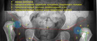
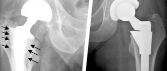
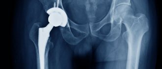
X-ray diagnosis of osteoarthritis of the hip joints
The accuracy of assessing the width of the joint space in the hip joints is influenced by 3 factors: the position of the patient, the rotation of the limb and the correct centering of the X-ray beam when radiography of the joint. A comparison of OA changes in the hip joints in the same patients in 2 positions (standing and lying) showed that when the patient was standing, the width of the joint space was significantly smaller than when the patient was lying on the table. The joint space is narrowed to a greater extent when the foot is directed inward. Shifting the X-ray tube away from the center of the joint can significantly change the width of the joint space. It is recommended that the central x-ray beam pass through the center of the femoral head. However, it is necessary to note the fact that separate radiography of the hip joints leads to an increase in radiation exposure to patients.
In the initial stages (stages 1–2 according to Kellgren) OA of the hip joints is determined by X-ray examination:
slight narrowing of the joint space, mild subchondral osteosclerosis, pinpoint calcifications in the area of the outer edge of the roof of the acetabulum (primordium of osteophytes), sharpening of the edges of the fossa of the femoral head in the area of attachment of the round ligament of the femoral head (Fig. 1).
Rice.
1. Overview Ro-graphy of the hip joint in a direct projection. Deforming osteoarthritis stage II. by Kollgren. Large osteophytes on the edges of the articular surfaces. Mushroom-shaped deformity of the femoral head In the later stages of the disease (corresponding to stages 3–4 of OA according to Kellgren) the following are noted:
• progressive narrowing of the joint space
• the formation of osteophytes of various shapes and sizes on the edges of the articular surfaces of the acetabulum, femoral head, which is why it acquires a mushroom shape over time. In the middle part of the acetabulum, a wedge-shaped osteophyte may form, which can cause lateral displacement of the femoral head
• deepening of the acetabulum may be associated with the development of osteophytes; its protrusion is possible against the background of osteoporosis or thinning of the bones that make up the bottom of the acetabulum
• pronounced subchondral osteosclerosis. Appears primarily in the area of the roof of the acetabulum, then in the upper part of the femoral head
• in advanced cases – a decrease in volume and flattening of the articular surface of the femoral head against the background of pronounced cyst-like restructuring of bone tissue, alternating with areas of subchondral osteosclerosis. Bone cysts can be single or multiple. They occur in the upper part of the acetabulum or in the area of greatest load on the articular surface of the femoral head
• aseptic necrosis of the femoral head
• subluxations of the femur, more often upward and laterally, less often upward and medially
• compaction of bone tissue and shortening of the femoral neck.
Free intra-articular bodies in coxarthrosis are rarely detected.
With secondary dysplastic coxarthrosis, all radiological symptoms develop early (in young or middle age) and can result in aseptic necrosis of the femoral head and subluxation or complete dislocation of the hip.
Ischemic coxarthrosis is described with rapid narrowing of the joint space, restructuring of the bone structure in the head and neck of the femur, early osteosclerotic changes, but without significant osteophytosis, with fairly rapid development of destruction of the femoral head.
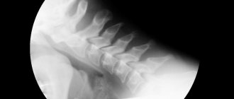
X-ray diagnosis of osteoarthritis of the knee joints
The knee joints are one of the most difficult joints to properly x-ray due to their structural complexity and wide range of motion. OA in the knee joints can be limited in extent even in a certain part of the joint, which also makes it difficult to diagnose changes in the joint. Recent clinical and epidemiological studies have confirmed the importance of examining the patellofemoral joint in the assessment of OA of the knee joint, since a joint examination of these joints reveals the disease in approximately 50% of all examined patients and proves that it is imperative to add a targeted image of the patella to direct radiography of the knee joints lateral or axial projection. The knee joint slightly bent in a standing position and in a straight projection is the most acceptable position for an objective assessment of the width of the joint space. Each knee joint is flexed so that the bearing surfaces of the tibial articular surface are horizontal, parallel to the central x-ray beam, and perpendicular to the cassette. The center of the joint (joint space) should coincide with the center of the x-ray beam. X-ray examination of the femoropatellar joint can be carried out with the patient lying on his stomach with the leg maximally bent at the knee joint or using the Ahlback method, when the patient is standing and the knee joint is bent at an angle of 30° from the vertical position. In this position, the joint is under functional load, it is ensured that the surfaces of the joint are clearly visible, and a more accurate assessment of cartilage tissue is provided than when the patient is examined in the supine position.
X-ray diagnosis of arthrosis of the femoropatellar joint in the lateral and axial projections is characterized by: narrowing of the joint space between the patella and the thigh; osteophytes on the posterior corners of the patella and femoral condyles; subchondral osteosclerosis of the patella; single subchondral cysts with a sclerotic rim.
This arthrosis is almost always external, sometimes external and internal, rarely only internal (diagnosed only by an axial image).
Early radiological signs (correspond to stages 1–2 of arthrosis according to Kellgren):
1. Stretching and sharpening the edges of the intercondylar eminence of the tibia (at the site of attachment of the cruciate ligament).
2. Slight narrowing of the joint space (usually in the medial part of the joint).
3. Sharpening of the edges of the articular surfaces of the condyles of the femur and tibia, more often in the medial part of the joint (associated with a greater load on this part of the joint), especially in the presence of varus deformity of the joint; less often - in the lateral part or simultaneously in both halves of the articular surface (Fig. 2).
Rice.
2. Ro-graphy of the knee joints in the lateral projection. Arthrosis of the femoropatellar joints (more on the left). Arthrosis of the knee joints (grade I according to Kollgren on the right, stage IV according to Kollgren on the left) With the progression of arthrosis of the knee joints (corresponds to stages 3–4 of arthrosis according to Kellgren):
• narrowing of the joint space increases
• subchondral osteosclerosis develops in the most loaded part of the joint
• multiple large osteophytes appear on the lateral, anterior and posterior edges of the articular surfaces
• subchondral cysts are rarely found
Secondary synovitis with the development of a subpatellar or popliteal Baker cyst:
• the articular surfaces of the femur and tibia flatten, become uneven and lose their anatomical and functional differentiation
• the sesamoid bone (fabella) takes on a multifaceted irregular shape
• calcified chondromas may be detected
• rarely possible development of aseptic necrosis of the condyles of bones.
Ostearthrosis of the proximal and distal interphalangeal joints
Standard radiography of the hands is carried out in a direct projection. The fingers are placed together, the hands lie flat on the cassette in line with the axis passing through the forearms and wrists.
Initial manifestations (correspond to stages 1–2 of arthrosis according to Kellgren):
Slight sharpening of the edges or osteophytes with mild subchondral osteosclerosis; small, subchondral cysts with normal or slightly narrowed joint space, small calcifications in the soft tissues in the area of the lateral edges of the articular surfaces of the bones.
Pronounced changes (correspond to stages 3–4 of arthrosis according to Kellgren):
Moderately pronounced or large osteophytes, deformation of the edges of the articular surfaces, significant narrowing of the joint spaces, osteosclerosis (Heberden's nodes in the distal interphalangeal joints and Bouchard's nodes in the proximal ones), cysts with a sclerotic rim, marginal defects of the articular surfaces, while the bony protrusions on one side may become wedged to another. Typically, marginal defects are surrounded by a zone of osteosclerosis (Fig. 3).
Rice.
3. Overview Ro-graphy of brushes. Multiple arthrosis of the distal and proximal interphalangeal joints. Multiple Heberden's and Bouchard's nodes. Severe arthrosis of the 1st left carpometacarpal joint Standard and microfocus radiography of joints
Methods for assessing the progression of OA are based on identifying changes in radiological symptoms in the joints. Long-term studies of radiological changes in patients with OA in the knee joints who received non-hormonal anti-inflammatory treatment showed the absence of radiological progression of the disease after 2 years of observation and minimal differences between the treatment groups and the control group. The lack of significant changes in these and other long-term studies suggests that radiographic symptoms with standard joint radiography remain relatively stable over long periods of time in OA and suggest that more sensitive technology such as microfocal joint radiography
, should be more widely used in assessing the dynamics of change.
.
Long-term studies of radiological changes in patients with OA in the knee joints who received non-hormonal anti-inflammatory treatment showed the absence of radiological progression of the disease after 2 years of observation and minimal differences between the treatment groups and the control group. The lack of significant change in these and other long-term studies suggests that radiographic symptoms on standard joint x-rays remain relatively stable over long periods of time in OA and suggests that more sensitive technology, such as , should be more widely used in assessing changes over time. . Microfocus X-ray machines use special X-ray tubes with a point source of radiation. Quantitative microfocus radiography with direct image magnification shows sufficient sensitivity to detect small changes in bone structure. With this method, the progression of OA and the effect of treatment can be recorded and accurately measured in a fairly short time between studies. This is achieved through standardization of the study and the use of a radiographic measuring procedure, improving the quality of the obtained radiographs of the joints with direct image magnification, which makes it possible to register structural details of the bone invisible on standard radiographs. WHO/ILAR recommends manually measuring joint space width using the Lequesne method using a magnifying lens and calculating the joint width at various points. Such measurements show that with repeated measurements the coefficient of variation is 3.8%. The development of microcomputer and image analysis technology provides a more accurate method of measuring changes in joint anatomy than manual methods. Digital processing of x-ray images of the joint allows the width of the joint space to be automatically measured by a computer. The researcher's error is practically eliminated, because the accuracy of repeated measurements is established by the system itself.
Ultrasound in the diagnosis of arthrosis
Ultrasound diagnostics for suspected arthrosis is used infrequently, since it does not provide as accurate results as X-rays or MRIs. Using the method, you can see all the tissues and cartilage, evaluate their degree of wear, thinning, and quantitative changes in the synovial fluid. Efficiency largely depends on the qualifications of the specialist, since deciphering what is seen is often subjective.
Ultrasound of the knee determines the degree of preservation of the menisci, uric acid crystals, Baker's cyst
What is the difference between ultrasound, CT and MRI? In what cases is this or that study indicated? The expert answers these questions:
Synovial fluid examination
To perform the analysis, a joint puncture is performed. The main parameters of synovial fluid are subject to study:
- macroscopic indicators - color, volume, viscosity, turbidity and mucin clot;
- number of cells;
- cytology of the stained specimen;
- microscopy of the native preparation.
Normally, synovial fluid has a straw-yellow color and a transparent consistency. With arthrosis, its viscosity increases, the mucin clot is formed well, the number of cells is normal or slightly increased (maximum up to 5000/μl). Other indicators remain mostly normal. With reactive synovitis, the number of neutrophils is reduced by half.
A serious change is observed during inflammatory processes accompanying various forms of arthritis. One way or another, the interpretation of the results is carried out only by an experienced rheumatologist, taking into account the medical history, laboratory and instrumental studies.
Analysis of synovial fluid makes it possible to clearly distinguish arthrosis from arthritis
Additional instrumental methods
Sometimes other methods are prescribed to clarify the diagnosis. They have a number of contraindications due to their invasiveness, are quite expensive and are rarely practiced.
- Arthroscopy. An early diagnostic method that allows you to detect changes in cartilage tissue, even if they are not yet visible on an x-ray. It is performed under local anesthesia for 30-60 minutes. It has high diagnostic accuracy and minimal risks of complications.
- Chondroscopy. A type of arthroscopy, the purpose of which is to study the condition of the cartilage, namely the degree of its damage. Most often used to evaluate the effectiveness of treatment with chondroprotectors for arthrosis.
- Osteoscintigraphy. A type of radionuclide diagnostics that involves the introduction of a radiopharmaceutical into the body to detect bone pathology.
Arthroscopy is a highly informative visual diagnostic method using an arthroscope
Diagnosis of arthrosis is often difficult because external symptoms are not so obvious and overlap with signs of other joint diseases. There are a number of instrumental and laboratory methods that allow you to make an accurate diagnosis. You should not ignore the specialist’s recommendations regarding the examination, because the correct choice of treatment tactics, and therefore the result, depends on this.
Clinical picture of the development of arthrosis
The main feature of the development of arthrosis is significant pain syndromes, while morphological metamorphoses can be practically invisible on an X-ray image. The opposite clinical picture also occurs; the patient experiences virtually no unpleasant sensations, but numerous changes are visible on the image.
Osteoarthritis of the knee joint
The main reasons for such opposite clinical pictures are:
- The absence of vasculature and nerve endings in the articular cartilage explains the absence of symptoms until the disease spreads beyond the joint.
- The presence of nerve endings in the synovial membrane, joint capsule, as well as in tendons and muscles are not always damaged evenly.
- The rate of development of arthrosis is individual for each patient and the slower it affects the joints, the less pronounced the manifestations of the disease will be.
It should be noted that most often arthrosis affects the shoulder, hip, knee, and wrist joints. It is when the disease begins that it is important to monitor the condition of all joints in order to begin drug therapy on time.
Arthrosis on x-ray