Detection of pathology of the musculoskeletal system at the initial stage in children in most cases seems to be a complex process. The problem is that it is important to diagnose such diseases at an early stage, when there may be no clinical manifestations. This issue is especially acute for the hip joint, dysplasia of which is detected in every seventh child. In such cases, radiography comes to the rescue. In the article we will look at what symptoms of pathology and indicators of normal hip joints in children can be found on x-rays.
What is hip dysplasia and its manifestations
Hip dysplasia (synonym - congenital hip dislocation) is a congenital pathology that is characterized by its inferiority. The cause of the disease is a violation of the formation of the joint during intrauterine development of the fetus. Factors that influence the course of the disease after birth also play an important role: tight swaddling of the baby and rickets.
Due to the inferiority of all components of the joint, the head of the femur can sometimes extend beyond the acetabulum (hence the second name - “dislocation”). Therefore, symptoms appear that make the doctor suspect the disease:
- asymmetry of skin folds on the buttocks;
- shortening of the thigh;
- muscle hypertonicity of the limb;
- limitation of hip abduction;
- a characteristic “click” when the leg is moved to the side (Marx-Ortolani symptom).
X-rays for hip dysplasia are performed to confirm the pathology. The indication for an X-ray examination is the presence of one or more risk factors: breech presentation, large fetus, foot deformity, the presence of dysplasia in one of the parents, toxicosis during pregnancy.
It is also worth mentioning that congenital hip dislocation in the vast majority of cases (more than 80%) occurs in girls.
Thanks to timely x-rays of the hip joints, the doctor has the opportunity not only to determine the presence and degree of dysplasia, but also to begin appropriate correction of the pathology.
Contraindications for radiography in children
X-ray diagnosis of hip dysplasia in children is allowed from the age of three months. Before this, active adaptation processes take place in the baby’s body, primarily hematopoiesis and the development of the nervous system. In addition, the ossification nucleus of the femoral head is formed between 2-5 months in girls and 3-6 months in boys. The X-ray radiation dose can inhibit these processes, so any X-ray examination before the specified age is carried out only if absolutely necessary.
In addition to this limitation, the following conditions are contraindications:
- immunodeficiency diseases;
- juvenile idiopathic osteoporosis.
Indications
X-rays of the hip joint are performed according to the following indications:
- joint deformity;
- shortening of the leg;
- swelling and redness in the hip area;
- pain during movement, after physical activity and at rest;
- suspicion of fractures of the bones forming the joint;
- before and after surgery (localization and comparison of fragments, reduction of dislocation, endoprosthetics, monitoring the effectiveness of drug treatment);
- suspected tumors, joint metastases, cysts, degenerative and metabolic diseases.
How is an X-ray examination of the hip joint performed in children?
X-rays of children's hip joints are performed using a standard digital or analogue machine. No special preparation required. In some cases, a cleansing enema may be performed before the x-ray so that the contents of the intestines do not distort the picture.
X-rays are taken in the supine position, in two states - with the legs straightened and with the knees apart. Both hip joints must be included in the image to allow comparison behavior.
Together with all the preparation of the equipment, the procedure takes about ten minutes, of which the child will be exposed to x-ray radiation for about a second.
During the X-ray itself, it is important that the child does not move. To do this, the mother or laboratory assistant holds him by the knees.
Contraindications and restrictions
There are no direct and obvious contraindications for x-rays of the hip joints. To protect the patient from scattered residual radiation during the examination, special personal protective equipment (aprons, collars, gonad covers) is used, which is supplied with all X-ray rooms. When performing radiography of the hip joint, the X-ray machine provides the ability to adjust the irradiation field to accurately target the area of study without affecting the rest of the body. Also, the X-ray machine itself has settings for both adults and children. Even for pregnant women and the fetus, x-rays of the hip joints do not pose any danger if protective equipment is used.
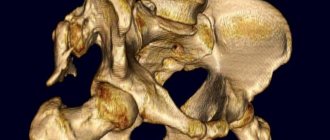
X-ray of a child’s hip joint, normal and with dysplasia
The examination and description of the x-ray image should be carried out by a qualified specialist - a radiologist or orthopedist. Determining normality and pathology based on X-ray results is a complex task and requires a lot of experience and knowledge.
To evaluate a photograph of the hip joints, the doctor measures certain angles and lines that he “draws” on the radiograph. To begin, draw a vertical line through the middle of the sacrum. Next - a horizontal straight line through the lower points of the ilium (Hilgenreiner line). Perpendicular to it, along the superolateral edge of the acetabulum, a vertical line is drawn (Perkin's line). Next, a tangent line is drawn through the edges of the acetabulum until it intersects with the Hilgenreiner line. The resulting angle is called “acetabular” and its assessment is important for identifying dysplasia. In newborns, its value is about 25 degrees and decreases with age. At five years old it is about 15 degrees.
An important role is played by the ossification nuclei of the femoral head, which normally appear on average at 4-6 months, depending on the sex of the child. Normally they are located below Hilgenreiner's line and medial to Perkin's line.
Two more important parameters are the “h” and “d” values. The “h” value shows the vertical displacement of the femoral head - from the Hilgenreiner line to the middle of the head. Normally it is 9-12 mm.
The “d” value is the displacement of the lateral edge of the femoral head relative to the acetabulum. The distance is measured from the bottom of the depression to the middle of the “h” value. Normally it is 15 mm.
X-ray signs of hip dysplasia
An X-ray of hip dysplasia in children allows us to determine differences in all of the above parameters. The magnitude of the acetabular angle in the disease is greater than the average age norms. It is not always possible to directly assess the angle itself, because with dysplasia, part of the acetabulum cannot be determined due to its immaturity. In this case, the radiologist has to “build” the angle along additional lines.
In addition, dysplasia on X-ray is determined by the later appearance of ossification nuclei. Even if they are present on the x-ray, they are smaller than normal and displaced higher and laterally. The value of “h” with dysplasia decreases, and “d” increases.
There are many additional and even more complex parameters that can only be assessed by experienced doctors.
When should the examination be carried out?
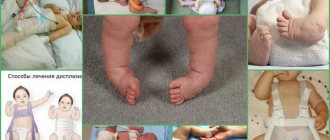
While the child is not walking, it is difficult to identify symptoms of dislocation or pre-dislocation. However, there is one external manifestation: free dislocation in the hip area and a click during reduction, which is also very easy. When a child begins to walk, asymmetrical skin folds and different leg lengths become noticeable. In sleep, when the muscles are relaxed, the leg takes a slightly outward position.
Important!
If a newborn baby's hips are spread apart, they should lie on the changing table. At a slightly older age, the deflection angle should be 60 degrees. If this figure is lower, this is a reason to consult a doctor.
Alternative research methods for suspected dysplasia
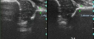
A child up to three months old can undergo an ultrasound examination. Ultrasonography of the hip joints is a fairly accurate diagnostic method and is absolutely safe for a small patient. The information content of ultrasound in the case of detecting a congenital dislocation is not inferior to x-rays for children under six months of age, since thanks to this method the structure of the cartilage is visible. Indications for its implementation are the presence of risk factors for the development of dysplasia.
Today, a program to conduct ultrasound for all newborns as a screening method is being actively implemented.
Advantages of contacting SM-Clinic in St. Petersburg
We employ some of the best specialists in the Northern capital. The examination is carried out using high-quality Siemens Sireskop equipment. This device is specially designed to allow radiography of the youngest children, and it also minimizes the radiation dose - depending on the area of study, it is 0.03-0.1 mSv.
There are no queues in our medical center, patients feel comfortable and calm. Call to find out prices for the services you are interested in or to sign up for a procedure. Registration is also possible through the website.
Pathology correction methods
A pediatric orthopedist deals with correction of hip dysplasia. After the most informative diagnosis and understanding of the degree of pathology in a particular patient, it is important to prescribe an appropriate treatment method.
The basic principle of any type of treatment is early initiation. Mild forms such as subluxation can be treated conservatively. The wide swaddling technique, exercise therapy, massage, and Pavlik stirrups are used. All techniques involve long-term holding of the legs in abduction and flexion positions and active movements within the permitted range.
For more complex or advanced forms, surgical treatment is used. It consists of correcting defective components of the hip joint - either the socket itself or the head (the operation never concerns the neck of the femur).
Preparing for an X-ray of the pelvic bones
Preparation for the procedure requires the patient to exclude from the diet foods that promote gas formation, perform a cleansing enema or take enterosorbents. Preparatory activities begin 72 hours before the diagnosis. For this period of time, dairy products, legumes, fresh vegetables and fruits, as well as black bread are excluded from the diet. Diagnosis is carried out on an empty stomach: at least twelve hours must pass between the last meal and the procedure. A cleansing enema is done in the evening before the day when the examination will be carried out, and, if necessary, two hours before it.
How are pelvic bones x-rayed?
The patient is asked to remove metal objects from the body and report whether there are grafts or other foreign objects in the body. Pictures are taken in frontal and lateral projection:
- The direct projection requires the patient to assume a supine position with his legs straightened and feet turned inward. A special cushion is placed under the knees. If this position causes pain, the patient is placed on his stomach, raising the pelvis from the healthy side;
- The lateral projection requires the patient to lie on their side with the lower limb flexed at the hip. If pain symptoms are present, it is allowed to bend both legs.
What does an x-ray of the pelvic bones show?
Deciphering the results requires not only clear images, but also relevant knowledge from the doctor. This is due to the fact that changes of the same type have different interpretations. That is why in the process it is necessary to take into account the patient’s history and complaints: each disease or anomaly has certain clinical manifestations. So:
- Displacements in the hip joint noticeable in the image indicate its dislocation;
- Anomalies in the structure of the femoral head and acetabulum are signs of dysplasia;
- Narrowing of the joint space and the presence of osteophytes indicate a degenerative disease such as osteoarthritis;
- Darkening is a sign of malignant neoplasms, shadows are metastatic foci;
- Bone fragments confirm the suspicion of a fracture;
- Clearly visible regeneration of bone structures is a symptom of aseptic necrosis.
You can make an appointment for an x-ray of the pelvic bones at the CELT clinic right now through our website or by phone.