Ultrasound of the hip joints in infants is held at the Health 365 clinics at the address: st. Bazhova, 68, Ekaterinburg.
An ultrasound of the hip joints is recommended when a newborn (age from birth to 4 weeks) becomes an infant (from 4 weeks to 1 year), i.e., when the baby is 1 month old. Ultrasound of the hip joints is a study that allows a baby to detect the presence of dysplasia (impaired formation), subluxation or dislocation of the hip joints using harmless ultrasound. Ultrasound diagnostics is highly informative, as it makes it possible to obtain an image of the cartilaginous structures of the hip joint, make a diagnosis at the right time, draw a precise line between neurological and orthopedic pathology, and determine treatment tactics, which in each of these cases has its own specifics.
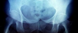
Until recently, the leading role in the diagnosis of developmental disorders of the hip joints was given to the x-ray method. However, the radiation exposure that accompanies the X-ray method and the difficulty of correctly positioning the child for examination are its disadvantages.
The modern approach to the early diagnosis of developmental disorders of the hip joints involves the active identification of infants with this pathology using ultrasound, to solve the main problem - making the diagnosis as early as possible and starting treatment before 6 weeks of age.
Such an early start of treatment allows you to use the high growth potential of the structures of the hip joint, achieve complete anatomical and functional restoration in a short time, prevent possible complications such as necrosis of the head, early arthrosis, and eliminate the possibility of surgical intervention.
In what cases is ultrasound of the hip joints prescribed?
Diagnostics can be prescribed by a traumatologist, surgeon or orthopedist. Typically, ultrasound examination of the hip joints is indicated in the following cases:
- Limited mobility, difficulty getting out of bed after sleep;
- The appearance of stiffness when moving;
- Painful sensations;
- Suspicion of a dislocation or dislocations in the past;
- Change in skin color in the hip joint area;
- The appearance of a distinct crunch in the joint during physical activity;
- Frequent muscle spasms in the buttocks and thighs;
- Different lengths of the lower limbs.
Advantages and disadvantages of ultrasound of the hip joints
Today, there are many different diagnostic procedures, such as x-rays, computed tomography or magnetic resonance imaging. Of course, these studies provide more detailed information, but such methods have a higher cost. Ultrasound examination, in turn, is cheaper, completely safe and allows you to identify pathological processes even in the early stages. In addition, the diagnostic result can be obtained within 10 minutes after the end of the examination.
Let's look at a few more benefits:
- The procedure is non-invasive;
- It is painless;
- It is also possible to study the condition of the blood vessels in the area under study.
How is the procedure carried out?
The patient removes clothing below the waist (except underwear) and lies down on the couch. Diagnosis is made using a sensor, with which the doctor reads information about the condition and density of bone, cartilage and soft structures. To obtain a complete clinical picture, the doctor uses several access options.
Anterior approach
The ultrasound doctor asks the person to lie on his back, and a small cushion is placed under the femoral surface. This position helps to better examine the structural elements of the hip joint. By observing the image on the screen, the specialist receives information about the condition of the ilium, femoral head, ligaments, muscles and lymph nodes of the groin.
Rear access
For such an examination, the specialist asks the patient to lie on his side and bend his knees slightly closer to his chest. During the study, it is possible to identify the functioning and characteristics of the sciatic nerve, obtain information about the surface of hyaline cartilage, and examine the superficial and internal structure of muscle fibers in the gluteal region.
Medial access
The person lies on his back, bends the lower limb and moves it to the side. This position allows for a detailed examination of the pelvic flexor muscles and articular ligaments if a pathological process is suspected.
Lateral access
The person lies on his side, and the specialist asks him to rotate his hip inward. In this position, the protruding elements of the tibia and the trochanteric bursa are clearly visualized.
On average, this manipulation lasts no more than 30-40 minutes. The patient is given the results of the examination; in some cases, additional images are attached so that the doctor can independently assess the clinical picture.
Dr. Komarovsky about dysplasia in children
In order to avoid unpleasant consequences and not to miss the development of the disease, the child should be shown to an orthopedist at one month of age, who will refer the baby for an ultrasound examination .
Children at risk include those who:
- at birth there was a breech presentation;
- the mother and immediate family have been diagnosed with dysplasia;
- During pregnancy, my mother suffered from toxicosis, severe infectious diseases, and did not eat well.
Until now, the exact cause of developmental disorders of the hip joint has not been identified, but the symptoms of a dangerous disease of the musculoskeletal system have been identified.
The following manifestations may be a warning sign for parents:
- when the baby’s legs are spread, their movement is limited;
- the joint “clicks”, crunches;
- the folds of the buttocks are uneven and different in depth;
- If the child's legs are bent while lying on his back, then one knee looks higher than the other.
Decoding the results
An orthopedist, surgeon, or orthopedic surgeon can interpret the results. The research protocol usually contains the following information:
- the presence of pathological processes, a detailed description of the deviation.
- congenital or acquired changes inside the joint;
- condition of ligaments and nerve bundles;
- increase in the thickness of the joint capsule;
- functioning of adjacent muscles;
- features of cartilaginous structures;
- the amount of synovial fluid inside the joint.
- the presence of neoplasms, if any, their shape, structure, size and individual characteristics;
- information about the ilium, labrum and femoral head;
- metastasis of nearby tissues;
- presence of hematomas.
Please note that the result of an ultrasound examination is not a basis for making a diagnosis. If any inconsistencies or pronounced pathological processes are detected, it is additionally necessary to perform a computed tomography scan and pass various tests for laboratory research. In some cases, a biopsy may be necessary (for example, if the doctor suspects a malignancy). The final diagnosis and necessary treatment are prescribed only after additional studies.
Normal hip joints according to ultrasound
If the patient does not have various pathological processes, the protocol indicates that the hip joints have a clear and smooth surface, without deformations and bone growths. The joint capsules should have folds and branches; their structure is normally hypoechoic.
The following information is also usually indicated:
Right joint:
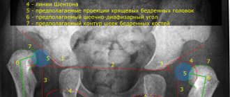
The bony part of the acetabulum is normal. No major changes. Rectangular bony protrusion. The overlap of the cartilaginous part of the roof of the femoral head is sufficient. The limbus is projected laterally from the femoral head, is presented, and has a normal angle of inclination. The head of the femur in the acetabulum is located correctly. The ossification nucleus is located. Angle a is less than 60 degrees. Angle b is greater than 55 degrees.
Left joint:
the roof of the acetabulum is moderately flattened, without structural changes. The femoral head is centered correctly. The ossification nucleus is located. Angle a is more than 60 degrees. Angle b is more than 55 degrees. The angular parameters of the joint are not changed. When performing functional tests, the joint is stable.
Please note that these indicators may vary depending on age - deviations from them do not always indicate the presence of diseases or some problems.
Diagnostic criteria
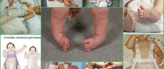
Symptoms of the dysplastic process of the hip joint with the formation of anomalies in the development of the pelvic bones can often develop in infants. If the examination and treatment are carried out correctly, upon reaching the age of one, the child becomes absolutely healthy and begins to move independently.
If the disease is severe or advanced and shows clinical and radiological signs of congenital dislocations or subluxations in the joint, serious treatment will be required before surgery and subsequent rehabilitation of the joint functions. In such a situation, clothing for children with dysplasia is selected taking into account the possible wearing of plaster or special splints and orthoses. It is necessary to swaddle the baby freely, without restricting movements.
When assessing the state of the musculoskeletal system in a newborn, one should take into account the fact that many parts of the musculoskeletal system in a newborn child are not mature and continue to form under the influence of a number of factors, both external and genetically determined. This phenomenon is a physiological norm. It takes several years for the structure of the hip joints to fully develop. Even at such early stages of a child’s development, characteristic signs are identified to assess the condition and functionality of the joints, providing an opportunity to determine whether the development and formation of the tissues of the hip joint is occurring correctly.
What pathologies can be identified
Most often, during an ultrasound examination of the hip joints, the following diseases are detected:
- Arthrosis (it develops as a result of congenital dislocation of the hip, a disturbance in the circulatory system, or as a result of a fracture of the femoral neck);
- Arthritis (may occur due to infection of joint joints by pathogens);
- Synovitis, which is characterized by inflammation and fluid accumulation inside the joint;
- Necrosis of bone tissue (can occur due to frequent fractures, dislocations, bruises);
- Tears and spasms of muscle fibers;
- Swelling of the joint.
The hip joints are most susceptible to various pathological processes. Diseases can be either congenital or acquired. If you visit a medical facility as soon as the first symptoms appear, you can hope for a favorable outcome and complete recovery. Otherwise, your doctor may recommend surgery.
Benefits of Sonography
When conducting an ultrasound examination, high-frequency acoustic waves are reflected from surfaces encountered in their path. All tissues in the human body have their own special structure. They differ in density, contain a certain amount of liquid, and therefore differ significantly in the speed of reflection. These values are read by special sensors and then visualized on the monitor in the form of images. An experienced diagnostician, when interpreting an ultrasound of the hip joints in infants, identifies ligaments, tendons, bone, and cartilaginous tissues, and then assesses their condition. Ultrasound has many advantages over other methods of studying the hip joint in young children:
- there is no aggressive and extremely undesirable radiation when examining newborns;
- one of the parents is present during the diagnostic procedure, hold and calm the baby;
- there is no need for the child to be in a stationary position, so the doctor can assess the state of the joint in dynamics.
3% of newborns are diagnosed with congenital dysplasia of varying degrees. This is the name for defective one- or two-sided development of the hip joints, which is characterized by a decrease in the functional activity of the joint. The disease is highly treatable, especially if diagnosed early. The resulting images make it possible to determine the shape and degree of dysplasia, and differentiate it from other congenital pathologies. What can be diagnosed using ultrasound:
- improper development of the acetabulum;
- underdevelopment of the cartilaginous rims surrounded by the acetabulum;
- broken ligament structure;
- degrees of dysplasia - preluxations, subluxations, complete dislocations.
If any form of dysplasia is detected, doctors immediately begin therapy. To assess its effectiveness, frequent ultrasound examinations of the hip joints in infants are required. This is another advantage of the diagnostic technique. Unlike radiography, sonography can be used to constantly monitor the baby’s condition and the rate of tissue regeneration. The diagnostic procedure has only one contraindication - the child’s hypersensitivity to the ingredients of the gel used to conduct acoustic signals.
X-ray examination is also less informative due to the structural features of the hip joint in newborns. There is still little bone tissue in it, but a lot of cartilaginous tissue. And in the images obtained during radiography, only the pathological state of the bone structures is clearly visible.