until October 31
We're giving away RUR 1,000 for all services per visit in October More details All promotions
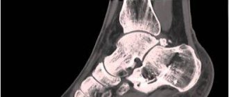
X-ray of the spine (x-ray of the spine)
is a simple and effective diagnostic method that allows you to identify pathologies, primarily associated with spinal deformity.
The human spine is adapted to upright posture: it connects various parts of the skeletal system, maintains the vertical position of the body and acts as a shock absorber when walking. That is why the spinal column has an S-shaped, curved shape and is not a rigid rod, but a complex structure consisting of elements attached to each other - vertebrae. Between the vertebrae there are intervertebral discs that absorb shocks and shocks.
parts of the spine
The spine is divided into sections. Highlight:
- cervical spine, consisting of 7 vertebrae (designated C1-C7);
- thoracic spine: 12 vertebrae (designated D1-D12);
- lumbar: 5 vertebrae (designation L1-L5);
- sacrum or sacrum: 5 fixed vertebrae that form the sacrum;
- coccyx.
X-ray of the spine with functional tests
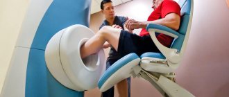
The most informative is considered to be radiography of the spine, which is performed in a vertical position.
To obtain maximum information, radiography is performed with a functional load - with bending, maximum flexion or extension in a vertical, sitting or horizontal position. It provides additional information to clarify the diagnosis. Functional tests are strictly individual in each specific case. They allow:
- assess the mobility of the spine: determine the full range of movements, functional blocks (limitations of mobility) in the selected segment;
- more accurately identify the degree and nature of vertebral displacement;
- plan orthopedic surgery.
How is the examination carried out?
Each part of the spine is examined differently and requires a different level of preparation. It is worth considering the conduct for each area separately.
X-ray of the cervical spine
Radiation doses during X-rays of the spine do not exceed the norm, no matter what part we are talking about. To examine the cervical spine
, the patient may be asked to lie down on a special table or remain standing. When you need to get an image of vertebrae 1 through 3, you will need to take a targeted shot through the mouth. The patient is asked to open his mouth so that the shadow of the lower jaw does not cover the necessary vertebrae, after which an x-ray is taken. Functional tests may also be necessary - the patient needs to bend his neck as much as possible and touch his chin to his chest to take the first picture, and also tilt his head back as much as possible to take the second.
X-ray of the thoracic spine
X-rays of this area are not as frequent, which is associated with limitations in the mobility of the thoracic region
. If the procedure is still necessary, it can be performed standing or lying down, and the picture is taken in two projections - lateral and direct. Occasionally, oblique tests may be prescribed, as well as functional tests, which are very rarely necessary due to limited mobility. In the latter case, the doctor himself determines the optimal position and asks the patient to take it.
X-ray of the lumbosacral spine
You need to think not only about how often you can do the X-ray procedure of the spine
, but also how such research is carried out. After careful preparation, the patient is asked to lie on the table. AP and lateral views may be necessary, and the legs may need to be flexed.
Expert equipment
The study is carried out on the latest generation X-ray system DIGITAL DIAGNOST (PHILIPS, the Netherlands). This is a completely digital x-ray station. Its advantages:
- Maximum data transfer and processing speed
- Obtaining the highest resolution (quality) image possible
- Lowest radiation exposure comparable to a short flight
The results of radiation diagnostics of the Yauza Clinical Hospital are accepted anywhere in the world, which is important for patients planning to undergo certain stages of treatment abroad.
Preparation the day before the examination
Preparation for an x-ray begins several days in advance, but it is most important on the day of the procedure. You will need to come to the office on an empty stomach, regardless of the appointed time. Therefore, many advise signing up for the morning hours. In the morning you need to follow the following rules:
- Completely avoid breakfast, coffee, tea or even juice.
- Drink a bag of laxative if there are more than 6 hours before the appointed time or give a cleansing enema.
- Give up bad habits, do not smoke on the day of the event.
- Drink 15 drops of valerian tincture or other sedative to reduce possible stress.
- Take water and food with you so that you can eat immediately after the procedure if it takes place in the afternoon or evening.
- Before undergoing the examination, you must remove any jewelry, especially piercings.
Preparatory procedures are extremely important when examining the lower back or lower back. The described measures will make it possible to more effectively cleanse the intestines of the accumulation of gases and feces, improve visualization, and increase the accuracy of diagnosis.
[node:field_similarlink]
Alternative research methods
Sometimes patients wonder: what is better – CT, X-ray or MRI of the spine? Each method has its own capabilities, advantages and disadvantages.
- X-ray of the spine is a method that allows for examination with functional tests, as well as standing, with axial load and imaging along the entire length of the spinal column. The radiation dose with it is less than with CT. Limitations of the method - there is no way to assess the structure of the spinal cord and muscle corset.
- MRI is a method that allows you to evaluate the structure of the spinal cord, intervertebral discs and their herniated protrusions, joints, cartilage, nerve roots emerging from the spinal canal, soft tissues, muscle corset and identify inflammatory changes in the vertebral bodies.
- - an informative method for assessing bone structures with a higher resolution, but also with a higher radiation dose than with radiography. CT allows for 3D reconstruction of bones. Indispensable when planning operations. Compared to MRI, CT is significantly inferior in assessing soft tissue and cartilage structures.
Contraindications
Features of the procedure
Depending on the expected diagnosis, the doctor in the prescription indicates the position that the patient will take in the process. This will allow you to take pictures in different planes. In the case of x-rays in the lumbar region, two projections are used: frontal and lateral. They are represented, in turn, by the following variations:
- Front. It involves placing the patient with his back to the device tube and facing the fluorescent screen.
- Rear. The patient is positioned facing the X-ray tube and with his back to the screen.
Additionally, oblique projections can be performed to identify vertebral displacements, since the lower back is a fairly mobile area. In this case, the patient is positioned at an angle of 45 degrees to the protective screen. A certain position of the body increases the information content of the image and allows the doctor to accurately diagnose the pathology, its stage and degree of development.
No ads 2
Spine
For examination, the patient should be placed with his back to the light source. The person being examined should stand straight, with relaxed muscles, barefoot, with his arms hanging freely along the body. In a normally built adult, the spine has physiological curvatures in the form of two lordoses in the cervical and lumbar regions and one kyphosis in the thoracic region. In children in the first months of life, the spine is in the form of uniform kyphosis. In a one-year-old child, the spine approaches a straight line and remains this way until approximately 7 years of age. The final shape of the spine is established by adulthood and remains until 45-50 years of age, after which the thoracic region again begins to gradually round, approaching senile kyphosis. In adult women, lumbar lordosis is more pronounced than in men.
In practice, in addition to the normal structure of the spine, it is customary to distinguish the following types of postures: flat back, round back, stooped back. In the thoracic region, a slight deformation is enough for kyphosis to become clearly noticeable. The appearance of kyphosis in the cervical or lumbar regions indicates the presence of serious pathological changes: the protrusion of one or more spinous processes with angular kyphosis forms a hump (gibbus), which can be observed with partial or complete destruction of the vertebral bodies. The lateral curvature of the spine is called scoliosis. It is detected by the deviation of the line of the spinous processes from the vertical axis of the body, drawn through the intergluteal fold. On the convex side, the shoulder girdle and shoulder blade are raised.
Functional scoliosis, caused by significant shortening of one leg, appears when the patient is standing and disappears when lying down. With thoracic scoliosis, a rib hump is formed on the convex side, which is especially visible when flexed. Tension of the long muscles of the back is noticeable in the form of protrusions on the sides of the spinous processes; this symptom is especially often observed with discogenic radiculitis.
Indications for radiography of the lumbosacral region
Only a doctor can prescribe x-ray diagnostics, based on the patient’s complaints, initial examination and other data. Indications for the procedure are: congenital anomalies in the spinal column; injuries and the presence of complications after injuries; suspicions of the presence of neoplasms; rachiocampsis; loss of sensation in the legs; pain in the limbs and back.
During the study, only the conditions of the vertebrae themselves are considered; anomalies of ligaments, intervertebral discs, blood vessels, etc. can be judged through the use of other research methods, such as MRI or CT. At the same time, the study will help determine the presence of infectious and inflammatory processes of the spine, tumor formations and degenerative diseases of the spine through the analytical abilities of the doctor.
Content:
- Indications for radiography of the lumbosacral region
- Contraindications to undergoing the study
- Preparing for the study
- Algorithm for X-ray diagnostics
- Decoding the results
Contraindications
Not everyone can have an X-ray of the lumbar region; it is contraindicated for some patients. This group includes small children, pregnant and lactating women, and overweight people. Manipulation is especially dangerous in the first trimester, when all the organs and internal systems of the embryo are formed. Children under 15 years of age are recommended to use a protective film during the procedure if there is no alternative to diagnosis.
It is also not recommended for people suffering from neurological pathologies or mental disorders. It should be noted that the radiation received during operation of the device does not accumulate, so there is no point in taking any measures to remove it.
X-ray is the most informative, painless and inexpensive way to find out about the condition of the musculoskeletal system. It is carried out only as prescribed by a doctor in a district clinic or hospital. In this case, you do not need to pay for the procedure if you have a compulsory medical insurance policy with you. In private medical centers, the referral service is not required, but it is not provided free of charge. In the case of examining the lumbar region, special preparation will be required to increase the information content of the image.
Identification points of the spine
In the cervical region, the identification point is the superior spinous process of the seventh cervical vertebra, especially clearly visible when the upper limbs are lowered. The line connecting the inner ends of the scapular spines passes through the spinous process of the third thoracic vertebra. The line connecting the angles of the shoulder blades passes through the spinous process of the seventh thoracic vertebra. The line connecting the highest points of the iliac crests passes through the spinous process of the fourth lumbar vertebra. When examining a normal spine, very limited sections are accessible by palpation - only the ends of the spinous processes. When running the palmar surface of the second finger along the spinous processes downwards, starting from the cervical region, you can even detect a slight protrusion of the spinous process posteriorly or to the side.
The localization of painful foci is determined by pressing the distal phalanx of the first finger on the spinous processes of the vertebrae from vertebra to vertebra, from top to bottom. The transverse processes are palpated away from the spinous processes. To determine the localization of the pathological process, tapping on the spine is sometimes used; the shock causes pain in the affected area. The same can be achieved by applying certain pressure on the head or shoulders along the axis of the spinal column. It should be noted that these techniques are quite crude and are not always applicable.