Computed tomography (CT) of the ankle joint is an x-ray type of study based on the transfer of information about the state of the internal structures of the ankle, followed by sectoral digital processing. The research results are analyzed by examining detailed fragments - sections. The Siemens CT machine installed in our center allows us to obtain submillimeter sections with a thickness of 0.5 mm and detect the smallest changes in the structure of the joint under study.
What does a CT scan of the ankle show?
The ankle is a rather fragile connection of a number of small joints. Some bones are relatively small and may not fit well in certain views on conventional x-rays. Computed tomography of the ankle joint does not have this drawback - thanks to post-program processing, it is possible not only to examine in detail the condition and structure of the bone joint component from all sides in different planes, but also to build a three-dimensional image of the joint - a 3D model.
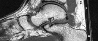
Tomogram of the ankle joint
Injuries and various injuries to the ankle require immediate examination. The ankle joint, along with its fragility, bears enormous loads and is often subject to damage of varying degrees of severity. The timeliness of detection and analysis of damage largely determines the further success of treatment and restoration of impaired function. Before taking remedial measures, it is necessary to understand and analyze the problem in detail. For this purpose, modern medicine actively and successfully uses computed tomography of the ankle joint.
Benefits of X-ray
Radiography is one of the main diagnostic methods. It has a large list of indications, high reliability and information content, comparative ease of implementation, and there is no need for preparation. During the x-ray, the integrity of the skin is not damaged, everything is painless. The examination will take 5-10 minutes.
Fears about the dangers of X-rays are unfounded if the examination is carried out on a digital machine. It has a minimal load level, and the images are highly accurate and detailed. With such a device, you can take several pictures in a row (in different projections) in one procedure and undergo examination several times a year as needed. Digital X-rays are not only safer than analog ones, but also more convenient. There is no need to wait until the pictures are printed, all the results are stored on digital media, they are convenient to use, enlarge while viewing, and store. Therefore, when choosing a clinic where to have an ankle x-ray done, check the type of equipment used for the procedure.
Computed tomography of the ankle joint, safety of the method
The device on which computer examination is performed is called a tomograph. It consists of several interconnected parts:
- the X-ray machine tube is the direct source of rays, directing them through the thickness of the human body;
- Gantry frame – a frame that receives the return flow of processed signals. It is inside it that there is a mechanism due to which one or more X-ray tubes move during the examination of the patient;
- system for converting and analyzing the received data - a set of devices that evaluates the received x-ray encryption and converts it into data about the state of the body fragment being studied;
- a movable computer operating table (transporter) is the immediate place where the patient is placed for examination. During the CT scan, the table shifts the patient’s body relative to the simulated information field required by the device;
- computer program for decoding the final analysis and output of data.
Obtaining information about the state of health or the presence of pathology in the ankle joint is based on the ability of different types of tissue to absorb and transmit a certain dose of X-ray radiation, which is a stream of particles with a high energy and frequency charge.
Tomograph used in our center
Specialized sensors during the procedure actively record the levels of decrease or increase in the number of particles as they pass through different environments of the human body. The bone tissues of the body have the greatest degree of absorption of high-frequency energy, and the least - organs containing single or multiple air cavities.
The technique has been used for many decades and has proven to be highly informative in diagnosing many diseases, as well as absolute safety with a reasonable approach - CT scan of the ankle is performed only if indicated, you should not do the study just like that.
Indications for X-ray
Main indications for ankle x-ray:
- Fractures, cracks of bones that enter the joint: fibula, tibia, talus.
- Dislocations, cartilage injuries, sprains.
- Bone tumors, metastases.
- Vascular calcification.
- Bone necrosis.
- Deformation of the feet, clubfoot, flat feet.
- Arthrosis, arthritis, osteoporosis and other diseases.
The main symptoms of diseases and long-term consequences of injuries are:
- Swelling of the joint area, foot.
- Pain that may intensify or appear with changes in weather, in damp and cold weather.
- Stiffness of movements.
- Poor circulation of the foot, colder and paler skin on it.
- Changes in gait, uneven wear of shoes.
How is a CT scan of the ankle performed?
After completing the documents, the patient is invited to the room with a tomography unit and is placed on the tomograph table. The ankle is securely fixed in special supports to avoid motion artifacts, and the rest of the body is necessarily covered with a lead apron. The patient spends approximately 10-15 minutes in the office, depending on the complexity of the clinical case. If there is a suspicion of vascular pathology or an oncological neoplasm, a contrast agent is administered intravenously to clarify the diagnosis.
To achieve maximum data accuracy, our center uses post-program processing of the received images. As a result of scanning, all images are taken in the axial plane, and then computer programs build horizontal and frontal projections so that the doctor can evaluate the structure of the ankle from all sides.
For a more accurate CT scan of the ankle joint, it is preferable to perform the study on multislice tomographs - this is the one installed in our Magnit center - Siemens Somatom Emotion 16. This type of tomograph is optimal for CT scans of the ankle in terms of price and quality of the images obtained.
Procedure for performing a CT scan of the ankle joint
If the tomographic picture is complex, especially if an oncological tumor is suspected, the use of a contrast agent during the study is allowed. The drug is administered intravenously and allows you to more accurately analyze the stage of formation and localization of metastases, as well as assess the condition of blood vessels and detect disturbances in blood flow in the area of study caused by atherosclerosis or thrombosis.
Contraindications
There are no absolute contraindications to digital radiography.
Relative ones include:
- Pregnancy – radiation exposure can have a negative impact on the developing baby.
- A situation where a patient needs emergency care.
- A highly agitated state or mental illness that makes it difficult to remain still while the X-ray machine is operating.
- Age up to 14 years.
In these cases, the doctor decides whether an x-ray is necessary. He may prescribe a different diagnostic method or perform x-rays with additional precautions. To minimize training, a lead apron is used to cover the genital area of an adult, or the entire body of a child.
Computed tomography of the ankle joint: indications and contraindications for the study
The ankle joint belongs to a group of anatomically complex joints. Due to the fact that its constituent parts include not a pair, but several bone joints at once, pathologies can also be different. Basically they can be grouped into three groups:
- tissue pathologies, without affecting the bone structure: sprains and tears of ligaments, external and internal damage to muscle layers and blood vessels;
- pathology of the skeletal system without affecting the superficial soft tissues (most often these are oncological processes);
- combined pathology of soft and bone tissues: fractures, displacements, open injuries, dislocations and subluxations.
In the first option, computed tomography of the ankle joint is not used; preference is given to magnetic resonance imaging, which has a greater affinity for soft tissue. But with the second and third options, it can be very difficult to do without computer research.
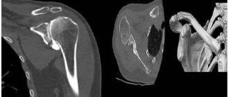
3D model of the ankle (front and back views). Arrows indicate fracture sites
Indications for computed tomography of the ankle joint
The main indications for this type of study will be:
- open and closed fractures of the ankle joint;
- ankle sprains and subluxations;
- suspicion of a bone fracture caused by a blow or injury;
- rupture of soft tissues and blood vessels accompanying the fracture;
- suspicion of an oncological tumor in bone or cartilage;
Computed tomography can also be used:
- as a comprehensive study for complex injuries received during a disaster;
- as an additional study before the upcoming operation in neighboring areas.
Contraindications to computed tomography of the ankle joint
Since X-rays are used for diagnosis, there are a number of contraindications:
- pregnancy in any trimester;
- breastfeeding period - you need to temporarily stop feeding and express milk at least twice;
- children under 5 years of age;
- radiation sickness;
- recently performed other X-ray examinations (fluorography - less than 4 months ago, other types - less than 6 months ago), provided that a CT scan of the ankle is planned independently, without medical recommendations and indications;
- mental illness of the subject: autism, severe claustrophobia, epilepsy, schizophrenia - only with the consent of the appropriate doctor;
- the presence of metal inserts, prostheses or other elements after metal osteosynthesis in the scanning area - it is important to agree on these points before the study, as they may complicate the description.
Alternative diagnostic methods
- — obtaining high-precision and high-quality images to identify the pathology of the bone structures of the joint and determine the pathological accumulation of fluid in the joint cavity (pus, blood). Provides a certain radiation exposure.
- MRI - there is no ionizing radiation, it allows you to evaluate the condition of the cartilage and soft tissues that make up and surround the ankle joint (muscles, tendons, ligaments, cartilage) and the blood flow in them.
- Ultrasound - reveals the pathology of soft tissues adjacent to the joint, an absolutely safe and inexpensive research method.
Carrying out a CT scan of the ankle joint in St. Petersburg.
The Magnit Medical Center offers to perform a CT scan of the ankle joint using a modern tomograph from Siemens. To make an appointment, just call or leave a request on the website.
This month there is a special price for CT scanning of the ankle - the procedure can be completed for only 2,450 rubles.
The results of the study can be obtained in the form of a digital recording on a CD provided by our center or, if desired, printed on film. The doctor who conducted the study will advise after receiving the results and will tell you whether the identified changes require further treatment or if everything is in order.
We are waiting for you at 6th Krasnoarmeyskaya Street, 5-7.
What pathologies can be diagnosed using x-rays?
As a result of the x-ray, high-quality images will be taken and analyzed. Based on the information received, the doctor will determine which diseases the detected changes correspond to.
Congenital and acquired deformities (clubfoot, flatfoot)
This problem is most often faced by parents of young children. Many factors contribute to the occurrence of deformations. These include: heredity, malpresentation of the fetus, lack of amniotic fluid, improper development of the nervous system. Also a reason why you may need to have an ankle x-ray
, is improper fusion of bones after a fracture.
Fractures and cracks
Foot and limb injuries are some of the most common reasons why x-rays are needed. The images clearly show even the smallest violations of the integrity of the bone tissue, which allows the doctor to prescribe a plaster cast and prescribe restriction of mobility until the bones heal.
Arthritis
Arthritis is characterized by joint pain, restricted movement, swelling of the limb and fever. These symptoms are usually more pronounced after a night's sleep and can also cause it to worsen. In the photographs, this process is expressed by changes in the structure of bone tissue and inflammation.
Arthrosis
Arthrosis is characterized by degeneration of cartilage tissue and is irreversible. It often occurs after a person has been injured, but persistent increased loads on the lower extremities can also be the cause. With an X-ray of the lower leg, the doctor can fully examine the cartilage and joint, and then draw a conclusion about the pathological processes in them, prescribe treatment and achieve stable remission.
Gout
Gout is a type of arthritis characterized by the deposition of uric acid salts. As a rule, gout affects the big toes and is more common in men. The disease is easy to cure; it is enough to determine the cause of the unpleasant symptoms using an X-ray examination.
Osteomyelitis
It is a purulent infectious disease that affects not only the joint, but also the periosteum and bone marrow. The disease is serious and can only be treated in a hospital. To confirm the diagnosis, it is often necessary to take an x-ray of the lower leg and evaluate the condition of all bone structures.
Neoplasms
One of the reasons why an x-ray may be needed is neoplasms of various types. However, x-rays are prescribed only if a formation on the bone is suspected, since it does not allow examining soft tissues.