There are contraindications. Specialist consultation is required.
Description Advantages Indications Contraindications Preparation for surgery Progress of surgery Rehabilitation
The most common causes of elbow problems—trauma, overuse, or age-related wear and tear of the intra-articular structures of the elbow joint—can be treated with arthroscopy, which also helps eliminate the painful manifestations of many conditions that damage the articular cartilage and soft tissue around the elbow joint.
Description
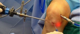
Arthroscopy of the elbow joint is a minimally invasive therapeutic and diagnostic medical operation, which is carried out using thin devices inserted into the joint cavity through small punctures. During the operation, an arthroscope, a type of endoscope, is used.
With the help of arthroscopy, it is possible to determine the cause of joint stiffness, limitation of movements when flexing and extending the arm, and pain when loading the joint. This type of intervention is used in the complex treatment of bursitis or arthritis, as well as to remove overgrown bone tissue and cartilage of the elbow joint
When is arthroscopy prescribed?
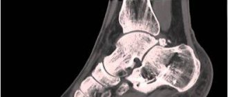
Ankle arthroscopy is prescribed for severe injuries, failure of conservative treatment, or lack of effect from taping or elastic bandages for a long time. The procedure is prescribed by the surgeon. The reason for the operation may be:
- development of deforming arthrosis of the joint or its chronic, advanced stage;
- damage to cartilage tissue, its detachment;
- unstable joint condition;
- pain of unknown etiology;
- synovitis of various nature;
- chondromatosis;
- arthritis;
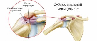
- impingement syndrome.
The procedure can be both a diagnostic method and a therapeutic one. Diagnostics using an arthroscope allows you to assess the condition of the joint and its structures, determine existing pathologies and the state of their development, take tissue samples for analysis, and clarify the existing conclusion.
Surgical intervention is necessary to excise individual fragments, remove adhesions, tissue or bone growths, synovial membrane (full or partial), replace damaged areas, and compare pieces of destroyed bones.
Indications
Arthroscopy may be recommended to remove loose intra-articular bone and cartilage fragments or to remove scar tissue that is limiting joint movement. The most common diagnoses that are indications for arthroscopic interventions on the elbow joint:
- so-called “tennis elbow” (lateral epicondylitis);
- the need to remove loose intra-articular bodies (cartilage fragments or bone fragments);
- intra-articular adhesions;
- osteoarthritis (arthritis due to wear and tear of articular cartilage);
- rheumatoid arthritis (arthritis of inflammatory causes);
- osteochondritis dissecans (damage to the capitate eminence of the articular end of the humerus due to physical overload, found in javelin throwers, shot throwers and gymnasts).
If the diagnosis is established, surgery is performed to:
- clarification of the severity of damage;
- detection of hidden defects;
- treatment planning and preparation for surgery;
- assessing the effectiveness of treatment and the progress of recovery processes;
- clarification of why repeated or new symptoms occur after therapy.
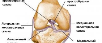
Shoulder ligament surgery
- Surgeries on the shoulder capsule
- Muscle plastic surgery
- Bone grafting operations
- Tenodesis operations
- Tenosuspension operations
- Combined operations
- Skin plastic surgery types
- Shoulder alloplasty operations
- Auto-, homo-, heteroplasty operations
- Beach chair. This is a semi-sitting position, similar to sitting in a reclining chair.
- Transverse position. In this position the patient lies on his side
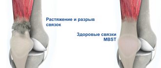
- To repair compressed muscles, diseased or damaged tissue is removed, the coracobrachial ligament that may be causing the compression is cut, and bone or part of a bone spur is cut away
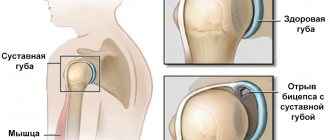
- A labral tear can be repaired by reattaching the labral cartilage and adjacent ligaments to the shoulder
In our country, the most common operations are those of Weinstein, Rosenstein, Drobotun, Shtutin, Khitrov and Sverdlov. The operations of Putti-Platt, Magnusson, Nicolas and others have found recognition abroad. None of the existing operations can currently be considered radical.
In our center, shoulder joint arthroplasty (ligament plastic surgery) is performed arthroscopically, using endoscopic techniques.
If it is necessary to repair the rotator cuff muscle by connecting its tendons to the shoulder joint, the surgeon may use special sutures to securely fix the tendons into the shoulder joint. If necessary, special metal or plastic brackets can be installed.
Operation process
Positioning and preparation
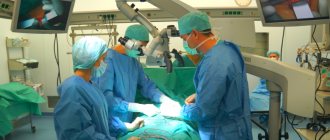
In the operating room, the surgeon will position you to adjust the arthroscope and have a clear view of the inside of the shoulder. The two most common patient positions for shoulder arthroscopy are:
Each position has its own advantages. Surgeons choose a position based on the procedure as well as their individual training.
Procedure
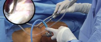
First, fluid is injected into the shoulder to inflate the joint (this is to make it easier to see all the structures of your shoulder through the arthroscope). Next, the surgeon will make a small incision in the shoulder for the arthroscope. Images from the arthroscope are projected onto a monitor so the surgeon can see everything inside your shoulder.
Once the problem is clearly identified, the surgeon will insert other small instruments through separate incisions. Specialized tools are used for shaving, cutting, removing and suturing. For this purpose, a cold plasma installation is indispensable, the electrodes of which replace an entire arsenal of mechanical surgical instruments. In many cases, special devices are used to secure the suture into the bone.
The Magnum 2 implant is an innovative device that, together with the Magnum 2 insertion handle, allows for reliable fixation in the bone. The trigger mechanism (TensionLock) moves the tissue of the rotator cuff of the shoulder joint to the prepared site, provides tension on the threads and produces knotless intraosseous suture fixation.
Designed for use on the glenoid, the knotless Mini Magnum handle implant provides secure fixation in a small size.
Fast, easy and reliable recovery
- The TensionLock mechanism secures the labrum tissue to the glenoid and secures it securely and quickly
- TensionLock mechanism allows you to adjust thread tension for perfect tissue-to-bone contact
- Knotless technology eliminates the hassle of tying knots
After the operation, you will remain in the ward for 1-2 days. Nurses will monitor your progress. Although recovery from arthroscopy is faster than from open surgery, it may still take several weeks. Recovery from shoulder arthroscopy may take some time. It is usually recommended to give up an active lifestyle and undergo a course of physical therapy and exercise therapy to fully restore mobility of the shoulder joint.
You may experience some pain and discomfort for a week after surgery. If you have had major surgery, the pain may be present for several weeks before it subsides. Ice will help reduce pain and swelling. Your doctor may prescribe pain medications if necessary.
Rehabilitation
Rehabilitation plays an important role in returning to daily activities. An exercise program will help you regain strength and ease of movement. If you had more complex surgery, your surgeon will assign a physical therapist to provide supervision and an exercise program.
Complications
Most patients do not experience complications after shoulder arthroscopy. Although, as with any operation, there are some risks. They are usually minor and treatable. Potential problems after arthroscopy include infections, excessive bleeding, blood clots, and damage to blood vessels or nerves. Your doctor will discuss possible complications with you before surgery.
Long term results
Since patients have different health conditions, the time to full recovery varies from person to person.
If you had minor surgery, you will not need a sling and will recover after a short recovery period. You can return to work or school within a few days after the procedure.
More complex procedures will result in a longer recovery period. Although arthroscopy incisions are small, there can be significant internal damage to the joint. Full recovery may take several months.
In rare cases, if the surgeon sees significant damage during arthroscopy, he may be forced to move to open surgery.
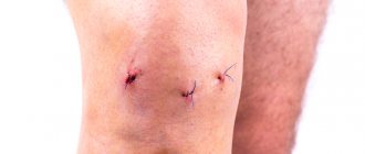
The arthroscopy techniques used in our clinic make it possible not only to restore the function of the damaged organ from the first days after surgery and to soon return the patient to their normal lifestyle. These techniques make it possible to reduce the likelihood of developing post-traumatic complications to a minimum, and, among other things, achieve a good cosmetic effect.
Contraindications
Joint arthroscopy is not performed if:
- inflammation of periarticular tissues
- exacerbation of chronic pathologies,
- mental disorders,
- bleeding or purulent processes in the joint area,
- osteomyelitis,
- bone tuberculosis,
- ligament rupture or disruption of the integrity of the joint capsule.
Since some of these contraindications are relative, the surgeon makes an individual decision about the possibility of arthroscopy or prescribes additional treatment to eliminate them.
Possible complications
Complications can occur in very rare cases, and even more so due to an incorrect diagnosis or very late and poor-quality treatment. For example, if for some reason the stitches come apart, creating an open wound. An infection can easily get into the wound and cause an inflammatory process. But in order for this to happen, you probably need to escape from the hospital on the day of the operation.
One of the real threats is improper fusion of the ankle. This leads to the development of arthrosis and joint deformation. The process is accompanied by regular swelling, as blood circulation is impaired and, as a result, lameness appears. Another scenario: hemorrhages into the joint cavity, sprained ligaments and the effects of anesthesia (nausea, dizziness). All sensations should be reported to the doctor.
Complications occur in 0.1% of cases out of a hundred. But the most important thing to remember is that the tendons, nerves and blood vessels remain intact. And it is very easy to avoid any complications: you need to strictly follow the recommendations of your doctor, especially in the first weeks of rehabilitation.
Preparing for surgery
Before the operation, the patient undergoes a comprehensive examination, which includes:
- consultations with a traumatologist-orthopedist and anesthesiologist;
- general blood analysis;
- blood chemistry;
- analysis for blood group and Rh factor;
- coagulogram;
- analysis for hospital infections;
- general urine analysis;
- fluorography;
- ECG.
Two weeks before surgery, you need to minimize or completely eliminate alcohol intake and smoking, and stop eating and drinking 12 hours before.
Types of intervention
Types of surgical intervention differ in the technique of making incisions. In knee orthopedics, arthrotomy is based on 3 traditional approaches:
- anterior internal/external parapatellar (according to Ollier, Langenbeck);
- transverse (according to Textor);
- posterolateral (according to Voino-Yasenetsky);
There is another principle for creating access, which combines the advantages of typical techniques. This is a paracondylar arthrotomy (according to Kornev), which makes it possible to widely expose the arch of the superior inversion on both sides, the joint cavity along the entire line of the joint space, and two posterolateral chambers. The Kornev incision provides access to all potentially dangerous areas, where often during pathological processes pus, effusion, and blood are concentrated and retained. Of course, the doctor will determine which technique is most rational to use only on the basis of reliable diagnostic results on the exact localization of the pathological focus.
Possible difficulties and problems
All types of arthrotomies of the knee joints, like any deep invasions of the anatomical parts of the body, can cause complications. Although not as common, consequences do occur in some patients, these include:
- development of infectious processes in the wound area;
- injury to neurovascular formations with instruments;
- formation of a blood clot in the veins of the limb (phlebothrombosis);
- separation and migration of a thrombotic clot with blockage of the pulmonary artery;
- reflex dystrophy syndrome (pain reaction to extensive invasion);
- inflammation of the synovium with accumulation of effusion;
- hemorrhage in the knee joint;
- allergy to the anesthesia used.
Technique for knee arthrotomy
As we have already said, arthrotomy is based on several tactics for exposing the articulation. Therefore, we will try to briefly outline the essence of each technique.
- Anterior parapatellar capsulotomy
. The beginning of the skin incision is above the kneecap, approximately 8 cm from it, at the junction of the external thigh muscle and the quadriceps tendon. From this point, the skin is incised downward, following the outer line of the patella with a scalpel. The incision ends 20 mm below the tibial tuberosity. After the surgeon dissects the hypodermis and fascia along a given trajectory, the fibrous layer and synovial capsule are opened. Next, the necessary manipulations are carried out. This technique is not often used because it carries a high risk of injury to the common peroneal nerve. - Transverse method according to Textor
. A wider horseshoe-shaped incision is made, covering both sides of the joint. During its implementation, the patella's own ligament is crossed and the ligaments are dissected along the lateral surfaces of the articulation. A bilateral incision is made from a bent knee position 6 cm above the patella. It is guided down the end structures of the rectus femoris tendon and to the side of the kneecap. Having reached the lower pole of the patella, the incision is continued along the rounded protrusion of the condyle to the site of attachment of the collateral ligaments. A U-shaped incision is used to dissect the skin and fibrous joint membrane down to the synovial membrane. The synovial membrane is dissected parapatellar, after which the superior inversion is opened. The capsule is dissected retrocondylarly (downwards and backwards) together with the posterior inversions. Next, the main therapeutic actions begin; they usually involve large-scale resection of the joint. - Posterolateral arthrotomy
. It is used more often for purulent lesions (gonitis, epiema) of the joint. The osteochondral junction is opened using 4 incisions. First, two anterior parapatellar incisions are made, parallel to the two sides of the patella. Then the posterior inversions are opened using two longitudinal-lateral incisions. The posteromedial inversion is entered between the femoral condyle and the inner head of the gastrocnemius muscle, and the posterolateral inversion is entered through an incision above the biceps femoris tendon. Following strictly the operating procedure, the specialist pumps out the pus, introduces and installs drainage systems.
Surgical interventions are completed with careful hemostasis. The dissected areas of the synovial membrane and soft structures are sutured layer by layer with catgut threads. The skin edges are sutured with silk suture material. Finally, the leg is immobilized in the correct position with a plaster cast. A “window” is cut in the applied plaster, through which the surgical wound will be treated and bandaged.
Postoperative rehabilitation
Recovery can take up to 12 months. The duration of rehabilitation is higher if arthrodesis of the joints of the lower extremities was performed. The patient must use crutches to move around and avoid putting any weight on the operated leg. You can speed up recovery with the help of physiotherapy, exercise therapy, massage and electrophoresis. Physical therapy can only be performed after the cast has been removed and diagnostics have been carried out.
The rehabilitation period reaches 3-4 months if the operation was performed on the metatarsophalangeal, ankle, or knee joint. Recovery can take up to 8-12 months if hip arthrodesis was performed.