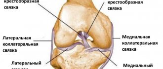
Labral tears are a fairly common injury and are usually caused by falls that result in a sprained arm; or from repetitive movements or sports that place major stress on the arms (tennis, volleyball, swimming, weightlifting). The labrum is an important part of the cartilage surrounding the glenoid (glenoid socket of the shoulder). The glenoid itself has a fairly flat surface, the humerus fits into the socket of the shoulder, and the labrum gives the glenoid a curved shape, improving shoulder stability. The tight, but nevertheless flexible attachment of the humerus to the glenoid allows for a huge amount of movement that a healthy shoulder can perform.
A SLAP tear is a type of labrum tear that occurs at the top of the labrum, where the long head of the biceps tendon attaches to the glenoid. These types of injuries are quite common among athletes who participate in sports that actively involve the shoulders. Injuries to this area can be extremely painful, and in severe cases can even cause a tear or avulsion of the biceps tendon. When assessing the likelihood of an injury such as a labral tear or SLAP, it is important to determine whether a labral tear is associated with any type of shoulder instability. If instability is present, some procedures will need to be performed first to stabilize the shoulder. X-rays and MRIs reveal any pre-existing conditions or identify problems that may exist, such as impingement or fracture. If instability is not present or is not a problem, surgery may be aimed at repairing the labral tear itself.
Rupture of the labrum can occur with regular stress on the shoulder, as well as with unsuccessful falls.
Content
- 1 The main signs of acetabular labral tears
- 2 Prevention
- 3 Clinical picture 3.1 History and complaints
- 3.2 Physical examination
- 3.3 Radiation diagnostics
- 3.4 Special methods
- 3.5 Comparison of diagnostic methods
- 5.1 Conservative treatment
The main signs of tears of the acetabular labrum[edit | edit code]
- Acetabular labral tears are similar to meniscus tears in the knee.
- Pain in the groin or buttock, or pain that arcs around the outside of the hip joint.
- The pain is often accompanied by clicking or a feeling of obstruction in the joint.
- Ruptures can be traumatic or dystrophic.
- Dystrophic tears are common in ballet, as well as in sports that require strong hip flexion (football, mountaineering) or frequent hip rotation (golf, figure skating, martial arts).
- The abnormal structure of the hip joint (dysplasia, femoroacetabular impingement syndrome) predisposes to dystrophic ruptures.
- With hip dysplasia, the acetabular labrum is hypertrophied and prone to rupture.
- Femoroacetabular impingement syndrome involves flattening of the superior aspect of the femoral neck, which increases friction of the acetabular labrum and predisposes to labral tears and osteoarthritis of the hip joint.
- Surgical treatment of acetabular labral tears produces less predictable results than treatment of knee meniscal tears.
- The prognosis depends on the location and severity of the rupture, its nature (traumatic or dystrophic), the presence of bone abnormalities and the severity of chondromalacia.
Prevention[edit | edit code]
The best way to prevent it is to avoid movements that cause the femoral neck to strike the acetabular labrum. This, of course, is impossible in those sports that involve swinging movements of the legs or strong rotation of the hip, in particular in golf, figure skating, martial arts, dance sports and artistic or rhythmic gymnastics.
Acetabular labral tears often occur in poorly trained athletes during leg swing movements, for example, in high school dancers performing a step jump, or in athletes who press heavy loads with their legs without a full warm-up. The correct organization of the training process can prevent such gaps.
Many female athletes experience acetabular dysplasia, accompanied by increased mobility of the hip joint. This helps gymnasts and ballerinas in their activities, but it also reduces hip stability and predisposes to labral tears. Tears of the acetabular labrum are often associated with osteoarthritis of the hip joint, especially developing against the background of hip dysplasia, and with femoroacetabular impingement syndrome.
Clinical picture[edit | edit code]
History and complaints[edit | edit code]
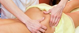
Pain from a labral tear is usually felt in the groin or upper thigh. Often, athletes, showing a sore spot, clasp the thigh with their thumb and forefinger in front and outside the hip joint so that these two fingers form the contours of the letter C. Pain can also be felt behind the hip joint, in the gluteal region. It is possible that anterior tears tend to present with pain in front of the joint (in the groin), and posterior tears tend to present with pain behind the joint (in the gluteal region).
Pain from a labral tear is often accompanied by mechanical symptoms: clicking or a sensation of obstruction in the joint. As with other diseases of the hip joint, pain can radiate down the leg, usually along the front leg, less often along the inner surface of the thigh into the knee.
The pain can be of different nature and range from mild, dull, provoked by physical activity and persisting during rest, to severe and constant, seriously limiting daily activities. Few patients have a significant limp or require crutches, but they try to avoid certain positions and movements (mainly hip flexion, abduction, and rotation) that cause pain. This usually affects their athletic performance.
Traumatic labrum rupture is primarily caused by an external force applied to a fully extended and externally rotated hip. Often an athlete can point to a specific injury, such as a fall or twisted ankle, that preceded the onset of pain.
Less commonly, the disease develops gradually, and the patient cannot definitely indicate the time of its onset. Sometimes groin pain remains after a “groin sprain,” which actually turns out to be a torn labrum.
Physical examination[edit | edit code]
Examination can usually distinguish a labral tear from the internal type of snapping hip syndrome. The patient is placed on his back and his sore leg is bent, bringing the thigh into a position of flexion, adduction and internal rotation; the pain of this movement indicates a tear of the acetabular labrum.
Combining the principles underlying the Thomas test (for hip flexion contracture) and McMurry test (for knee meniscus tear), Joseph McCarthy proposed his own test to assess the condition of the hip joint. The athlete is placed on his back with his legs bent to fix the pelvis, after which the affected leg is extended, rotating the thigh outward, and then the same movement is repeated, rotating the thigh inward. When a painful click appears, the test is considered positive and indicates a tear of the acetabular labrum. A tear in the acetabular labrum may also be indicated by pain in the groin when lifting a straight leg against the doctor’s resistance, but this is too nonspecific a sign that can also appear with other diseases of the hip joint.
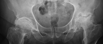
Radiation diagnostics[edit | edit code]
Confirming an acetabular labral tear using existing radiological diagnostic methods is quite difficult. A plain radiograph of the pelvis in the direct posterior projection is studied (to compare the hip joint of the diseased and healthy leg) and a radiograph of the affected hip joint in the position of flexion and abduction of the hip (in the so-called frog position), but if the athlete does not have hip dysplasia, these radiographs are more likely In all, they will be normal. Subchondral cysts that arise against the background of an old rupture of the acetabular labrum indicate chondromalacia or detachment of the acetabular labrum from the articular cartilage. These cysts are most often located in the superolateral part of the acetabulum.
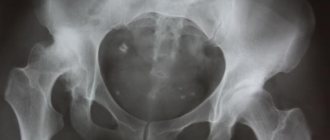
Femoroacetabular impingement syndrome
- one of the congenital anomalies of the hip joint, accompanied by dystrophy and tears of the acetabular lip. Its most characteristic radiological sign is the so-called pistol grip sign, that is, flattening of the anterosuperior part of the femoral neck, which is why it appears abnormally convex and, together with the femoral head, resembles a pistol grip. Bone cysts may be visible in this convex part of the neck. A study from the Mayo Clinic found that 87% of patients with acetabular labral tears had at least one abnormality on radiographs. This study included all patients with acetabular labral tears seen in the clinic over a 6-year period and was not limited to sports-related injuries.
CT and bone scintigraphy are usually uninformative. Conventional MRI cannot reliably confirm or refute a rupture of the acetabular labrum (although the detection of cysts adjacent to the labrum can be considered a fairly convincing indirect sign of its rupture). Magnetic resonance arthrography is much more informative, and since its results depend on the technique used, it must be carried out in accordance with a specific protocol. Magnetic resonance arthrography is performed in two stages: the first stage is hip arthrography with gadolinium, and the second stage is MRI of the hip using surface coils and a powerful tomograph that produces high-resolution images. It is also useful to use specially developed and computer-based protocols for scanning the acetabular labrum: they allow you to obtain a series of images in an oblique sagittal projection, in which the neck of the femur lies.
The high rate of false-negative results when using radiological diagnostic methods, including magnetic resonance arthrography, gives reason to recommend arthroscopy of the hip joint if the clinical picture is consistent with a tear of the acetabular labrum, even if no pathological changes were detected during magnetic resonance arthrography or if it was not performed .
Special methods[edit | edit code]
For differential diagnosis of pain in the hip joint, a local anesthetic can be injected into the joint under fluoroscopic control: temporary pain relief after this procedure indicates intra-articular pathology.
Comparison of diagnostic methods[edit | edit code]
The main methods for diagnosing intra-articular pathology remain questioning and physical examination - in comparison with arthroscopy (as a diagnostic standard), they allow a correct diagnosis to be made in 98% of cases. In comparison, MRI produces false-negative results in 42% of cases and false-positive results in 10% of cases, and magnetic resonance arthrography produces false-positive results in 8% and 20% of cases, respectively. Pain relief in response to local anesthetic injection into the hip joint indicates intra-articular pathology with 90% confidence, but this test is not specific for acetabular labral tears, but covers all diseases of the hip joint.
Historical reference.
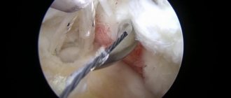
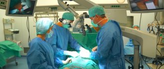
Arthroscopy allows you to see the internal condition of the joint on a monitor in great detail.
Severin Nordentoft, a doctor from Denmark, first reported in Berlin at a surgical congress in 1912 about the possibility of examining the knee joint without opening its cavity. For the first time, the scientist Kenji Takagi (Japan) managed to examine the knee using a device with a diameter of 7 mm in 1919. At the same time, the Tokyo “school of arthroscopy” was founded. Takagi subsequently reduced the diameter to 4 mm and was able to perform an intra-articular biopsy under vision guidance. In 1931, the first photographs of the formations from the inside were obtained. Scientists continuously worked to improve the device and methodology.
The Swiss Eugen Bircher worked on the creation and development of arthroscopy for about 10 years, and in the 1920s he was able to carry out diagnostic operations on the knee joint using a laparoscope. His numerous studies and attempts to improve the device made a huge contribution to the development of endoscopic surgery. Dr. Masaki Watanabe developed the first arthroscope to be mass produced in 1960. He performed operations and created the first atlas on arthroscopy. Later, Dr. Heshmat Shahriaree began performing arthroscopic meniscal resections. Over time, thanks to the development of fiber optics, the instruments were significantly improved and became widespread. And a new method of surgical treatment has become available in advanced clinics.
Treatment[edit | edit code]
Conservative treatment[edit | edit code]
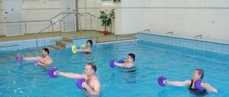
A labral tear usually cannot be treated conservatively. Exercise therapy can relieve muscle spasms and correct gait, and non-steroidal anti-inflammatory drugs reduce inflammation and alleviate symptoms to a certain extent, however, neither exercise therapy nor non-steroidal anti-inflammatory drugs can eliminate the source of inflammation and completely relieve the patient of symptoms. Only arthroscopy can achieve complete cure.
Surgical treatment[edit | edit code]
Hip arthroscopy allows the doctor to see the labral tear and thereby confirm the diagnosis. The goal of the operation is to remove the torn part of the lip that is hanging loose in the joint cavity and causing symptoms, while preserving the remaining intact part of the lip as best as possible. During arthroscopy, you can also examine other structures that may be causing the patient's symptoms: the articular cartilage of the acetabulum and femoral head, the ligament of the femoral head, and the joint capsule.
Special methods[edit | edit code]
Sometimes athletes with persistent severe pain in the hip joint, indicating inflammation, are injected into the joint, but this usually provides only temporary results.
Benefits of arthroscopy.
The advantages of this manipulation include:
- High diagnostic accuracy.
- Only one procedure is performed for both diagnosis and treatment.
- Possibility of technologically advanced targeted biopsy
- Cosmetic effect - after the operation there is no large scar left, only small, barely noticeable marks on the skin.
- Low level of trauma - tissues in the area of endoscopic intervention are minimally affected.
- The operation does not require anesthesia. In our clinic, arthroscopy is performed under local anesthesia.
- There is no need to wear a cast.
- The operation does not involve taking strong painkillers in the postoperative period.
- Easy and quick rehabilitation - the patient can return to normal life in just a few weeks, and athletes, accordingly, can return to training.
- Minimal risk of complications (scar formation, adhesions, infections).
- There is no need for a long hospital stay. Most often, you can go home on the day of surgery.
- The result is lower costs (no need to pay for a long stay in the clinic).
Arthroscopy as a diagnostic procedure is carried out on large joints: knee, hip, shoulder. Somewhat less frequently, arthroscopic treatment is performed on the ankle, elbow, wrist, intervertebral joints, and foot.
The effectiveness of the technique.
Arthroscopy makes it possible to diagnose the location of the problem, the degree of its prevalence, and identify all, even the smallest, injuries that accompany the injury.
Unfortunately, not all diagnostic methods make it possible to identify pathology at the initial stages of development - some tissues are not clearly visualized on X-ray images; computed tomography and magnetic resonance imaging also do not always provide answers to the questions the doctor has. Moreover, an incorrect diagnosis can lead to the wrong direction of treatment. That is why today it is difficult to imagine diagnosing articular deformities without such a diagnostic procedure as arthroscopy. It is rightfully the best way to assess intra-articular pathologies and injuries.
Forecast[edit | edit code]
The results of arthroscopic treatment of single traumatic ruptures of the acetabular labrum are very good: 80-90% of patients experience a complete recovery, after which they can return to professional sports. Even after successful surgery, clicking in the joint (especially in certain positions) will usually remain, which should be warned about in advance.
With dystrophic tears caused by frequent repetition of movements that are traumatic for the joint, the prognosis for returning to sports is quite poor, especially if chondromalacia is detected during arthroscopy.
For some time after arthroscopy (from 2 days to 2 weeks), the patient is prohibited from leaning on the operated leg. In the first 2-6 weeks after surgery, exercise therapy is carried out aimed at developing the joint, and after 6-12 weeks it is usually possible to resume training.
The literature provides very little information regarding the prognosis of arthroscopic operations in patients with bone anomalies of the hip joint. In cases of severe hip dysplasia, acetabular retroversion, or femoroacetabular impingement syndrome, symptoms should be expected to persist. Surgical treatments for these anomalies have been described, including supracetabular osteotomy and femoral neck osteoplasty, which requires trochanteric osteotomy and dislocation of the femur from the hip joint during surgery. The same dislocation of the femur allows access to and repair of complex tears of the trochanteric labrum like a watering can handle.
What is arthroscopy?
Professor Kuznetsov Igor Aleksandrovich before the operation.
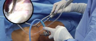
Arthroscopy is the name of a minimally invasive surgical technique that is used to examine and treat large (shoulder, ankle, knee) human joints. For many years, it has been successfully used by orthopedic traumatologists in leading clinics around the world, already gaining enormous experience. Surgery is performed using an arthroscope (a fiber-optic light device - a thin tube with a camera at the end), which is inserted into the joint cavity together with instruments through small incisions in the skin. The camera produces a high-resolution image displayed on the monitor. Studying it, the specialist examines the joint in detail from the inside and makes an accurate diagnosis. Then, if necessary, the doctor immediately carries out therapeutic procedures.
Today, this method is the “gold standard” for the treatment of most joint pathologies - a high therapeutic effect is achieved thanks to improved technologies and a decent safety profile. More and more clinics around the world are mastering arthroscopic equipment and switching to this method. Observation of patients after arthroscopy shows a rapid restoration of function, as well as a return to their usual lifestyle, which is most important for people involved in sports.
SportClinic took first place in the “Traumatology and Orthopedics” in the ranking of the best private clinics in St. Petersburg in 2021.