The doctor studies the patient's medical history. Then, asks for his complaints. Only after this he conducts an examination and, if necessary, prescribes x-rays, blood and synovial fluid tests.
During the examination, the doctor pays attention to: swelling, redness and temperature of the joint; sensitivity of the knee; range of passive and active movement; stability of the joint; crepitus during movement; pain when the bulk of the body weight is on the knee; problems with gait; any signs of damage to muscles, tendons, ligaments; symmetry of the disease and how many joints are affected by the disease (a sign of rheumatoid arthritis )
TREATMENT
There is no cure for arthritis. But, there are a number of procedures that help relieve pain. No surgery
Lifestyle changes: Reducing the load that worsens the condition. For example, climbing stairs. Switching from one sport to another, in which the intense load is less. For example, replace jogging with swimming or cycling. Losing weight helps reduce the load on the knee joint. As a result, pain is reduced and the functioning of the joint improves.
These tips can help reduce stress on the knee joint and slow the progression of arthritis.
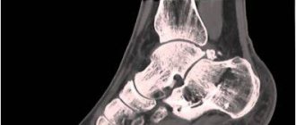
Ultrasound of the knee (knee joint)
Duration of the examination
: 30 min.
Preparing for the examination
: No
Contraindications
: No
Preparation of the conclusion
: 20 minutes.
Restrictions
: No
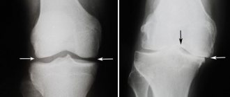
Ultrasound of joints is an informative way to examine parts of the musculoskeletal system. The method is based on the use of high-frequency sound waves that do not pose a danger to humans. Ultrasound examination is a necessary procedure in diagnosing pathologies, choosing treatment, and monitoring surgical intervention. Today we will talk about ultrasound of the knee (knee joint) - what it is, how the procedure is performed, indications and contraindications.
The essence of the method
Ultrasound of the knee is an innovative way to study pathologies of the knee area under the influence of high-frequency ultrasonic waves. In the process of interacting with the joint, the waves form high-quality images and display them on the monitor screen. Ultrasound of the knee is a fairly simple procedure that does not require special preparation from the patient.
Diagnostics allows us to identify inflammatory and infectious changes, degenerative and atrophic lesions, rheumatoid and traumatic pathologies. Ultrasound examination makes it possible to timely detect the disease, find out the causes and extent of pathological changes, and prescribe therapy.
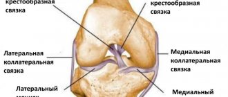
The knee joint is complex in structure. This is one of the most important elements of the musculoskeletal system. Every day the knee is exposed to a lot of stress - walking, running, squatting, lifting. Lifting heavy weights also affects the knee joint. Pathologies and injuries to the joint lead to limited mobility. It is important to identify the disorder at an early stage in order to prevent the disease in the future.
Advantages of ultrasound of the knee (knee joint):
- Fast diagnostics. Ultrasound technology takes a maximum of 30 minutes. Within half an hour, the specialist examines the joint in several planes.
- High information content. Pictures are much more effective than radiography, since not only the bone structure is visible, but also soft tissues.
- Safety. The study does not expose the body to radiation. Ultrasound of the knee is safe for expectant mothers, children, and patients with acute pain. Diagnostics can be completed an unlimited number of times.
- Non-invasive. During the session there are no injections or punctures of the area being examined.
- Availability. You can undergo an ultrasound anywhere in our country at reasonable prices.
Ultrasound of the knee joint allows you to visualize the condition of the muscles, tendons, ligaments and meniscus. Diagnosis is carried out not only for pathologies, but also for preventive purposes.
For what diseases is it prescribed?
Damage to muscles, tendons, menisci, and knee ligaments occupy a leading place among pathologies of the knee area. Untreated injuries cause the development of serious diseases:
- Osteoporosis of the knee joint is a chronic progressive disease of bone tissue caused by a lack of calcium, resulting in a violation of the architecture and density of bones.
- Knee bursitis is an inflammation of the joint capsule caused by the accumulation of fluid in the joint cavity.
- Arthritis is a joint disease of inflammatory etiology.
- Tendinitis is a pathology of tendon tissue caused by damage to the muscular-ligamentous system.
- Ligamentitis of the knee joint is a disease associated with the onset of destructive pathological processes in the area of cartilage, tendon, and synovial tissue of the large joint of the bones of the lower limb.
- Synovitis is a pathological condition of the synovium of the joint, accompanied by the formation of fluid in the cavity of varying consistency.
- Chondromatosis is an inflammatory process characterized by the formation of loose bodies in the joint.
- Koenig's disease is a pathology caused by the separation of a small section of cartilage and its movement into the joint cavity.
- Arthrosis is a chronic joint disease associated with thinning and destruction of cartilage tissue.
- Gout is a disease of tissues and joints caused by metabolic disorders.
Diagnostics helps to identify abnormal and genetic pathologies, the presence of malignant and benign neoplasms, the causes of inflammation of the knee joint, and prescribe adequate treatment.
Indications for diagnosis
Athletes, dentists, hairdressers, massage therapists, loaders - all people whose professional activities involve stress on their legs - are most often exposed to knee joint injuries. A referral for an ultrasound is issued by a traumatologist, surgeon, therapist, oncologist, neurologist. When patients come for an appointment, they complain of the following symptoms:
- persistent pain in the knee area;
limited mobility;
- inability to step on your foot;
- difficulties with flexion and extension of the joint;
- acute pain syndrome in the knee area;
- swelling and redness of the joint;
- suspicions of the presence of neoplasms;
- domestic and professional injuries.
The presence of these symptoms may indicate not only a pathology of the knee area, but also diseases of the musculoskeletal system, endocrine and nervous systems. If you experience any of the signs of knee problems, make an appointment with your doctor.
Preparation for the procedure
Before the session, it is recommended to wear such clothing so that you can easily free your knee. There is no need to go on a diet or give up medications that are prescribed for constant use.
Carrying out the procedure
An ultrasound of the knee is performed using a sensor that reads information from the area being examined and sends images to a computer screen. The patient takes a lying or sitting position. The surface of the joint is lubricated with a special gel so that the sensor slides over the surface without irritating the knee.
During the procedure, the doctor will ask the patient to change position several times. When examining the anterior and lateral parts of the knee joint, the patient lies on his back with his legs straightened. When examining the menisci, the patient must flex his limbs. The posterior section is examined while lying on the stomach.
Diagnostics are carried out in several projections for maximum accuracy. To adequately assess the changes, a study of the affected and healthy areas is carried out. The duration of an ultrasound scan of the knee (knee joint) is 15-30 minutes, depending on the purpose of the examination and diagnosis.
results
Issuing a report takes on average 30 minutes. The specialist needs to describe the data obtained in detail. What the doctor pays attention to:
- condition of joint contours;
- the presence of abnormal and organic changes;
- homogeneity of hyaline cartilage;
- presence or absence of bone osteophytes;
- the presence of fluid in the joint capsule;
- swelling of soft tissues;
- presence of neoplasms.
The specialist groups the data, compares it with standard indicators and records the information in a file. You will be given a paper document with a photo of the area being examined. If you do not have medical training, you should not interpret the report notes yourself. Go to the attending physician who issued the referral for an ultrasound scan or make an appointment with a specialized specialist at the clinic where you were examined.
Contraindications
There are no contraindications or restrictions for ultrasound of the knee joint. The image may be distorted if there are wounds and abrasions on the surface of the knee.
Return to list
ALTERNATIVE TREATMENTS
The benefits of alternative therapies have not been proven. But you can try. You need to find a qualified practitioner and inform your doctor of the decision. Alternative methods can help relieve pain. These include acupuncture and magnetic therapy.
Acupuncture is acupuncture. The procedure uses thin needles to stimulate certain areas of the body, which can relieve pain or restore sensitivity if a certain area of the body is numb. There are even several scientific studies on the effectiveness of acupuncture. The evidence collected suggests that this technique helps relieve arthritis pain. It is used in many parts of the world. Before starting the procedure, the patient should find out about the qualifications of the specialist, familiarize himself with his certificates and inquire about the sterility of the instruments.
Magnetic therapy is a painless procedure. The inflamed joint, which is located in the electromagnetic field, is exposed to a pulse signal. But it is worth remembering that the benefits of magnetic therapy have not yet been proven.
MRI using contrast
Magnetic resonance imaging (MRI) using contrast is used to determine the presence or absence of tumor, inflammation, or blood flow problems in the knee joint. A contrast solution can increase magnetic resonance in the part of the body being examined. The composition is injected into a vein, from where it enters the circulatory system and nearby tissues, thereby increasing image clarity.
Blood circulation in the knee joint in the presence of inflammation, injury or hematoma is very different from normal blood circulation, which becomes noticeable in the photographs. MRI with contrast can also detect the presence of tumors due to the fact that it highlights the network of blood vessels of the tumor. Most often, a composition with gadolinium is used for contrast.
The day before the diagnostic examination, the patient must be tested for allergies to the components of the contrast drug. Normally, the solution is eliminated from the body on its own within two days and does not cause any harm.
You should not perform MRI diagnostics with contrast if you have kidney failure or inflammation of the genitourinary system.
Preparing the patient for surgery
Preparation consists of a thorough examination of the patient, collecting anamnesis, and conducting basic instrumental and laboratory tests. Until the doctor is convinced that this is a safe operation and weighs the pros and cons, the operation is impossible. Therefore, at first, a round of some specialists of a narrow profile is always prescribed, for example, in addition to an orthopedist or traumatologist, depending on the situation, also a cardiologist, endocrinologist, pulmonologist, allergist, etc.
It is extremely important to take into account absolutely all the results of the preliminary diagnosis. They will influence the choice of anesthesia; a special role is given to the selection of the optimal type of anesthetic that will not cause harm to the body.
In addition to medical rounds, the patient is given directions for an ECG and fluorography, and for laboratory tests of blood and urine. Having made sure that it is advisable to undertake this operation, they conduct a consultation on preparation. During the consultation, you are warned that you will need to stop eating 12 hours before the procedure, and stop taking anticoagulants (aspirin, heparin and medications based on them) a couple of weeks before the procedure.
Patients who left reviews report cautionary recommendations from doctors regarding smoking and drinking alcohol. You will have to give up bad habits 10-14 days before the intervention and, of course, try not to return to them afterward or at least minimize the dose. If you intend to resort only to diagnosis, preoperative preparation is carried out according to similar rules.
Description of the microsurgical process
In reviews, patients tell how quickly and painlessly they underwent the procedure, without general anesthesia. This is quite likely, but do not forget that clinical cases are not the same for everyone, and the physiological status of each patient is different. Therefore, it would be more correct to say that pain, thanks to strong local anesthesia, is almost impossible. However, post-operative discomfort in the area of the operated area will most likely bother you a little for some time.
Disintegration of ligaments when they rupture.
The video allows you to understand all the intricacies of the process. There are plenty of them on the Internet. But due to the fact that not everyone will be able to watch specific stories to the end, we suggest that you simply and calmly read about the important points.
Positioning of the patient on the operating table.
Based on the etiology and degree of pathological changes, the patient’s condition and concomitant diseases, the anesthetic agent is selected individually. Most often, local anesthesia is used, that is, regional anesthesia (epidural or conduction). The regional anesthetic contains lidocaine, ropivacaine or bupivacaine. To enhance the anesthetic effect, a medicine from the opiate category can be added to the main solution in a mini-dose. In some situations, however, general intravenous anesthesia is appropriate.
First, regardless of the surgeon's plans, a diagnostic arthroscopy will be performed. It involves inserting an endoscopic probe in the form of a rod into the cavity of the bone joint. When anesthesia takes effect, then diagnostic surgery begins. To do this, the limb, half bent at the knee, closer to the thigh, is twisted to reduce blood flow, after which a puncture (6 mm) is made, through which a contrast agent is injected to enhance the visibility of the articular and periarticular structures. Next, a tube is inserted through this operating port. The specialist thoroughly examines every millimeter of the problem organ.
The surgical field, you can see a glow inside.
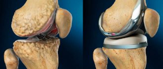
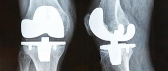
The video is broadcast in real time on the screen and reveals any existing defects in the diseased joint. If the doctor considers it necessary to perform regeneration of pathological elements, he will create an additional incision (6-8 mm), through which he will eliminate the detected defect using the necessary instrument from the microsurgical set. He can perform partial resection of osteochondral tissues, plastic surgery of ligaments and tendons, suturing or removal (partial, complete) of the meniscus, expropriate chondromic bodies, extract effusion and pus, administer anti-inflammatory drugs, take a fragment of tissue or synovium to study their composition, etc.
At the end of the surgery, the surgical field is washed and all instruments are removed. The doctor treats the wound with disinfectant compounds, installs drainage and makes a small suture, which he closes with a sterile adhesive plaster. On the operated knee, in order to give it maximum immobility, a tight fixing bandage is applied from the foot to the middle of the thigh.
The entire session takes an average of 1 hour, in some cases up to 3 hours. Restorative measures are simple, but they must be strictly followed so as not to provoke dangerous complications. Negative phenomena that can develop will be discussed further.