directions
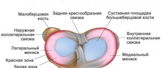
In the structure of the musculoskeletal system, the knee joint is the largest and most complex joint. The knee joint includes menisci - cartilaginous layers that perform the function of shock absorption in the knee and uniform distribution of loads on the knee. It is the knee menisci that are considered the most vulnerable areas subject to injury and mechanical damage. Tears, degeneration, excessive mobility - this is an incomplete list of meniscus diseases. The knee itself, while withstanding daily stress in the form of physical activity and strenuous work, is also subject to wear and tear. X-ray examination of the knee joint helps to identify the cause of the disease, see pathological changes, and diagnose injury. In traumatology, knee radiography is the most common non-invasive diagnostic method.
Our clinics in St. Petersburg
Structural subdivision of Polikarpov Alley Polikarpov 6k2 Primorsky district
- Pionerskaya
- Specific
- Commandant's
Structural subdivision of Zhukov Marshal Zhukov Ave. 28k2 Kirovsky district
- Avtovo
- Avenue of Veterans
- Leninsky Prospekt
Structural subdivision Devyatkino Okhtinskaya alley 18 Vsevolozhsk district
- Devyatkino
- Civil Prospect
- Academic
For detailed information and to make an appointment, you can call +7 (812) 640-55-25
Make an appointment
We draw your attention to the schedule of technological breaks in the CT and X-ray rooms.
You can get high-quality and quick x-rays of the knee joint at a multidisciplinary medical clinic in St. Petersburg. In the trauma department of the Medicenter clinic network, you will have an X-ray examination of the knee joint using a safe, modern high-tech digital Clinomat device. The Medicenter’s equipment allows for complete effective diagnosis of the patient, analysis of X-ray images in various processing modes, and quickly and accurately prescribing treatment based on the results obtained.
What is x-ray diagnostics
X-ray diagnostics is based on special X-rays that the device emits. Soft tissues allow them to pass through, while hard tissues absorb them, which is why in the picture the former are painted dark, and the latter light. Bone tissue is most clearly visible in the photographs, so the method is used to examine the condition of bones and joints. The results are provided on paper or digital media and saved on the computer’s hard drive.
Today you can get an X-ray image on digital media
The most common injuries and diseases of the knee joint
Ailments of the musculoskeletal system are a whole group of diseases that affect and damage joints, cartilage, muscles, and periarticular tissues. These diseases are varied, can appear at any age and have different symptoms and signs. Ailments of the knee joint are very painful, affect a person’s motor activity, limit his movements, complicate life and sometimes lead to a complete loss of the ability to walk and disability. Diseases of the knee joint most often develop as a result of injuries, inflammation, infections, and degenerative changes. The knee joint is most often susceptible to fractures, dislocations, sprains and other injuries. The most common knee diseases are arthritis, arthrosis, osteoporosis, osteophyte, meniscopathy, cysts and tumors.
Interpretation of the radiograph
Despite the fact that the average person is unlikely to be able to independently understand the features of the resulting black-and-white “photo,” he can recognize the symptoms of the most common ailments. Signs of their development are indicated by a radiologist, who makes a conclusion transmitted to the treating doctor.
So, if a patient is suspected of arthrosis, then in the official conclusion its confirmation can be found by reading the following theses:
- unevenly narrowed gap;
- deformation of the gap according to the degree of neglect of the process;
- bone growth along the articular edge;
- osteophytes of different sizes;
- compaction of bone tissue at the border with cartilage;
- limestone deposits on ligaments.
But it is much easier to recognize fractures even without proper medical skills. They can be seen upon detailed examination of the photographs, where the damage is clearly visible. It is more difficult with fresh cracks and depressions, which are better visible several days after the injury.
Arthritis, unlike arthrosis, causes widening of the joint space. Anatomically, the phenomenon is explained by the presence of an inflammatory effusion in the joint cavity. In some cases, to establish the exact extent of the lesion, a magnetic resonance imaging scanner is still necessary.
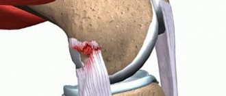
It is relatively easy to recognize a dislocation. It is characterized by bone displacement, where one part does not match the surface of the second. The main victim of dislocations is the kneecap.
With sprains or destabilization of the functioning of ligaments, it is a little more difficult due to the structure of tissues classified as soft. But even such anomalies can be found if you pay attention to the possible increase in the distance between the surfaces of the tibia and femur bones.
A somewhat similar principle is used to look for a violation of the integrity of the patellar tendon. The image will demonstrate the kneecap displacement. When the tendon has additionally undergone a process of sclerosis, its outline can be seen more clearly. To create an image of increased accuracy, you can use an artificial technique by adjusting the hardness of the radiation.
Since people usually seek advice from a surgeon or traumatologist at the “I can’t stand it anymore” stage, they develop a pronounced degenerative-dystrophic process. An X-ray examination will reveal it due to the proliferation of bone tissue formed on the sides of the articular surfaces. Growths in the form of osteophytes gradually lead to irreversible deformations, significantly worsening the quality of life of the victim.
Another common reason for visits to the radiography office is referrals from oncology clinics. Tumors of a benign or malignant nature are located not only in the joint, but also in the periarticular soft tissues of the knee. You can find them by marks without a clear shape with destroyed cells around them. The more affected the area around the pathological focus, the higher the chances that metastases have formed nearby.
The final common disease is osteoporosis. This is where only an experienced doctor can figure it out, who will pay attention to the lack of calcium in the bones. The image will confirm the fears if transparent inserts become visible there when the borders are sealed.
Causes of knee diseases
Most often, athletes (hockey players, figure skaters, light and heavy athletes, gymnasts) are susceptible to injuries and diseases of the knee joint. Falls, impacts with the knee joint on hard objects, unsuccessful jumps and incorrect foot positioning while running can cause ailments. Diseases and injuries of the knee joint can also occur due to metabolic disorders and age-related changes. In these cases, wear of the cartilage tissue occurs.
Thus, radiography helps to identify the cause of the disease or clarify the diagnosis. A referral for an X-ray of the knee joint is given by an orthopedist, surgeon, rheumatologist, and even an oncologist.
Is it dangerous
X-rays are often prescribed not only at the diagnostic stage, but also during the treatment of arthrosis of the knee, hip or other joint. Many fear that radiation will harm the body and trigger irreversible processes in it, for example, the degeneration of cells into malignant ones, and weaken the already weak immunity of an elderly person. Is it possible to take x-rays often?
The harm from radiation on modern devices is minimal if all safety rules are followed. The dose of radiation is comparable to that which we receive daily from television or while flying on an airplane. Therefore, you should not refuse the examination if the doctor insists. The main thing is to take precautions.
How dangerous is x-ray and who is strictly contraindicated to undergo this examination? The answers are in the video below:
Indications for radiography of the knee joint
The following diseases and injuries may be indications for X-ray examination of the knee joint:
- dislocations, displacements;
- sprains, ligament tears;
- fractures, cracks;
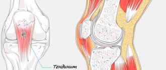
- tendon damage;
- patellar tendon damage;
- osteosclerosis, osteoporosis;
- osteophyte;
- cysts;
- arthritis, arthrosis;
- tumor processes;
- gout;
- bursitis;
Pain when bending the knee, when walking, a characteristic crunch, swelling of the knee and other signs may also be a reason for an X-ray of the knee joint.
Restrictions
X-rays are not performed on children under three months of age, who are prescribed an ultrasound if necessary. Doctors also do not recommend excessively irradiating infants in the pelvic area, as this can lead to infertility, blood disease, and a tumor process in the future. Children are examined strictly according to indications and in a standardized manner, no more than once every six months.
X-rays are not performed on pregnant women to avoid negative effects on the fetus. It is also contraindicated in people with metal prosthetics or implants in the area being examined and in people with schizophrenia (and other mental disorders) who are unable to remain still. Other people, including older people, can undergo the examination.
The procedure for performing an X-ray of the knee joint
In case of severe pain, injury, or fracture, the specialist helps the patient lie down, because The photograph is taken in a supine position. X-rays of the knee joint are usually done in two projections - frontal and lateral. The patient is accordingly placed in a position either lying on his back with his leg extended, or on his side with the leg also fully extended.
If necessary, after conducting the examination in standard settings, the traumatologist or radiologist recommends additional x-rays using special settings. The Schuss position involves an x-ray of the knee joint with the leg bent at an angle of 30 degrees, and the Fick position involves an x-ray of the knee with the leg bent at an angle of 45 degrees.
In general, the procedure is painless and takes about 20 minutes.
Features of x-ray of the upper extremities
X-rays of the elbow are also prescribed after injuries - severe bruises, dislocations, fractures - or if various pathologies are suspected. It allows you to obtain information about the joint space and its narrowing, about the condition of the ends of the bones of the humerus and forearm. The specialist also receives data about the areas adjacent to the elbow joint, which facilitates diagnosis. After all, the cause of pain is not always arthrosis of the elbow, arthritis or bursitis: it often has a spreading nature and a completely different source.
Shoulder pain often occurs against the background of neurological and vascular diseases, but the cause may also be shoulder arthrosis, as well as systemic inflammatory diseases of the shoulder joint. X-rays are mainly prescribed if a dislocation or fracture is suspected. The image also shows adjacent formations - the collarbones and shoulder blades. It is also informative for arthrosis and arthritis, necrosis of the humeral heads, tendonitis and other diseases.
Osteoarthritis of the upper extremities is often discovered by chance on an X-ray
X-ray is a simple, quick, painless diagnostic method that provides information about the condition of bones and joints. When arthrosis is detected, additional instrumental methods are often prescribed to examine the deep structures of soft tissues and study the condition of the cartilage. However, MRI and CT scans are expensive and not always necessary. Therefore, if the orthopedist insists on an x-ray, you should not refuse.
Gonarthrosis and the army
Many young male patients wonder: are they drafted into the army with such a complex pathology? To clarify, we suggest you read the information that we have specially prepared for you. So, first of all, your military suitability will be assessed:
- severity of clinical symptoms of the disease;
- degree of deformation;
- the presence of concomitant pathologies (systemic pathologies, congenital anomalies of the musculoskeletal system, flat feet, kyphosis and scoliosis of the spine, etc.).
Suitability to become a recruit is determined by a military medical commission on an individual basis based on x-rays of the joint, personal medical history and laboratory test results. It is impossible to say unequivocally whether you will be released from service or not without seeing your examination results. However, you can use information from regulatory documents, which contain diagnoses and their criteria for which exemption or deferment from military service is allowed. Arthrosis of the knee is regulated by Art. 65, which lists possible options that allow exemption from the army:
- Stage 2 , in which there are osteophytes larger than 1 mm, the joint space is narrowed 2 or more times from normal values, there is impaired joint congruence and subchondral osteosclerosis. The young man most likely will not serve, even though he is marked “fit” in the relevant document.
- 3-4 degrees allow you to fully count on exemption from military service. This diagnosis is accompanied by severe pain, while X-rays clearly show large spiky growths, significant osteochondral deformations, and a complete or almost complete absence of joint space. In fact, a disabled person, it’s hard to even say where one can work with such a sentence, let alone the army. Therefore, category “D” is assigned with the note “unfit”.
Regarding stage 1 of the disease, we note that it does not provide grounds for exemption from the army; the conscript is only given privileges to mitigate the conditions of service.
What is the difference between gonarthrosis and arthrosis?
It might be worth starting the article with an interpretation of these medical terms. But if you read it carefully, you probably guessed that there is no fundamental difference in the concepts of these two names, except in pronunciation and spelling. Gonarthrosis is also no different from osteoarthritis of the knee joint. The essence, features of the initial manifestation and later periods of development, prevention and therapeutic measures of all these pathologies are equivalent. All of them indicate the same direction of degenerative-dystrophic pathogenesis, however, with only one difference: the diagnosis “gonarthrosis” cannot be used in relation to other bone joints. Why?
Gonarthrosis, which means degeneration and dystrophy of the articular cartilage of the knee joint (translation from the Greek knee + arthrosis), provides information about the specific localization of deforming arthrosis (osteoarthrosis), namely in the knee. But, for example, the term “coxarthrosis,” which you also repeatedly came across when reading medical Internet sources, also means deforming arthrosis, but of the hip joint (hip + arthrosis). Arthrosis and osteoarthritis are general concepts that are simultaneously suitable for any joint.