What is knee arthroscopy?
Overload and injury to the knee joint are the most common causes of knee dysfunction. People who actively and professionally engage in sports such as skiing, running, basketball, volleyball and football are considered especially vulnerable. Some injuries can be treated with joint immobilization, physical therapy, and painkillers and anti-inflammatory medications. However, there are some lesions that may require surgery.
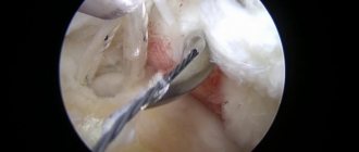
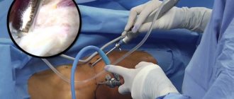
In addition to imaging tests such as X-rays, knee ultrasound, or magnetic resonance imaging, orthopedists often perform diagnostic arthroscopy. Thanks to this examination, it is possible to accurately assess the condition of intra-articular structures: muscles, ligaments, articular cartilage, meniscus and synovium. The arthroscope is inserted into the joint through a small incision. Saline solution is injected inside to make it easier to view the pond through the camera. The device allows you to reach hard-to-reach places without the need to make large incisions. As a result, the patient recovers faster and less burdensome. If treatment is required, surgical instruments may be inserted into the joint. This procedure is known as operative arthroscopy.
For what diseases is arthroscopy indicated?
- Arthrosis of various joints.
- Arthritis.
- Trauma and hemarthrosis.
- Chronic inflammation of the joints.
- Knee ligament rupture.
- Meniscus damage.
- Meniscus cyst.
- Dislocation and subluxation of the patella.
- Removal of growths, adhesions in the joint, foreign bodies and excess fluid.
Arthroscopy is not performed if there is an infection in the problem joint, with bone or fibrous ankylosis, or with purulent inflammation around the joint. There are also relative contraindications, at the discretion of the doctor, for example, massive hemorrhage in the joint or extensive damage can also interfere with the manipulation.
Severe general condition of the patient is a reason to postpone arthroscopy
What are the benefits of treatment?
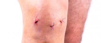
Compared to other methods that require full opening of the knee joint, arthroscopy is relatively less invasive, which is a great advantage for the patient. The incisions made during the procedure are painless, and the scars after them are small. After such a procedure, the patient can be discharged from the hospital within 1-2 days. Orthopedists choose to perform arthroscopy because it is more accurate than other imaging methods. In addition, arthroscopy allows you to evaluate the functioning of the joints during movement.
Advantages of arthroscopic surgery.
The entire operation is carried out through several punctures in the knee.
Significant advantages of meniscal arthroscopy include:
- Low level of trauma (since access to the joint is through small holes).
- The risk of complications is reduced to a minimum compared to classical surgical access (infections, bleeding, various restrictions of movement, scar formation).
- There is no need for postoperative immobilization (plaster), as well as the use of hard-tolerated painkillers.
- Material savings. The cost is comparable to that of open access, but the monetary costs are lower, since you do not need to stay in the hospital for a long time.
- Local anesthesia - during arthroscopy, anesthesia is not required (which is most important for patients with pathologies of the cardiovascular system).
- Fast rehabilitation.
- There are a couple of small marks left on the skin that are barely noticeable.
- Diagnostics and therapeutic manipulations are carried out simultaneously - within the framework of one intervention.
You can read more about joint arthroscopy, types, advantages and costs in this section:
How to prepare for knee arthroscopy? How is the procedure done?
Before undergoing an arthroscopy procedure, the patient should undergo an orthopedic consultation, as well as a series of imaging studies (ultrasound, magnetic resonance imaging) and laboratory blood tests (including morphology, blood type, glucose levels, coagulation system, liver tests). In addition, the patient should be vaccinated against hepatitis B. To do this, it is advisable to consult a family doctor. All concomitant diseases, as well as the fact of taking medications, must be discussed in advance with the attending physician.
The arthroscopy itself does not last long and is usually performed under spinal anesthesia, less often under local or general anesthesia. During the procedure, after an incision is made, specialized cameras and instruments are inserted into the joint, thanks to which the orthopedist will be able to assess the intra-articular structures and the extent of possible damage, and then carry out treatment. The most common treatments include treatment of the meniscus, synovium, and treatment of articular cartilage and ligaments.
Features of different types of anesthesia
For intraoperative pain relief
use different techniques of general, regional and
local anesthesia
, as well as their combinations. Each of them has a number of disadvantages and advantages.
Local anesthesia
From a technical point of view, local anesthesia is the simplest. Even a traumatologist can perform local administration of anesthetics without the help of an anesthesiologist. However, such pain relief
does not always provide a sufficiently strong analgesic effect. And its quality largely depends on the practical skills of the traumatologist.
Conduction anesthesia
It is one of the best options for pain relief during arthroscopic interventions on the knee joint. It is technically more complex than local, and therefore requires great skill of the anesthesiologist. The doctor must have a good knowledge of the structure of the peripheral nervous system, master the technique of blocking nerves at different levels, and be able to use special instruments to perform a femoral / sciatic nerve block.
Conduction anesthesia provides long-term pain relief during movement of the lower limb and thereby improves the course of the early postoperative period. However, due to technical difficulties, it is still less common than spinal.
Spinal anesthesia
Guarantees adequate postoperative pain relief and early restoration of muscle tone. However, a number of complications can occur, including spinal cord or root injuries, radiculopathy, cauda equina syndrome, seizures, etc. The high incidence of side effects (back pain, headaches, hypotension) often reduces patient satisfaction with the quality of such pain relief.
Anesthesia
With arthroscopy
used rarely, usually in one-day hospitals. Modern intravenous and/or inhalational anesthetics can be used for anesthesia. During surgery, the patient often breathes independently through a laryngeal mask. For special indications, he may undergo artificial pulmonary ventilation (ALV).
Anesthesia has its advantages:
- safety;
- good handling;
- minimal number of side effects;
- no effect on the muscle tone of the limb in the postoperative period;
- possibility of rapid discharge from hospital.
At the same time, anesthesia does not always provide adequate postoperative pain relief. Therefore, in the early stages after arthroscopy, the patient may be additionally administered anesthetics.
Rehabilitation after knee arthroscopy
For the procedure to be successful, the patient must strictly follow the orthopedist's recommendations. It is important to ease the operation on your leg for a few weeks, which will likely mean you will need to use crutches. It is advisable to practice walking on crutches at least a little before surgery.
During recovery, the patient should avoid physical activity. However, this does not mean a complete renunciation of activities. After arthroscopy, it is important to perform exercises under the supervision of a physical therapist to strengthen and stretch the muscles and improve joint stabilization. Recovery from the procedure may take up to 6 weeks. Full recovery takes about 12 weeks, but this depends on the individual.
Following your doctor's instructions may protect against complications after arthroscopy. Pain, swelling and bruising are the most common symptoms after the procedure, but deep vein thrombosis or bacterial infections may occur in some cases. The reason for such side effects is, as a rule, too early stress on the operated joints.
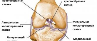
The structure of the meniscus.
Synthetic collagen implant.
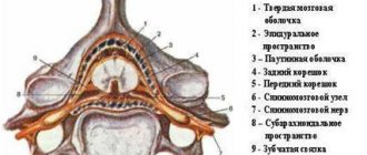
The meniscus is a crescent-shaped cartilaginous formation of the knee located between the surfaces of the knee joint. Performs a shock absorption function, helps to evenly distribute the load between the articular surfaces, promotes better gliding, providing freedom of movement. In each knee, the lateral (facing the outer) and medial (facing the inner) menisci are located respectively on the lateral surfaces. They take on most of the load, which puts them at high risk of injury. Due to the nature of the blood supply, the closer to the central zone the damage is located, the lower the likelihood of complete recovery. Over time, especially in athletes or people with vigorous physical activity, cartilage tissue becomes thinner, and it can subsequently become torn. In addition, ruptures often occur during acute injuries. As a rule, the highest incidence of lesions is at the internal meniscus. Deformations are often accompanied by violations of the integrity of the ligaments, since these anatomical formations are closely connected.
The manifestation of pathology is accompanied by the following symptoms:
- Sharp pain that becomes more intense when the knee is loaded.
- Increasing swelling.
- Intra-articular hemorrhage.
- Stiffness and motor dysfunction.
- Pain in the popliteal region, sudden blocking of the joint when flexing or extending.
- The appearance of a click at the time of injury and a crunching sound during movement.
Who will benefit from this technique?
Arthroscopy is usually performed in cases where the diagnosis is unclear and other diagnostic methods do not clarify the clinical picture. They may prescribe a similar manipulation to evaluate and monitor the effectiveness of the therapy just performed. In rare cases, arthroscopy is prescribed when a patient complains of pain, complications and discomfort in the joint after previous interventions.
A direct indication for this procedure is damage to the meniscus of any nature. In this case, diagnostic or therapeutic arthroscopy is prescribed, depending on the nature of the complaints and the course of the disease. In addition, the procedure may be prescribed for a combination of disorders in the menisci and ligaments, for any inflammatory processes in the joint, for recent injuries, as well as for developing arthritis and osteoarthrosis. In general, any change in the knee joint, whether traumatic or inflammatory, is an indication for arthroscopy.
Contraindications to arthroscopy
Despite the usefulness of the procedure, as with any other manipulation, there are contraindications for arthroscopy. It can be:
- Infections of a ongoing or chronic nature, especially localized near the damaged joint;
- Inflammatory processes in the skin near the intended site of intervention;
- Pathology of internal organs in serious condition;
- Adhesions and ankylosis, in which the joint cannot bend normally;
- Knee injuries in which the ligaments are exposed and the knee is damaged;
- Heavy bleeding due to injury, purulent processes in the wound.
How to prepare for the procedure
Initially, as with any other interventional procedure, the patient is sent for a series of standard diagnostic tests. These include general blood and urine tests, fluorography, blood tests for pathologies, ECG, and a general examination by a doctor. Next, after passing all the tests, the patient is given a referral for the procedure. In this case, the doctor must clarify with the patient a number of nuances, the presence of which may complicate or reduce the effectiveness of the manipulation. This includes taking anticoagulants and other medications, as well as menstruation in women. Since the procedure as a whole does not involve dissection, intervention still takes place, so it is important to exclude the occurrence of thrombosis and bleeding during arthroscopy .
Before the procedure itself, the patient undergoes a short consultation with the surgeon and an appointment with the anesthesiologist. If general anesthesia is planned, then the day before the patient adheres to a light diet, prepares clothes for the operating room, and shaved the joint area. It is advisable to take care of the postoperative period in advance, that is, purchase crutches or a wheelchair, and consider transportation home.
Stages of arthroscopy
In general, all stages of the procedure are the same for both diagnostic and therapeutic interventions. Only some nuances may vary. The general picture of the procedure is as follows:
- The patient is placed on the table with his back, the operated knee is bent at a right angle and fixed;
- The patient is given anesthesia or general anesthesia;
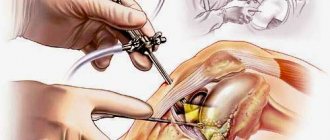
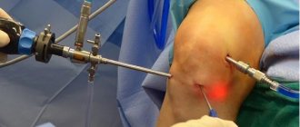
- The operated area is treated with antiseptics, then symmetrical punctures are made on both sides of the knee. A camera is inserted into one puncture, and instruments are inserted into the second through a trocar;
- To improve visibility, saline may be injected to expand the field of view. After these manipulations, the surgeon begins an examination or medical work; the latter may require additional tools in the form of a scalpel, forceps, and scissors. If complete removal of the meniscus is required, instruments can be inserted into two punctures at once;
- After surgical procedures, another solution is injected to wash away any remaining tissue. If the meniscus has been excised or parts of it have been removed, sutures are applied to it;
- Next, the instruments are removed, and sutures are applied to the punctures. The wounds are washed with an antiseptic and the knee is bandaged.
The procedure generally takes no more than 3 hours, even in difficult situations. Usually the patient can be discharged from the clinic the next day.
Postoperative period
Often this procedure is quite simple and does not require much time or effort for recovery. But a number of recommendations for rehabilitation still exist:
- Physical activity must be prescribed for the entire recovery period, and aimed specifically at the operated leg;
- On the first day, cold may be applied to the affected joint to reduce swelling;
- Painkillers may be prescribed to relieve pain after surgery;
- For the first few days, you should not heat or overcool the joint or sunbathe, as this can cause the development of an inflammatory process;
- You should not squat or kneel until the joint has fully recovered, otherwise this may result in re-injury of the meniscus;
- The doctor prescribes various procedures for faster joint restoration, including current stimulation, massage, magnetic therapy, and also prescribes complexes of vitamins and minerals for tissue restoration;
- Physical therapy is prescribed with an emphasis on the operated limb;
- The patient can be discharged the next day, but they are issued a sick leave certificate for 1-2 weeks. The overall recovery period varies within a month.
If all recommendations are followed correctly, the knee joint quickly recovers and allows the patient to soon return to their normal lifestyle.
General information
Arthroscopy is a minimally invasive surgical procedure that allows for an accurate diagnosis of diseases of the knee joint, examination of intra-articular structures in real time,, if necessary, a biopsy of the joint capsule, and also a number of therapeutic surgical procedures in the knee cavity.
Arthroscopy of the knee joint is carried out by introducing an arthroscope (this is a special type of endoscope) into the joint cavity through a micro-incision in the skin in the knee area. Arthroscopic manipulation of the knee joint can be used both for the diagnosis of articular diseases and for the treatment of a large number of orthopedic diseases of the knee, for example, resection of the meniscus, elimination of the articular mouse, restoration of the integrity of intra-articular ligaments, removal of damaged areas of cartilage, arthroscopic sanitation of the knee joint.
Causes of arthrosis
Deforming osteoarthritis is very common among older people, as well as among those who devote their lives to sports. Most often, the disease appears after an injury (especially if there is no treatment after it).
In addition, the following factors influence the occurrence of arthrosis:
- Age from 50 years.
- Overweight.
- Frequent loads on the lower limbs.
- Intensive workouts in the gym (working with heavy weights).
- Old injuries.
- Joint diseases (rheumatism, gout, arthritis).
- Genetic predisposition.
- Joint instability.
- Various metabolic disorders.
- Alcohol abuse, smoking, systematic use of drugs, potent medications.
Doctors note that one of the main reasons for the increasing number of cases of the disease is associated, oddly enough, with the increase in life expectancy of the population. Most likely, in the future this problem will become even more pressing than it is today.
Rehabilitation
The minimally invasive nature of the procedure allows you to quickly return to your normal life. This is facilitated by therapeutic physical education. Exercise helps strengthen muscles and restore blood flow. The rehabilitation plan is purely individual, it is drawn up by the attending physician, giving a gradual load on the joint and prescribing the necessary medications.
It is possible to return to full-fledged sports after 3-6 months, and any risks of relapse can be eliminated only after a year. You yourself can contribute to effective recovery by regularly consulting with a specialist and practicing at home.
Contraindications
The greatest number of contraindications exist for general anesthesia . When administered systemically, anesthetic drugs can negatively affect internal organs. Therefore, anesthesia
Contraindicated for arrhythmias, heart defects, bronchial asthma, bleeding disorders, renal, hepatic and cardiovascular failure.
Contraindications to spinal and epidural anesthesia
There may be an infection at the injection site and an allergy to anesthetics. These types of pain relief will not be used even if there are no contraindications. This is possible if the patient himself refuses them.
list
Symptoms
You should contact a specialist if you notice at least one of the following symptoms:
- Pain syndrome.
In the early stages it occurs periodically, usually after intense exercise. The danger is that often the pain is not too strong, passes after a while, and many people simply do not pay attention to it. - Swelling.
As a rule, it accompanies a period of exacerbation of pain. - Crunch.
Due to the gradual thinning of the cartilage layer and the decrease in the amount of synovial fluid, the bones begin to collide with each other, which is why a characteristic rough crunching sound becomes heard. - Limited movement.
Dysfunction and stiffness are observed. The more advanced the disease, the less ability to move. - External changes.
The appearance of the limb gradually changes.