- Ultrasound can be carried out not only according to indications, when there is a suspicion of pathology or it has already been identified - the study can also be carried out for preventive purposes in the absence of complaints. For example, examination of the knee joint is indicated for professional athletes, especially gymnasts, runners, basketball players, skiers, bodybuilders, etc. Their occupation can cause chronic trauma to the joint, when no discomfort is felt, but the tissues are gradually deformed. If this condition is not detected early, the changes can become severe and cause significant limitations in mobility.
- Change in skin color;
- The occurrence of swelling in the knee joint;
- Presence of osteochondropathy;
- Suspicion of deformation of joint tissues;
- And, of course, pain.
Also, a diagnostic procedure can be carried out to monitor the effectiveness of therapy. This technique allows you to monitor changes after the operation.
As you know, this examination is completely harmless, which means that not only adults, but also children can be referred for ultrasound.
This type of diagnosis is prescribed for children in the following cases:
Why is ultrasound of joints done?
An ultrasound of the joints is performed to study the structure and condition of the joint, while the tissues located around them are examined. The diagnostic technique falls into the category of common and popular procedures, since there are no negative consequences for the body and it is easy to carry out.
An examination is prescribed by a doctor if the patient experiences pain in the joint area, if inflammatory processes in this area are suspected, crunching occurs during movement, injuries, limited mobility and swelling. Ultrasound examination is carried out to identify rheumatic diseases; the peculiarity is that pathologies are detected in the early stages. Surgical interventions are performed under diagnostic control.
Indications for the procedure
The knee joint is one of the largest and most complex in the human body. It is responsible for the movement of the limb, connecting the patella, femur and tibia. Against the background of increased stress and degenerative processes, various pathologies can develop. Ultrasound helps to detect the slightest disturbances in the structure and functionality of cartilage, joints, and the functioning of blood vessels. Most often, specialists have to deal with meniscus tears or cruciate ligament injuries.
Main indications:
- knee pain;
- limited mobility;
- bone deformities;
- the appearance of local swelling, redness of the skin;
- the presence of a palpable formation;
- decreased muscle strength.
Difference between MRI and ultrasound
Before we look at how MRI of joints differs from ultrasound, it is necessary to give clear definitions of each procedure.
Ultrasound examination is a diagnostic technique; scanning is carried out using ultrasonic waves. As a result, the doctor examines the condition of soft tissues and organs. The procedure is harmless. In this case, the doctor reads information about the condition of the meniscus, periarticular soft tissues, and cruciate ligament, but is not able to scan the bones and lungs. MRI is used to study bone tissue.
An MRI is a large magnet that produces magnetic waves within a specific magnetic field. During an MRI scan, the doctor examines all tissues and organs. The procedure can be used repeatedly and allows one to study the dynamics or consequences of an injury or disease. The question of which is better: MRI or ultrasound is not relevant. The difference is in information content, reliability and price.
What diseases can be detected?
Ultrasound diagnostics of knees in adults is used to identify the following pathologies:
- arthritis - inflammation of the knee joint structures of a traumatic, infectious, reactive nature involving the capsule, cartilage and synovial membrane;
- synovitis - inflammation of the synovium with accumulation of effusion;
- tendinitis - inflammatory damage to tendon tissue due to prolonged increased load;
- complete or partial rupture of ligaments as a result of traumatic impact.
During the examination, it is possible to assess the extent and severity of damage, detect hematomas, loose bodies, tumors, signs of aseptic necrosis, osteophytes and cysts. Diagnostics makes it possible to determine specific methods of therapy, understand the need for intra-articular injections and therapeutic or diagnostic arthroscopic puncture.
Compared to arthroscopy, ultrasound of the knee joint is non-traumatic and absolutely safe for the patient’s health. This allows the method to be used an unlimited number of times, including as part of preoperative preparation and to evaluate the results of treatment procedures.
Difference between X-ray and ultrasound
X-ray scanning is used in traumatology; during the examination, bone tissue pathologies are identified. During the ultrasound scanning process, the condition of bones, ligaments, cartilage, synovial bursa, and articular surfaces is assessed.
The advantage of ultrasound diagnostics is that it is harmless to the human body. X-rays affect the body with ionizing radiation and are prohibited for pregnant women. The radiation exposure received during the examination is taken into account.
Restrictions on performing ultrasound of the knee joint
As such, there are no contraindications to this manipulation. But still, in some cases, diagnostics may not be informative enough. For example, this occurs when the joint is completely immobile. If the skin has serious damage, for example, severe burns or abrasions, the results may also be distorted, and the examination process may be unpleasant for the patient.
Also, ultrasound of the knee joint is not recommended for people suffering from varying degrees of obesity. In this case, experts suggest giving preference to other diagnostic methods, for example, magnetic resonance imaging or arthroscopy.
What knee diseases can be determined using this method?
Ultrasound is recommended for any complaints - pain, redness, swelling, crunching in the knee. With its help, you can confirm or refute the presence of injury, meniscopathy, hemorrhage, dysplasia, inflammatory pathologies, and neoplasms.
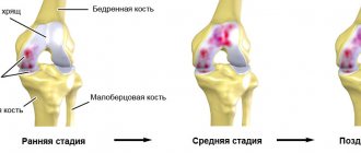
- Ultrasound allows one to predict the development of gonarthrosis by the height of the cartilage and the volume of intra-articular fluid.
- In case of injuries to the knee area, the examination will help differentiate between sprains and ruptures of ligaments, meniscal injuries and bone fractures.
- If a tumor is found in the knee, we can conclude that it is benign or malignant.
- The study reveals bursitis, synovitis, arthrosis and arthritis, tendonitis.
Ultrasound examination of the knee is important before or after surgery
Advantages of the method
- You can undergo an ultrasound of the joints at any age, including during pregnancy.
- The examination is safe and painless.
- It is allowed to go for the procedure several times in a short period of time, which allows you to monitor the clinical picture over time.
- Ultrasound machines are mobile and can be used if a person is immobile.
- No preliminary preparation is required.
- The gel is hypoallergenic in nature.
In complex cases, MRI is indispensable
Disadvantages of MRI of the knee joint
- MRI of the knee joint has a number of severe limitations. Tomography is contraindicated if the patient’s body has metal-containing implants, for example: a steel prosthesis, electronic pacemakers - neuro- or cardiac pacemakers.
- Disadvantages of MRI of the knee include the length of the procedure. It can last 30 minutes, and if the pathology is serious and contrast enhancement is required, then the scanning duration reaches 40-60 minutes.
- During the tomography of the knee joint, any movement is prohibited, as this will negatively affect the clarity and accuracy of the MRI images. Therefore, young children have to undergo MRI of the knee under anesthesia.
- MRI of the knee joint using a closed tomograph can be difficult for people suffering from claustrophobia, epileptic seizures and panic attacks.
Is it possible to undergo an ultrasound for those who do intra-articular injections?
Intra-articular injections of Noltrex synovial fluid prosthesis or another drug are not a contraindication to ultrasound, but there is one limitation. It is necessary to take a break of at least five days between the injection and the ultrasound examination. Otherwise, due to insufficient visualization, the results may be unreliable.
If there are indications, you should not refuse ultrasound of the joints - an informative and reliable method. However, it should be remembered that the accuracy of the study largely depends on the competence of the specialist. If you need to obtain information not about joints, but about bones, you cannot do without X-rays or CT scans.
Decoding the research results
The patient receives an interpretation of the ultrasound diagnostics within 10-15 minutes after the procedure. Normally, all surfaces should be smooth and clear, the joint capsules look like hypoechoic structures. The doctor pays attention to the uniformity of hyaline cartilage and the quality of tissue nutrition. There should be no inflammatory effusion inside.
The ultrasound examination method allows you to detect:
- signs of infectious and inflammatory processes;
- age-related and post-traumatic changes;
- hematomas;
- degenerative-destructive changes;
- deterioration of blood circulation and nutrition of cartilage tissue;
- cysts, tumors, metastases;
- dislocations, fractures, ligament tears, meniscus injuries.
To interpret the resulting images, the specialist must have knowledge of the anatomical features of the structure of the knees and the blood circulation of nearby tissues. The CONSTANTA Clinic employs experienced diagnosticians who are well acquainted with the common problems of this anatomical zone and successfully carry out differential diagnosis, providing reliable examination results.
Damage to tendons and ligaments
Injuries to the tendon-ligamentous system occur during sports activities, falls, rare jumps and awkward movements. Ultrasound makes it possible to assess the integrity of connective tissue and detect hematomas. With partial ruptures, the contours of the tendon are preserved, but a hypoechoic zone is noticeable at the site of injury. It can also be seen between bone fragments when the patella is fractured.
Meniscus injuries
The menisci can be damaged by sudden rotation of the tibia, excessive flexion or extension of the limbs. In the first days after injury, sharp pain occurs and movement is limited. The skin is swollen and hyperemic. In this condition, the patient needs urgent medical attention. If conservative therapy is ineffective, meniscectomy must be performed. When the menisci are damaged, their contours are disrupted, hypoechoic areas and extraneous stripes appear. The appearance of effusion inside the joint is characteristic. Diagnostics must be carried out as soon as possible in order to assess the severity of the condition and, for certain indications, carry out therapeutic and diagnostic arthroscopy.
Rheumatoid arthritis
This disease is accompanied by destructive changes, the formation of osteophytes, subcutaneous nodules and nerve damage. The disease is chronic. Patients complain of morning stiffness in the joints and constant pain. Inside the knee joint, exudate accumulates, contractures and characteristic deformities appear. During the examination, specialists detect osteophytes, effusion, narrowing of the joint space and thinning of hyaline cartilage.
Which joints can be examined using ultrasound?
Ultrasound can be used to examine:
Ultrasound of joints
- shoulder joints;
- elbow joints;
- knee joints;
- ankle joints with foot joint;
- hip joints;
- wrist joints with hand joints;
- mandibular joints.
Both joints are examined at once (for example, two elbows or two knees), even if only one hurts. In each person, the structure and development of tissues in the area of the right and left joints are approximately the same, and at the same time unique. Comparing a diseased joint with a healthy one allows us to evaluate the pathological changes that have occurred.
Description
Ultrasound of the knee is particularly useful for assessing quadriceps and patellar tendon injuries, medial and collateral ligament injuries, joint effusions, and fluid collections. Limitations of ultrasound include incomplete assessment of the deep structures of the knee, especially the cruciate ligaments, menisci, and most articular cartilage. An ultrasound of the knee joint usually takes no more than 20-25 minutes. The examination is carried out first in the supine position (anterior longitudinal and anterior transverse projections), and then in the prone position (posterior transverse, posterior lateral and posterior longitudinal medial projections).