Structure
The wrist has a complex structure due to its functional features. A small piece of the body connecting the hand and forearm consists of 8 bones. They have a peculiar shape, a rough triangular surface. By the appearance of the bones, the wrists fully reflect their name:
- The scaphoid bone (tarsus) is the smallest and most difficult to diagnose in fractures;
- The lunate bone got its beautiful name because of its resemblance to a crescent moon. In case of a fall, a dislocation or bruise of a bone is diagnosed, which is accompanied by swelling;
- The triquetral bone is prone to frequent fractures due to its location. When falling with emphasis on the hand, it bears the entire weight load;
- The pisiform bone looks more like a small chicken egg. The anatomical location protects against injury. Damage occurs only as a result of a direct impact and is excluded from a fall;
- The trapezius bone and flexor carpi radialis are in close contact, so when damaged, the tendons suffer;
- The trapezoid bone is located deep in the parts of the hand, so fractures in this area occur very rarely;
- The capitate bone is the largest. Rarely undergoes single fractures. The damage is accompanied by injuries to other bones due to the size of the capitate bone;
- The hamate bone is the trailing bone. Fractures occur in 2% of cases.
The full functionality of the joint depends on innervation and the flow of nutrients with blood that comes from the subclavian artery.
To find out where the wrist is, just look at the base of the palm. A small part of the arm connects the hand and the main limb.
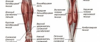
Muscles of the hand[ | ]
The musculature of the hand is a complex complex of approximately 33 muscles. Most of them are located in the forearm and are connected by tendons to the phalanges of the fingers through several joints. Two groups of muscles form two elevations on the palmar surface of the hand: thenar (thenar) - the elevation of the thumb and the hypothenar (hypothenar) - the elevation of the little finger. On the hand, the muscles are located only on the palmar side. Here they form three groups: the middle one (in the middle section of the palmar surface), the thumb muscle group and the small finger muscle group. The large number of short muscles on the hand is due to the fine differentiation of finger movements.
Middle muscle group of the hand[ | ]
Comprises:
- lumbrical muscles, which originate from the tendons of the deep flexor digitorum and are attached to the base of the proximal phalanges of the second to fifth fingers;
- palmar and dorsal interosseous muscles, which are located in the interosseous spaces between the metacarpal bones and are attached to the base of the proximal phalanges of the second to fifth fingers.
The function of the muscles of the middle group is that they are involved in flexing the proximal phalanges of these fingers. In addition, the palmar interosseous muscles bring the fingers of the hand towards the middle finger, and the dorsal interosseous muscles spread them apart[3].
Muscle group of the thumb[ | ]
Forms the so-called eminence of the thumb on the hand. They begin on the nearby bones of the wrist and metacarpus. Among them are:
- the abductor pollicis brevis muscle, attached to its proximal phalanx;
- flexor pollicis brevis, which attaches to the external sesamoid bone located at the base of the proximal phalanx of the thumb;
- the opponus pollicis muscle, which goes to the first metacarpal bone;
- The adductor pollicis muscle, which attaches to the internal sesamoid bone located at the base of the proximal phalanx of the thumb[4].
The function of these muscles is indicated in the name of each muscle.
Small finger muscle group[ | ]
Forms an elevation on the inside of the palm. This group includes:
- palmaris brevis;
- abductor digiti minimi muscle;
- flexor digitorum brevis;
- muscle opposite the little finger.
They arise from the nearby carpal bones and insert at the base of the proximal phalanx of the fifth digit and the fifth metacarpal bone. Their function is determined by the name of the muscles themselves.
Methods of treating wrist diseases
The special structure of the wrist plays an important role in determining the diagnosis to identify the disease and predisposition to various injuries:
- The accumulation of small bones leads to frequent fractures and dislocations. Knowledge of anatomy allows the specialist to determine the most likely location of the fracture. This will help make a quick diagnosis and begin treatment;
- The hand has greater mobility, which is a risk factor for the development of arthrosis;
- The hand joint has a developed blood supply system, which means an increased risk of developing arthritis.
The anatomy of the joint implies the presence of small bones that are grouped together. This structure allows you to make precise movements, grabs, and perform miniature work. When falling, a part of the body suffers greatly due to the person's attempt to instinctively put his arm forward to prevent the blow. But mechanical damage is not the only hand disease.
Notes[ | ]
- R.D.
Sinelnikov, Ya.R. Sinelnikov, A.Ya. Sinelnikov. ATLAS OF HUMAN ANATOMY, volume one. — 8th ed., revised. — Moscow: RIA “New Wave”. Publisher Umerenkov, 2021. - pp. 122-131. — 488 p. — ISBN 978-5-7865-0305-4 (New Wave) 978-5-94368-069-4 (Umerenkov Publishing House). - E.I.
Borzyak, L.I. Volkova, E.A. Dobrovolskaya and others. Human anatomy / M.R. Sapin. — 4th ed., stereotypical. - Moscow: Medicine, 1997. - P. 132-133. — 544 p. — ISBN 5-225-04443-3. - Anatomy: Carpal tunnels. Hand tendon sheaths. (undefined)
.
meduniver.com
. Date accessed: October 24, 2021. - E.I.
Borzyak, L.I. Volkova, E.A. Dobrovolskaya and others. Human anatomy / M.R. Sapin. — 4th ed., stereotypical. - Moscow: Medicine, 1997. - P. 310-313. — 544 p. — ISBN 5-225-04443-3.
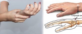
Hygroma of the wrist
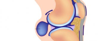
Hygroma is a neoplasm that is shaped like a ball with the material contents of a benign course. Inside the tumor capsule there is a liquid-like substance containing mucus. The cause is considered to be excessive stress involving the joint. It is subjected to unacceptable friction and damaged by compression.
Professions at risk of the disease:
- Athletes;
- Musicians;
- Office employees working on a computer;
- Garment workers.
Sometimes situations occur when the development of hygroma occurs without cause-and-effect relationships. In this case, additional tests are prescribed to identify hidden causes. The lump is usually located on the side of the center of the wrist, on the inside and outside.
At the onset of the disease, the tumor does not cause discomfort, it is difficult to notice, and the influence of the growing capsule does not affect motor function. The growth of hygroma is accompanied by compression of the surrounding nerves and tissues, causing pain. In extreme cases, hand movements become constrained. The tumor sticks out on the arm in the shape of an egg, which is an aesthetic defect. After detecting a characteristic lump, you should seek medical help from a specialist.
Causes:
- Point work done by hand;
- Heredity;
- Bruises, fractures, wrist injuries with serious consequences.
A slight fall with a certain probability can lead to the formation of a hygroma.
As the tumor grows, it makes simple movements difficult. This complicates life in everyday matters and puts professional activities at risk. When visiting a doctor, the stage of the disease and the structure of the tumor are determined. Timely treatment allows you to manage with conservative methods. In the advanced stage, surgical intervention is required.
To treat hygroma at an early stage, physiotherapy is prescribed. The wrist joint is warmed up using paraffin baths, and special compresses are made. The methods have contraindications in the presence of inflammation or damage to the capsule.
They often resort to the help of traditional medicine. Lotions are made based on compositions from various plants, pine needles, cabbage. Before use, consult a doctor to determine if you are allergic to the components of the tinctures. Compresses with decongestant ointments are applied to the wrist, where required. To achieve maximum effect, folk treatment methods are combined with traditional ones.
To determine an accurate diagnosis, puncture of the contents of the lump is prescribed. This is done to exclude cancer. The liquid is obtained by pumping out with a syringe. After the manipulations, the arm is fixed with an elastic bandage and physical activity is avoided.
In an advanced stage, hygroma is removed using laser burning. The operation is performed using local anesthesia. After the procedure, a bandage is applied that compresses the joint where the person’s wrist is.
Modern treatment methods make it possible to get rid of hygroma in a short time without relapse.
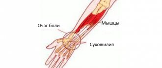
Carpal tunnel syndrome
The right hand most often suffers from painful sensations. This is easily explained by the increased workload of computerization of enterprises. The disease is called carpal tunnel syndrome.
As a result of increased load on the bones of a person’s wrist, the carpal tunnel nerve becomes pinched, which causes acute pain. People in certain areas of activity are at risk of contracting the disease.
Along with the hands, the elbow joint suffers from overwork. The patient cannot perform a basic action - pick up a pencil with his hands. A cold compress, rest, and painkillers help you return to a full life.
To prevent the disease, a method of exercises for the hands and their correct position during work has been developed.
How to position your hands to avoid illness:
- Hands cannot be held suspended on the table, in positions with increased load on the joints, so it is advisable to bend the arm at the elbow at a right angle;
- When working with the mouse, the hand remains straight;
- A computer chair should have sides for placing your hands on them while resting;
- It is recommended to purchase a useful mouse pad with a bump for the wrist, which will relieve the stress on the joint.
Gymnastics:
- Hands squeeze and unclench with force;
- Relax your fingers and shake them;
- Clasp your hands and tilt them alternately in different directions;
- Massage the wrist area with stroking movements;
- Place your arms in front of you and make circular movements in different directions.
After relaxing gymnastics, your hands rest for 2 minutes, after which you can start working. It is advisable to repeat the exercises as often as possible. This will protect against many diseases and relieve pain.
Literature[ | ]
- Human anatomy: textbook. for students inst. physical cult / Ed. Kozlova V.I. - M.: Physical culture and sport, 1978.
- Kazachenok, T. G.
Anatomical dictionary: Latin-Russian, Russian-Latin. — 2nd ed. - Minsk: Higher School, 1984. - Sapin, M. R.
Pocket atlas of human anatomy. / M. R. Sapin, D. K. Nikityuk. - M.: APP "Dzhangar"; Elista, 1999. - Sinelnikov, R. D.
Atlas of human anatomy: in 3 volumes - 3rd ed. - M.: Medicine, 1967.
Wrist fracture
When falling, a person puts his hand forward to protect himself from hitting the ground. This leads to fractures, which affect people with an active lifestyle. These include athletes, cross-country athletes, and motorcyclists. Often the cause of a fall is weather conditions - ice, snow, rain. The victim experiences acute pain, swelling and redness appear. An urgent visit to the hospital is required, as self-medication will lead to joint immobility and disability.
Fractures of the hand are common: in bad weather, the percentage of injuries increases. This is due to insufficient bone strength and heavy loads on them. A sharp blow forces a person to involuntarily slow down with his palm, which leads to a displaced fracture. A swelling forms in the upper part of the palmar surface. If the blow fell on the back side, then damage to the radius is diagnosed. In both cases, there is a danger of painful shock, so the person is immediately hospitalized.
People involved in traumatic sports are at risk:
- Boxing;
- Football;
- Cycling;
- Figure skating;
- Skateboard.
People involved in car accidents suffer fractures when they instinctively try to protect themselves from the impact.
Symptoms, diagnosis, treatment
The first symptom of a fracture is acute pain. The soft tissues begin to swell, touching the site of injury becomes painful, and hematomas appear. It is difficult to move your fingers because of the pain. A bulge appears in the area of the wrist joint; an attempt to move the hand is accompanied by a characteristic crunch; the bone on the wrist protrudes. First aid involves fixing the injured limb and applying a cold compress to the site of injury.
To diagnose a fracture, the doctor feels the area of injury and takes an x-ray. In severe cases of bone fracture, MRI diagnostics is performed. After identifying the presence of complications and identifying a broken bone, a treatment program is determined.
The first step is to remove parts of the bones. If possible, the surgeon carries out jewelry work to collect the remains of the joint for a full life for the patient. In milder cases, surgery is not required, and a plaster cast is applied until the bones are completely fused. During the treatment period, complete immobility of the limb is required.
After applying a plaster cast, the patient remains in the hospital for some time for observation. In case of severe swelling of the hand, the plaster is unclenched using forceps to avoid blocking the flow of blood to the fingers.
For severe pain, the doctor will prescribe painkillers. Elderly people are advised to take additional analgin, since in old age the pain threshold decreases. The cast is removed after 8 weeks of wrist fixation. Before removal, a control X-ray is taken to check the integrity of the bones.
The day after injury, rehabilitation activities are allowed. Exercises are aimed at strengthening muscles and preventing atrophy. It is allowed to massage using light stroking with a gradual increase in the intensity of pressure as you recover. The massage is done on the healthy and injured hand. In the first days after injury, more time is spent on the healthy arm. The hand being massaged should not hurt from touching, otherwise the session will be terminated. Massage improves blood circulation by stimulating soft tissues.
Joints of the hand[ | ]
X-ray of a dog's front paw
Wrist joint[ | ]
Additional information: Wrist joint
The formation of this joint involves the radius and bones of the proximal row of the wrist: scaphoid, lunate and triquetrum. The ulna does not reach the surface of the radiocarpal joint (it is “supplemented” by the articular disc). Thus, in the formation of the elbow joint, the ulna plays the largest role of the two bones of the forearm, and the radius plays the largest role in the formation of the radiocarpal joint.
In the radiocarpal joint, which has an ellipsoidal (ovoid) shape, flexion and extension, adduction and abduction of the hand are possible. Pronation and supination of the hand occur together with the same movements of the bones of the forearm. A small passive rotational movement is also possible in the radiocarpal joint (10-12°), but this occurs due to the elasticity of the articular cartilage. The position of the gap of the radiocarpal joint is determined from the dorsal surface, where it is easily detected through the soft tissues; in addition, its position is determined from the radial and ulnar sides. On the radial side, in the area of the inferior radial fossa, you can palpate the gap between the lateral styloid process and the scaphoid bone. On the ulnar side, a depression is felt between the head of the ulna and the triquetral bone, corresponding to the ulnar portion of the cavity of the radiocarpal joint.
Movements in the radiocarpal joint are closely related to movements in the midcarpal joint, which is located between the proximal and distal rows of carpal bones. This joint has a complex, irregularly shaped surface. The total range of mobility when flexing the hand reaches 85°, and when extending it is also approximately 85°. Adduction of the hand in these joints is possible by 40°, and abduction by 20°. In addition, circular movement (circumduction) is possible in the radiocarpal joint.
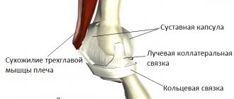
The radiocarpal and midcarpal joints are strengthened by numerous ligaments. The ligamentous apparatus of the hand is very complex. The ligaments are located on the palmar, dorsal, medial and lateral surfaces of the wrist, as well as between the individual bones of the wrist. The most important collateral ligaments of the wrist are the radial and ulnar ligaments. The first goes from the lateral styloid process to the scaphoid bone, the second - from the medial styloid process to the triquetral bone.
Between the bony elevations on the radial and ulnar sides of the palmar surface of the hand there is a ligament, the flexor retinaculum. It is not directly related to the joints of the hand, but is, in fact, a thickening of the fascia. Throwing over the carpal groove, it turns it into the carpal tunnel, where the flexor tendons of the fingers and the median nerve pass.
Carpometacarpal joints of the hand[ | ]
They are connections of the distal row of carpal bones with the bases of the metacarpal bones. These joints, with the exception of the carpometacarpal joint of the thumb, are flat and inactive. The range of movements in them does not exceed 5-10°. Mobility in these joints, as well as between the bones of the wrist, is sharply limited by well-developed ligaments.
The ligaments located on the palmar surface of the hand make up a strong palmar ligamentous apparatus. It connects the carpal bones to each other, as well as to the metacarpal bones. On the hand you can distinguish ligaments that run arcuate, radial and transverse. The central bone of the ligamentous apparatus is the capitate, to which more ligaments are attached than to any other bone of the wrist. The dorsal ligaments of the hand are much less developed than the palmar ligaments. They connect the bones of the wrist to each other, making up thickening capsules covering the joints between these bones. In addition to the palmar and dorsal ligaments, the second row of carpal bones also has interosseous ligaments.
Due to the fact that the bones of the distal row of the wrist and the four (II-V) bones of the metacarpus are inactive relative to each other and are firmly connected into a single formation that makes up the central bone core of the hand, they are designated as the solid base of the hand.
The carpometacarpal joint of the thumb is formed by the polygonal bone and the base of the first metacarpal bone. The articular surfaces are saddle-shaped. The following movements are possible in the joint: adduction and abduction, opposition (opposition) and reverse movement (reposition), as well as circular movement (circumduction). Due to the opposition of the thumb to all other fingers, the volume of grasping movements of the hand increases significantly. The amount of mobility in the carpometacarpal joint of the thumb is 45-60° during abduction and adduction and 35-40° during opposition and reverse movement.
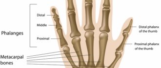
Metacarpophalangeal joints of the hand[ | ]
Formed by the heads of the metacarpal bones and the bases of the proximal phalanges of the fingers. All these joints have a spherical shape and, accordingly, three mutually perpendicular axes of rotation, around which flexion and extension, adduction and abduction, as well as circular movement (circumduction) occur. Flexion and extension are possible at 90-100°, abduction and adduction - at 45-50°.
The metacarpophalangeal joints are strengthened by collateral ligaments located on the sides of them. On the palmar side, the capsules of these joints have additional ligaments called palmar ligaments. Their fibers are intertwined with the fibers of the deep transverse metacarpal ligament, which prevents the heads of the metacarpal bones from diverging to the sides.
Interphalangeal joints of the hand[ | ]
Additional information: Interphalangeal joints
They have a block-like shape, their axes of rotation run transversely. Flexion and extension are possible around these axes. Their volume in the proximal interphalangeal joints is 110-120°, while in the distal ones it is 80-90°. All interphalangeal joints are strengthened by well-defined collateral ligaments.
Fibrous and synovial sheaths of the tendons of the fingers[ | ]
The ligaments, the flexor retinaculum and the extensor retinaculum are of great importance for strengthening the position of the muscle tendons passing under them, especially when flexing and extending the hand: the tendons rest on the named ligaments from their inner surface, and the ligaments prevent the tendons from moving away from the bones and with strong muscle contraction withstand significant pressure.
The sliding of the tendons of the muscles passing from the forearm to the hand and the reduction of friction are facilitated by special tendon sheaths, which are fibrous or osteo-fibrous canals, inside of which there are synovial sheaths, which in some places extend beyond these canals. The largest number of synovial sheaths (6-7) is located under the extensor retinaculum. The formation of the canals involves the ulna and radius bones, which have grooves corresponding to the passage of the muscle tendons, and fibrous bridges that separate one canal from the other, which go from the extensor retinaculum to the bones.
The palmar synovial sheaths belong to the flexor tendons of the hand and fingers running in the carpal canal. The tendons of the superficial and deep flexor fingers lie in a common synovial sheath, which extends to the middle of the palm, reaching the distal phalanx of only the fifth finger, and the tendon of the flexor pollicis longus is located in a separate synovial sheath, which passes along with the tendon onto the finger. In the palm area, the tendons of the muscles going to the second, third and fourth fingers are deprived of synovial sheaths for some distance and receive them again on the fingers. Only the tendons leading to the fifth finger have a synovial sheath, which is a continuation of the common synovial sheath for the flexor tendons of the fingers.
Bruised wrist
The wrist is susceptible to injuries of varying complexity. Often, a fall results in a bruise, which is confused with a fracture. The traumatologist prescribes an examination to exclude a fracture or dislocation. A bruise is a serious injury and requires timely treatment.
Causes:
- Falling onto an outstretched arm;
- A targeted blow to the wrist.
Symptoms:
- Pain immediately upon injury, which quickly subsides. After the formation of edema, the pain syndrome returns;
- Motor ability in the wrist joint is limited;
- Hematomas appear at the site of the injury.
Before arriving at the traumatology emergency room, the patient is given first aid:
- The wrist is fixed in one position;
- Ice is applied to the site of the bruise every 10 minutes;
- In case of severe pain, taking painkillers is allowed.
Treatment of a bruise allows you to avoid consequences for the vessels and nerves of the limb. The doctor recommends following certain rules:
- Avoid sports and physical activity;
- In case of serious bruises, the wrist must be secured with an elastic bandage;
- After five days from the date of injury, baths in warm water with the addition of sea salt are allowed.
To restore the supply of blood and nutrients to the joint, therapeutic exercises are prescribed. This helps eliminate stiffness and limited movement. It is allowed to apply creams and ointments with an analgesic and decongestant effect.
Self-massage is the main method of rehabilitation after a bruise. You need to start with stroking and move on to light kneading. Actions will help relieve swelling and restore limb mobility.
In case of a bruise, complications such as pinching of the ulnar nerve are possible. Acute pain pierces the fingers, the patient cannot fully straighten his arm. For treatment, massage and physiological measures are prescribed.
A hand bruise is an injury that requires medical examination. Connivance leads to serious consequences of limb atrophy due to vascular damage.
Types of pain in the hands
- Constant and periodic pain.
Pain may always be present or develop under certain circumstances. So, with injuries, discomfort is most often felt when pressing on the hand or moving the joint. And in case of inflammatory or degenerative processes, it can last for hours even after the cessation of exposure.
- The discomfort may be mild or severe.
The nature of burning unbearable pain is usually noted with gout. For carpal tunnel syndrome, dull pain is more typical.
- Painful sensitivity may appear on one hand or be felt symmetrically in both hands.
Pain in the left hand is often characteristic of coronary heart disease. With gout, painful sensations on the right hand will eventually spread to the left limb.
But it is impossible to determine the cause of pain only by its nature or location, since symptoms for various diseases can be similar. For example, pain in the bend of the wrist can be both a consequence of injuries and a symptom of pathologies of the nervous and cardiovascular systems. Because of this, it is difficult for a person to know which doctor to contact about pain. In such cases, you should start with a therapist.
Wrist Strengthening
A dislocation, bruise or fracture of the wrist leads to limited movement of the hand. Special exercises will help restore mobility and strengthen weakened muscles. The anatomical structure of the wrist is a joint, so it is not possible to pump it up. The training is aimed at using adjacent muscles. Exercises should be done every day using different strengthening techniques.
To restore motor flexibility to the wrist, it is recommended to jump on a children's jump rope. This exercise is used by athletes whose victory depends on arm strength. Jumps are carried out at a fast pace with maximum movements of the wrists.
Exercises with dumbbells strengthen the strength side of the wrist. To develop precise movements, use a thin sheet of paper. They place it on a flat surface and try to assemble it into the palm of their hand using only their fingers.
For guys and men, strengthening the wrist muscles is an integral part of training to pump up the entire arm.
The brush is studied by specialists and described by writers. People superstitiously tie a red thread on their right wrist. Athletes strengthen the joint with tight bandages. The anatomy of the wrist bones is the subject of research by scientists from all countries. A person is required to protect his hands to maintain motor skills.
Content
- 1 Bones and joints of the bones of the hand 1.1 Bones of the wrist
- 1.2 Metacarpal bones
- 1.3 Finger bones
- 1.4 Sesamoid bones of the hand
- 2.1 Wrist joint
- 3.1 Middle muscle group of the hand