Diseases accompanied by deformation of bone, tendon and ligament tissue often lead to changes in the shape of the fingers, hand, and large joints of the upper limb. If a bone in the arm protrudes, then first of all the possibility of traumatic exposure should be excluded. The upper limb includes three long tubular bones (humerus, ulna and radius). The phalanges of the fingers and metacarpal bones are also tubular in structure.
They all differ in that their deformation can be caused by tumors, exostoses and bone growths. Therefore, if a protruding bone structure is found in the middle or closer to the edge of the long bone, there is a high probability that a fracture or fracture has been suffered, which was not treated in accordance with modern standards of care for trauma.
If a bone in the arm protrudes in the projection of the interphalangeal, wrist or elbow joint, then deforming osteoarthritis must be excluded. Although when the tendon and ligament tissue of the upper limb is damaged, rough scars often form, palpated in the form of dense neoplasms. When they grow rapidly, they deform the joint and look as if a bone is protruding through the skin. In fact, it is scar fibrous tissue with a very dense structure. It is quite simple to distinguish them: upon palpation, they move freely from one place to another because they are not fused with the surrounding tissues.
If you have a protruding bone in the joint area or on the surface of the shoulder, forearm and hand, consult an orthopedist immediately. It is very important to diagnose as early as possible. This is how osteosarcoma or bone carcinoma can manifest - malignant neoplasms that can only be treated at an early stage. They very quickly metastasize to regional lymph nodes, mediastinum, lung tissue and spinal cord. Therefore, early diagnosis often becomes a real salvation of a person’s life.
You can make an appointment at our manual therapy clinic. The first appointment for each patient is completely free. During the consultation, the doctor will make an accurate diagnosis and talk about how the identified disease can be cured using manual therapy methods. If necessary, additional tests and examinations, consultations with specialists will be prescribed.
What is it?
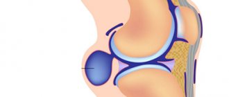
The word is formed from two parts: hygro- means liquid, the ending -oma is added if we are talking about a tumor. In principle, this term describes quite well the essence of what is happening, that is, a neoplasm filled with liquid. When this ball or lump is isolated, the surgeon receives a thin translucent shell with gel-like or liquid contents.
There are also other terms for hygroma:
- synovial cyst
- ganglion
- ganglion
- tendon ganglion
- ganglion cyst
- synovioma
- bursitis
With one amendment or another, all these words can be considered synonyms.
The causes are not entirely clear
We cannot say with absolute certainty why a synovial cyst appears in a particular place in a given person. From the point of view of structural changes in tissues, mucoid degeneration of the joint capsule or tendon sheath is described; microtubules are formed in this area through which fluid from the joint or tendon sheath is pumped into the formed cavity (this is how the “bubble” grows). The fact is that the connection between the joint and the hygroma occurs only unilaterally from the joint to the cavity of the neoplasm. The greater the load, the greater the pressure and the faster this hygroma can grow. In case of immobilization or rest, the volume of the tumor gradually decreases. I would compare this process to how a balloon is deflated.
Most often, young women come with such complaints with some tendency towards increased elasticity of the connective tissue (good stretching).
Where can it appear?
In principle, hygroma can appear on any joint or tendon sheath. There are characteristic localizations on the hand:
- Hygroma of the wrist joint can appear on the dorsal (most common) side. In this case, its source is the capsule above the lunate-scaphoid ligament.
- On the palm it comes out near the radial artery (this is where the pulse is felt) from the space between the radioscaphoid and long radiolunate ligaments.
- On the finger, it usually sits at the base on the palmar side in the projection of the skin fold. There it is very small and dense, like a bead. Less commonly, hygroma occurs on the finger in the projection of the proximal interphalangeal joint on the back.
- Mucoid cysts also form on the finger, which are also essentially hygromas, but grow from the distal interphalangeal joint, and not from the tendon sheaths, like the previous two “finger” variants.
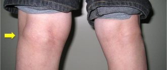
I was surprised when I learned that in France hygroma is a disease that we all know in Russia as bursitis of the elbow joint. Well, in fact, they are right, bursitis and hygromas are, in general, the same thing.
Diagnostics
It is best seen on an ultrasound. This is the most accessible and effective way for objective visualization of a tumor.
X-rays of hygroma do not show any changes. The exception is specific mucoid cysts, which may be associated with arthrosis and exostoses of the distal interphalangeal joints.
In rare cases, MRI may be used, especially to look for very small hygromas on the dorsum of the wrist and differentiate them from Kienböck's disease.
Why does the bone on my wrist hurt?
If a bone in your hand hurts, there can be many reasons for this.
For example, this condition is often caused by injury (bruise, dislocation, sprain or fracture). In this case, pain in the bone is usually accompanied by swelling of nearby soft tissues and their blue discoloration. Arthritis or arthrosis can also lead to discomfort in the bunion. The first is an inflammatory joint lesion caused by an infection, an allergic reaction, an autoimmune disease, the second is associated with degenerative changes, most often age-related.
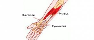
Pain in the bone is often caused by occupational diseases. So, those who sit at the computer for a long time may develop carpal tunnel syndrome. This is due to compression of the nerve in the wrist joint. Due to excessive stress, for example in athletes, tendonitis can develop - inflammation of the tendons of the wrist. Kienböck's disease (or avascular necrosis) is an occupational disease of mechanics, carpenters, crane operators, and woodcutters. The blood supply to the wrist area deteriorates, and the bone tissue begins to deteriorate. With this pathology, the bone on the wrist of the right hand hurts (if the person is right-handed); in left-handed people, the left wrist is affected.
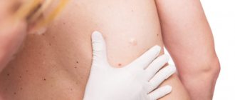
Hygroma of the hand (popularly “bump”) is a benign neoplasm. The patient often describes his condition as “a bone popped out on his wrist and it hurts.” A cyst forms on a joint bone (sometimes on several) - a dense capsule filled with fibrin threads, serous fluid, mucus; as it grows, it compresses the neurovascular bundles, which causes pain, sometimes quite severe. A cyst most often forms in people of certain professions: pianists, computer scientists, spinners, painters, teachers, professional tennis, golf, badminton players. Hereditary predisposition, trauma, inflammatory processes, and surgery on the wrist joint also play a role.
Carpal tunnel syndrome occurs in pregnant women, which is provoked by a sharp increase in body weight in the second and third trimesters. Pain in the bone appears in women mainly at night, accompanied by numbness of the limbs.
Treatment of hygroma without surgery
The safest thing you can do with a hygroma is to do nothing. This option often leads to success. The probability of spontaneous self-healing reaches about 40%.
One historical method is to crush the synovial cyst. A classic zemstvo doctor, with his strong finger, despite the grimace of pain on the patient’s face, can crush this bottle of gel-like liquid. Earlier sources describe the indispensable participation of a heavy and weighty object, namely the Bible, for the successful crushing of a hygroma. In principle, it can rupture on its own under any sudden movement or load. If the hygroma of the wrist has burst, then there is nothing to worry about in this situation, the hand has the right to ache for several days, instead of a neat round ball there will simply be widespread swelling under the skin. The likelihood of the hygroma returning after such manipulations is very high.
Protruding scaphoid bone in the wrist
A protruding bone on the wrist is a sign of displacement of the head of the ulna or deformation of the scaphoid. If a bone protrudes on the wrist from the little finger side, then pathological changes affect the head of the ulna. It can thicken for a number of reasons:
- consequences of traumatic exposure and bone tissue growth;
- proliferation of exostoses in the area of the end articular plates due to sclerotic and ischemic changes in tissue structure;
- deforming osteoarthritis;
- vascular pathology;
- disturbance of innervation (tunnel syndrome);
- cicatricial changes in tendons and ligaments.
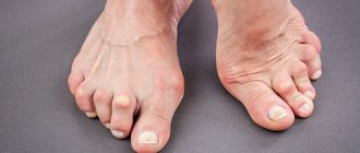
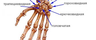
If the deformity is felt from the side of the thumb, then it is the scaphoid bone, located in the first row next to the wrist joint, that is protruding. It can be subject to deformation due to excessive physical stress on the wrist joint. Prolonged position of the hand in an unnatural position for it also entails a change in the position of the scaphoid bone. It begins to gradually shift and a rotational turn occurs. It begins to protrude through the skin. Gradually, compression of the nerve endings leads to inflammation and pain. Tunnel compression and even partial loss of sensation in the index and middle fingers may occur.
When the configuration of the carpal bones changes, degenerative deformation of the joint begins. It is destroyed, and exostoses and osteophytes form on the surface of the bones. These neoplasms impede mobility and have a negative pressure effect on the large nerve fibers passing through here (for example, the palmar and median nerves), blood vessels that provide blood supply to the soft fingers of the palm, hand and fingers.
Diagnosis begins with examination of the patient. Therefore, it is very important to contact an orthopedist immediately if such symptoms appear. During the examination, the doctor will make a preliminary diagnosis and prescribe additional examinations. An x-ray is required. It eliminates the possibility of bone tissue damage and tumor growth. If the image shows the preservation of the integrity of the bone tissue structure, the uniformity of the joint spaces and the absence of bone growths, then they begin to look for the cause of the appearance of the protruding bone effect in other tissues.
Ultrasound and MRI are prescribed for diagnosis. These examinations make it possible to study the structure of the neoplasm. In exceptional cases, a tissue biopsy is performed for the purpose of histological analysis. It eliminates the possibility of developing an oncological process at an early stage.
Puncture
Hygroma puncture is a quick and safe way to get rid of it. Also, a puncture can be considered as a diagnostic procedure: if you get a gel, it means it’s definitely a hygroma. The probability of relapse after puncture is about 80-90%. Injecting any medications into its cavity does not reduce the likelihood of relapse. It is worth remembering that the entry of steroid hormones (they are sometimes used in the hope of influencing the possible return of hygroma) into the subcutaneous fat or articular cartilage can cause very serious irreversible destruction of these structures.
When is surgery needed?
Most people with hygroma want a clear answer to this question. However, the doctor cannot answer it. A synovial cyst is not a dangerous disease; it has a chance to go away without any treatment; surgery (like any intervention) has its own possible complications and risks. Whether to operate on a hygroma or not is a patient’s informed decision. When a person understands the situation and is ready to share responsibility for the decision made with the doctor, then one can choose the time and place to perform the intervention.
How is the operation to remove hygroma performed?
I will describe the basic principles using the example of the most common dorsal hygroma on the wrist.
- The operation is outpatient. There is no need to stay in the hospital.
- Local anesthesia. Anesthesia means unnecessary risks, as well as excessive preoperative examination. I use a mixture of lidocaine and epinephrine, this avoids discomfort from the tourniquet, reduces the likelihood of a toxic reaction to the anesthetic and prolongs the effect of pain relief to 4-5 hours.
- Transverse access. Longitudinal access leaves rough, unsightly scars.
- It is imperative to excise the fragment of the joint capsule from which the hand hygroma grows. Only the skin is sutured.
- I don't use immobilization. For the first 2-3 days, a thick soft bandage is applied, which limits movement and makes the early postoperative period as comfortable as possible. Prolonged immobilization after surgery can lead to stiffness, but does not affect the likelihood of relapse.
- The skin heals in about 2 weeks.
How to deal with the problem
When the bone on your wrist hurts, the doctor decides what to do. Specific treatment methods depend on the disease causing the pain. In general, they are aimed at eliminating the disease itself and alleviating symptoms.
In case of injuries, as well as Kienbeck's disease, the organization of rest of the affected hand is of great importance: the hand must be freed from physical activity for some time.
To get rid of hygroma forever, they resort to surgical methods, in particular, laser evaporation. During the procedure, the tumor is excised along with the capsule. All other treatment (fluid suction, physiotherapy) gives only a temporary effect: after some time, the cyst forms again.
What modern methods of getting rid of hygroma exist?
Removal of this bubble with a gel-like liquid is possible both from the skin and from the joint. There is a thin camera and special instruments that allow you to look inside the wrist joint through small punctures of 2-3 mm. This procedure is called arthroscopy from the words “arthro” joint and “scopy” to look. Through these punctures you can not only look into the joint, but also perform useful manipulations inside, for example, remove a hygroma. The advantages of the arthroscopic method are smaller scars on the skin, faster rehabilitation, and a relatively lower likelihood of relapse.
What signs accompany bone pain?
Depending on the cause of the pathology, pain may be accompanied by other symptoms. In the case of a severe bruise, not only does the pisiform (or other) bone on the wrist hurt, but there is also swelling of the affected area and limited movement of the hand. When a dislocation or fracture occurs, the pain is especially acute and the joint becomes deformed.
Carpal tunnel syndrome is accompanied by numbness and weakening of the hand. Burning and tingling may occur, especially at night and in the morning, and swelling is likely after sleep.
Tendinitis manifests itself as a characteristic crunching sound in the joint. It becomes difficult for the patient to grasp objects with the affected hand.
With carpal tunnel syndrome, in addition to pain in the bone, a pregnant woman may experience itching, burning, tingling, and trembling in her fingers. These symptoms occur primarily at night.
In acute arthritis, the pain syndrome is pronounced, swelling and redness occur at the site of the lesion, and body temperature often rises. With chronic arthritis, the symptoms are smoothed out, which is why the patient may not see a doctor for a long time, leading to destruction of the joint. Arthrosis is characterized by gradually increasing pain in the bone, limited mobility, and painful sensitivity to pressure. A crunching or clicking sound is heard in the affected joint. Without treatment, the pain increases and the hand gradually loses mobility.
With Kienböck's disease, pain is felt around the clock and intensifies during manual work or when pressing on the affected bone.
In the case of hygroma, at first the person does not feel pain, but simply notices that a tumor is growing on the outer or inner side of the wrist (it seems that the bone is enlarging). The skin in this area gradually becomes darker, denser, and when moving the wrist, the bone begins to ache. When the hygroma reaches a large size, periodic tingling occurs in the palm, the patient cannot fully move the hand.