Spinal pathologies in the fetus occur in 1 case per 1000 pregnancies. Often disorders affect not only the spinal column itself, but also the spinal cord. Basically, a fetus with a spinal anomaly dies in utero or immediately after birth, so it is very important to identify the problem as early as possible so that the woman makes a thoughtful decision to terminate the pregnancy.
Causes of intrauterine spinal abnormalities
The content of the article
In most cases, the anomaly occurs in the lumbar region, less often in the cervical region, and very rarely in the sacrum and thoracic region.
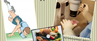
The reasons for such a complex pathology are still not clear. But among the factors that provoke disruption of the formation of the spinal cord, we can definitely highlight:
- TORCH infection;
- toxic effects of drugs;
- difficult working conditions;
- drug and alcohol use;
- many years of smoking experience.
Atheromas
Atheroma is a benign voluminous formation of skin and subcutaneous fat. Atheroma grows from the sebaceous gland, which, due to blockage of the excretory ducts, is gradually filled with dead organic contents. This can be caused by hormonal changes, poor diet, unfavorable environment, poor hygiene, use of cosmetics that are not suitable for the skin, etc. Most often, such formations occur in the location of large sebaceous glands: on the head, neck, face, upper back, chest, and genital area. Atheroma has a round shape and is soft to the touch.
Treatment
There are several approaches to treating atheroma. The method of treatment depends on the inflammation of the atheroma.
- The most ideal is to remove atheroma when there are no signs of inflammation. In this case, complete removal of the atheroma with the capsule is possible and the risk of relapse is much lower.
- If minor signs of inflammation are detected, it is possible to begin a course of anti-inflammatory therapy and relieve the inflammation completely. After inflammation subsides, surgical treatment is performed. However, it must be borne in mind that the inflammation can quickly progress into the phase of purulent inflammation and in this case it is necessary to undergo emergency surgery.
- If there are signs of obvious inflammation (pulsating severe pain, swelling, redness), urgent surgery is necessary. During the operation, an incision is made, purulent contents are released, and drainage is installed in the wound. After surgery in the phase of purulent inflammation, it is extremely rare to remove the tumor along with the capsule. Therefore, a relapse is possible in the future.
Laser removal of atheromas
The MEDI clinic actively uses carbon dioxide laser to remove atheroma. Laser removal is less painful and has a better cosmetic effect.
Myelomeningocele
Myelomeningocele (spina bifida cystica) accounts for 75% of all cases of spinal abnormalities in the fetus. The pathology is expressed in protrusion of the spinal cord beyond the vertebral arch. Sometimes the brain tissue is protected by the skin, but more often it comes out along with the nerve processes. In the latter case, the fetus is born with paralysis of the lower limbs, problems with the intestines and bladder. In 90% of cases, the fetus is diagnosed with hydrocephalus (water on the brain).
Characteristic signs of myelomeningocele on ultrasound:
- fluid is visualized on the back surface of the spine;
- a formation with fluid inside is visible through the vertebral fissure;
- the cranial fossa is smaller;
- cerebellar tissues are displaced;
- pronounced curvature of the spine;
- the size of the fetus is smaller than normal;
- hydrocephalus is diagnosed due to the low position of the spinal cord and blockage of the cerebrospinal fluid.
If a pathology is suspected, a woman donates blood for specific enzymes. Her alpha-fetoprotein levels are elevated, as with other fetal anomalies. Then the pregnant woman is sent for a 4D scan, during which a three-dimensional image of the fetus can be seen. However, this becomes possible only from the 20th week of pregnancy.
Myelomeningocele is a severe form of spina bifida, so if the diagnosis is confirmed by ultrasound, the woman is recommended to have an abortion. Even if the baby is born alive, he will not be able to walk, he will have problems with internal organs.
The operation to move the spinal cord and spinal nerves that have gone beyond the spinal column is carried out within 48 hours from the moment of birth. The protruding capsule is placed back into the spinal canal, and muscle and skin are sutured on top.
Such children require special treatment that will not significantly improve their quality of life. A very low percentage of children will be able to walk in the future, but most will need a wheelchair because the nerve endings become damaged and prevent the spinal cord from functioning properly.
Localization
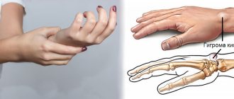
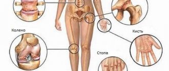
The main locations of the ganglion are:
- hand (palm, back of wrist);
- finger (flexor muscles);
- leg (knee, area under the knee, popliteal fossa);
- foot;
- metatarsophalangeal bones;
- joint (elbow, ankle, wrist, shoulder, knee);
- back of the head;
- neck;
- brain.
The reasons for the development of hygroma can be excessive loads, injuries, and hereditary factors.
Klippel-Feil syndrome
Klippel-Feil syndrome is very rare, occurring in 1 in 120,000 pregnancies, and tends to be inherited.
The pathology consists of an abnormal structure of the cervical spine, in which the vertebrae are fused together. The neck is practically absent and, depending on the type of syndrome, the pathology has other disorders. The most dangerous variety is KFS3, in which not only the cervical vertebrae are fused, but also the thoracic and lumbar vertebrae. Due to non-fusion of the spinal arches, additional ribs are formed.
In addition, the fetus visualizes extra fingers, their underdevelopment or fusion, kidney hypoplasia, fusion of the urethra, diseases of the cardiovascular system, absence of a lung, and disruptions in the functioning of the central nervous system. With any form of the syndrome, the fetus develops a curvature of the spine (scoliosis).
The cause of the pathology is a mutation of the GDF6 gene. The anomaly is detected no earlier than the 20th week of pregnancy. During an ultrasound examination, the following is noticeable on the ultrasound screen:
- shortening of the neck;
- the fetus does not turn its head;
- low hairline at the back of the head;
- facial asymmetry;
- absence of one lung;
- kidney hypoplasia;
- fusion or underdevelopment of the fingers;
- extra number of fingers;
- fusion of vertebrae in various parts of the spine.
The first two types of Klippel-Feil syndrome can be corrected. The child undergoes surgery and then undergoes complex rehabilitation therapy. To date, it will not be possible to completely restore the spine, but a person will be able to live a normal life, because mental abilities are not affected.
In the third form of pathology, the woman will be offered to terminate the pregnancy, because when the vertebrae fuse, the nerve roots are pinched, which causes various diseases of the internal organs to develop. Children with the KFS3 form require special care and still die at an early age.
Prevention
Various reasons lead to the development of fetal neck hygroma, and a woman can influence some of them. Of course, no one is immune from chromosomal abnormalities, and even geneticists often cannot predict their occurrence, but you can protect yourself from injuries and infections during pregnancy. To do this, you need to approach the issue of conception wisely and plan for a new addition to the family, and not rely on chance. Strong immunity, timely vaccinations against infectious diseases and careful attention to one’s own health are what can prevent the development of hygroma in the fetus.
In addition, when planning a pregnancy and subsequent bearing a child, a woman should eat properly and not drink alcohol or drugs. Smoking and other bad habits and addictions should be stopped at least six months before the expected conception.
Related Articles
Arthritis during pregnancy: types, symptoms, treatment methods
Wrist hygroma: symptoms, treatment and prevention methods
Folk remedies for the treatment of hygroma: the best recipes
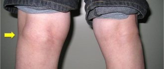
Hygroma of the knee joint in children and adults: ICD-10 code, symptoms and treatment
What does hygroma look like in a child and how to treat the tumor
Hygroma of the ankle joint: photos of symptoms, causes of development and methods of treatment
Spina bifida
Spina bifida or incomplete closure of the spinal canal, which occurs in early pregnancy due to improper formation of the nerve root. In addition to problems with the spine, with Spina bifida there is underdevelopment of the spinal cord. 95% of children with this pathology are born to completely healthy young parents.
The mildest form of pathology is Spina bifida occulta - a small gap in the spinal column, not accompanied by protrusion of the spinal cord and damage to the nerve roots. The defect is practically invisible from the outside, and from the outside it is difficult to say that there is some kind of problem.
Sometimes a newborn has problems with the intestines and bladder, scoliosis and weak leg tone. The only way to diagnose pathology is radiography. It is practically invisible on a screening ultrasound; only with a 4D ultrasound can one see incomplete fusion of the spinal arches in late pregnancy. The anomaly does not require correction, and the woman does not need to terminate her pregnancy.
Lipomas
Most often, lipomas (popularly called wen) grow in the subcutaneous fatty tissue, on any part of the body, including in the thickness of the mammary gland. When there are multiple lipomas, they speak of lipomatosis.
Lipoma is a relatively safe disease, because... the risk of progression to a malignant form (liposarcoma) is extremely low.
Diagnosis of the disease
An accurate diagnosis is established after surgery based on the results of histological examination. But, in the vast majority of cases, the diagnosis is established based on the results of an examination, ultrasound of the soft tissues where the tumor is located. The use of preoperative fine-needle biopsy (examination of the obtained cells under a microscope), as a rule, is not required, because this technique is used to exclude malignancy, therefore not in this case. Once diagnosed, treatment will be recommended.
Treatment of lipomas
Since lipoma is a benign tumor, surgical treatment is cosmetic in nature. In rare cases, when a large tumor or lipoma compresses vital organs and structures, it must be removed for medical reasons.
The operation to remove a lipoma is performed under local anesthesia , or in extremely rare cases under general anesthesia.
After preoperative marking, an incision is made above the tumor. At the MEDI clinic, it is customary to make all incisions along Langer’s lines of force, which subsequently contributes to a more even and less noticeable formation of the scar. After removing the lipoma, the wound is sutured. Very great importance should be attached to the technique of suturing the wound, since the better the quality of the suture, the less noticeable the scar in the future. We recommend using intradermal sutures. The specimen is sent for histological examination. The sutures are removed within 7-10 days.
Cystic hygroma of the neck
Hygroma on the fetal neck (lymphangioma) is a benign tumor that is formed as a result of disruption of the formation of the lymph system in the area of the cervical vertebrae during embryonic development. If the lymph flow is disrupted at the junction of the jugular sac with the jugular vein, a cyst or several cysts filled with fibroserous fluid are formed. The tumor is localized near the cervical vertebrae, affecting the development of the fetus.
Pathology on ultrasound can be seen already at the 1st screening for a period of 11-12 weeks. The main indicator will be an increase in the thickness of the collar space. Neck hygroma in the fetus occurs both as a result of chromosomal abnormalities and under the influence of external causes - mechanical intrauterine trauma, smoking and alcohol consumption by the mother, infections suffered during pregnancy.
At the 2nd screening, the hygroma is visualized as an asymmetric neoplasm with a dense shell, sometimes with septa inside, located in the projection of the cervical spine. The tumor itself does not pose a threat to the life of the fetus.
In the early stages, a chorionic villus biopsy is performed to identify chromosomal abnormalities. If they are confirmed, the woman will be offered to terminate the pregnancy. In the absence of genetic abnormalities, doctors take a wait-and-see approach. very often, by 18-20 weeks of pregnancy, the hygroma resolves on its own.
If this does not happen, then after birth the baby is likely to have the following abnormalities:
- paresis of the facial nerve - immobilization of the facial muscles due to prolonged compression of the nerve fiber by the hygroma;
- spinal deformity (the most common is torticollis - curvature of the cervical spine due to an inflammatory process in the neck muscles due to overexertion of the constant pressure of the tumor);
- deformation of the occipital bone and jaw;
- swallowing dysfunction;
- obstruction of the respiratory tract (obstruction of the respiratory canal due to blockage of the trachea by hygroma).
Hygroma is treated conservatively after the birth of the child. If the cause of the pathology is not chromosomal abnormalities, the prognosis for the baby is favorable.
Hygroma treatment
Hygroma is a benign cystic formation of the synovial bursa associated with the joint.
Clinically, this is a tumor of a round shape, dense consistency, covered with normal skin, with a diameter of 0.5 to 3 cm, inactive, as it is fixed at the base.
There is no clear view of the cause. There is a connection with injuries and excessive physical exertion, but in some cases, hygroma appears for no apparent reason. In this case, a slight bulge of the skin forms, as if there is a pea or cherry inside.
The favorite localization of the formation is the area of the wrist joint, and it is in this area that it often causes discomfort. Although it happens that it appears in other places.
Since the hygroma is associated with the joint, it happens that fluid flows into its cavity. Then for some time it may seem that the formation has disappeared, but, as a rule, after some time it appears again.
Hygroma can exist for a long time without causing any discomfort. Many people live with this education all their lives and do not pay any attention to it. Surgery should be considered in cases where the hygroma creates an unaesthetic appearance, causes inconvenience, causes pain during movement, or in case of its active growth.
Treatment of hygroma
Experience in treating hygroma indicates that conservative treatment methods are ineffective and in the vast majority of cases produce relapses.
An absolutely terrible and painful method is crushing the hygroma. In this case, the liquid is forced into the joint cavity, or the membrane of the hygroma ruptures and the contents pour into the tissue. Over time, at best, everything returns to normal. At worst, an inflammatory reaction may develop in the area of the injured hygroma, leading to suppuration. After crushing, sooner or later the membrane heals, restores its tightness, and the hygroma appears again.
They try to puncture the hygromas - suck out the contents with a syringe and inject various substances into it. In this case, the cavity collapses for a while, but the shell itself does not disappear anywhere, and the liquid sooner or later accumulates again.
The most effective surgical treatment of hygroma involves complete excision of the ganglion.
Radical surgery is the main guarantee of the absence of relapse.
The operation is performed under local anesthesia, lasts 20-30 minutes, the sutures are removed 7-10 days after the operation. For large sizes and complex localization, the operation is performed under anesthesia.
What happens if unqualified specialists undertake the operation?
Only the upper, most accessible part of the formation is isolated, which is removed after evacuation of the fluid that filled the hygroma. Most of the shell remains deep in the tissue. The first 2-3 months after the operation create the appearance of a favorable outcome of the treatment, but as the remaining part scars, the tightness of the hygroma is restored, and fluid begins to accumulate in it again.
In fact, instead of a good, beautiful operation, only the appearance of intervention is created.
Therefore, beware if you are told that the operation is “nonsense, for 5 minutes.” It takes much more time to thoroughly isolate the hygroma. The operation is very delicate and painstaking. If the hygroma is not completely isolated from the surrounding tissues to the very point where its pedicle communicates with the joint, the risk of relapse increases many times over.
Guaranteed treatment effectiveness is achieved with modern equipment and more than 15 years of experience of a highly qualified surgeon.
Wart removal
Warts are most often benign neoplasms that occur in most people, regardless of their age. In most cases, warts occur against a background of reduced immunity and are caused by various human papilloma viruses. The cause of the appearance of warts can be psychological trauma, stress, and vegetative neuroses. Often, warts develop in areas where there has been minor damage to the skin, such as cracks or cuts. Since warts most often appear on the face, hands and neck, they bring psychological discomfort to a person. Most often, neoplasms do not cause pain, with the exception of warts located under the nails or on the surface of the foot.
Technique of the procedure
Laser removal. Laser therapy is considered the most effective way to combat such tumors. After laser removal, there are no scars left on the skin, and the procedure is absolutely painless, so it is recommended even for patients with a low pain threshold.