They belong to one of the areas of the lower extremities, located in the middle between the foot and the knee. The lower legs are formed from the tibia and fibula, surrounded by muscle tissue on 3 sides, and are responsible for the movements of the fingers and feet. If any of them is damaged, a person experiences severe discomfort, pain, and movements become limited.
Anatomy
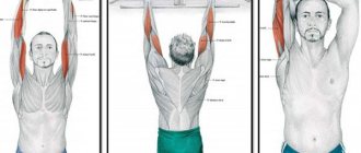
Anatomy of a hamstring
Start
- Long head: ischial tuberosity of the ischial ramus.
- Short head: linea aspera and lateral supracondylar line of the femur.
Attachment
- Lateral surface of the head of the fibula.
Innervation
- Long head: tibial nerve (L5-S2).
- Short head: common peroneal nerve (L4-S1).
Blood supply
Medial circumflex femoral artery, inferior gluteal artery, perforating arteries of the deep femoral artery.
Function
Movements
- Long head: flexes the knee, extends the hip, laterally rotates the tibia when the knee is slightly flexed, promotes lateral rotation of the femur when it is extended at the hip joint.
- Short head: flexes the knee, rotates the tibia laterally with the knee slightly flexed.
Functional contribution
- Due to the characteristics of its origin and attachment, the long head of the biceps femoris provides posterior stability to the pelvis.
- Both heads provide rotational stability to the knee joint by preventing anterior translation of the tibia relative to the femur during knee flexion.
- The connection with the arcuate popliteal ligament also provides varus and rotational stability to the knee joint.
Muscle tissue of the lower leg
The muscles form three groups. The anterior subgroup includes:
- Tibialis anterior muscle - is attached to the medial wedge-shaped and first metatarsal bones, runs along the entire length of the tibia. It can be easily felt under the dermis, especially in the transition area to the foot. The function is to extend and supinate the feet.
- Extensor digitorum longus – located on the outside of the tibialis anterior muscle, in the upper part of the lower leg. When moving to the foot, it is divided into 5 tendons, attached to the 2,3,4,5 toe, the last part is attached to the base of the fifth metatarsal bone. The function is to extend the foot and fingers.
- The extensor pollicis longus is weaker than the previous muscle formations between which it is located. Attached to the base of the distal phalanx, it is used for extension and supination of the feet and big toes.
The posterior subgroup is formed:
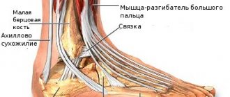
- The triceps surae muscle is located at the back and has three heads. Two of them are the gastrocnemius muscle, the last one is the soleus. Together they pass into the Achilles tendon, which attaches in the area of the tubercle on the heel bone. It is responsible for flexion at the knee joint of the leg and foot at the ankle.
- Flexor digitorum longus – responsible for supination and flexion of the foot and toes. It starts from the posterior part of the tibia, passes under the foot in the canal located under the retinaculum of the flexor tendons.
- Flexor pollicis longus is one of the strongest deep formations on the back of the lower leg. Used to flex the foot and big toe, partially for the 2nd and 3rd toes.
- Posterior tibialis muscle - located under the triceps muscle of the leg, in the lower area it is attached to the sphenoid bones and the bases of the metatarsal bones, the tuberosity of the scaphoid bone. Responsible for supination, adduction and flexion of the feet.
- The popliteus muscle is a flat and short muscle that is adjacent to the knee joint on the back side. Used for pronation and flexion of the lower leg.
The external third subgroup includes:
- The peroneus longus muscle has a feathery structure and is located on the outer portion of the bone of the same name. It starts from its head, the fascia of the leg and the lateral condyle of the tibia and the outer part of the fibula, in the region of its upper two-thirds. Attached to the lower surface of the first metatarsal, to the medial cuneiform and second metatarsal bones. Necessary for pronation, abduction and flexion of the feet.
- The peroneus brevis muscle originates from the outer portion of the peroneus brevis muscle and the intermuscular septa. It is attached to the tuberosity of the 5th metatarsal bone. Responsible for pronation, abduction and flexion of the foot.
Clinical relevance
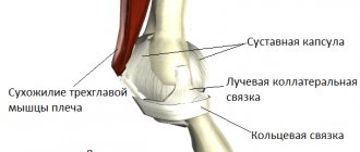
Thus, we know that the biceps femoris muscle has 2 heads, namely a short head and a long head. These two heads attach to the head of the fibula, where they are divided into two parts by the fibular collateral ligament. Any subluxation or dislocation of the biceps femoris tendon or its abnormal insertion, as well as any injury or instability of the meniscus, can lead to a rupture of the tendon of this muscle.
Friends, Dmitry Gorkovsky’s seminar “Myofascial release (scientific approach to increasing joint mobility)” will take place very soon. Find out more...
This is a fairly rare condition that is characterized by pain in the lateral aspect of the knee. In this case, the head of the fibula may be convex, and painful clicking sounds may be heard during active and passive flexion of the knee 90 degrees. Problems may also arise with activities of daily living and playing sports.
Since this is a rare phenomenon, not enough information has been collected about it. Additionally, the exact cause leading to rupture of the biceps femoris tendon is still unknown. However, according to research, the occurrence of this condition can be associated either with excessive use, prolonged and intense physical activity, or is congenital.
This can occur during sprinting, resulting in a sudden change in running speed and pain in the posterolateral thigh (due to overexertion of the biceps femoris and semitendinosus muscles). Injuries to the long head and semitendinosus muscles are the most common among football players.
Inspection
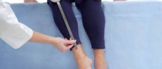
Palpation
- Position the patient in the prone position with the knee slightly flexed.
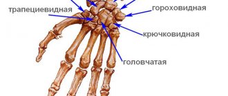
- Locate the head of the fibula to help locate the insertion of the biceps femoris tendon.
- Then palpate the biceps femoris muscle (lateral part of the hamstring).
- Move toward the ischial tuberosity to palpate the belly of the muscle.
Muscle length testing
- The patient is positioned in a supine position.
- With one hand, locate the anterior superior iliac spine (ASIS).
- Use your other hand to support the patient's leg just above the ankle.
- Flex the patient's leg so that it remains straight (knee extended) and add tibial internal rotation to tighten the hamstrings.
- Shortening of the muscle can be judged by posterior rotation of the pelvic bone (PVB).
Trigger points
- Pain associated with trigger points of the biceps femoris (long and short heads) is localized in the back of the knee and can spread up the posterolateral surface of the thigh, up to the gluteal fold.
Anatomy of the extensor girdle muscles of the human lower limb - information:
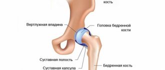
The posterior group (extensors, rotators and abductors) of the muscles of the lower limb girdle are attached to the trochanter major or in its circumference; it includes: m. gluteus maximus, m. gluteus medius, m. tensor fasciae latae, m. gluteus minimus, m. piriformis, m. obturatorius internus with mm. gemelli, m. quadratus femoris et m. obturatorius externus.
M. gluteus maximus, gluteus maximus muscle, a massive muscle layer lying directly under the skin and fascia in the buttock area. It starts from the outer surface of the ilium, from the fascia thoracolumbalis, from the lateral parts of the sacrum and coccyx and from the lig. sacrotuberale, descends obliquely downwards and sideways in the form of parallel muscle bundles, separated by thin connective tissue septa extending from the fascia covering the muscle. The most anterior part of the muscle bundles, turning into a wide flat tendon, bends around the side of the greater trochanter and continues into the fascia lata of the thigh (in its tractus iliotibialis). The back part of the muscle is attached to the tuberositas glutea of the femur. Between the muscle tendon and the greater trochanter lies the synovial bursa, bursa trochanterica m. glutei maximi.
Function. Being an antagonist of m. iliopsoas, extends the leg at the hip joint, turning it slightly outward, and with strengthened legs, it extends the body bent forward. In a standing position, when the weight falls in front of the transverse axis of the hip joints (military posture), muscle tension maintains the balance of the pelvis along with the torso, preventing it from tilting forward. (Inn. L5-S1. N. gluteus inferior.)
M. gluteus medius, the gluteus medius muscle, is covered in its posterior part by m. gluteus maximus, and lies superficially in front. It starts from the outer surface of the ilium with a fan-shaped abdomen and ends with a flat tendon at the lateral surface of the greater trochanter near the apex.
Function. When contracting, the hip abducts. Its anterior bundles, contracting separately, rotate the thigh inward, and the posterior bundles outward; when supporting the body on one leg, she tilts the pelvis in her direction. (Inn. L4-S1. N. gluteus superior.)
M. tensor fasciae latae, tensor fasciae lata , embryologically represents a split off of the gluteus medius muscle and is located immediately in front of the latter on the lateral side of the thigh between the two layers of the femoral fascia, fused with the beginning of m. gluteus medius, and with its distal end passes into a thickened strip of the fascia lata of the thigh, called tractus iliotibialis. This strip extends along the lateral surface of the thigh and is attached to the lateral condyle of the tibia.
Function. It stretches the tractus iliotibialis, through it it acts on the knee joint and flexes the thigh. Thanks to the connection with m. tensor fasciae latae gluteus maximus and gluteus medius muscles contribute to movement in the knee joint in the sense of flexion and outward rotation (P.F. Lesgaft). (Inn. L4-5 and S1. N. gluteus superior.)
M. gluteus minimus, the gluteus minimus , lies beneath the gluteus medius. It starts from the outer surface of the ilium and is attached to the anterior surface of the greater trochanter by a flat tendon. Under the tendon lies a bursa, bursa trochanterica m. Glutei minimi.
Function. Same as m. gluteus medius. (Inn. S1. N. gluteus superior.)
M. piriformis, piriformis muscle , begins on the pelvic surface of the sacrum lateral to the anterior sacral foramina, exits through the foramen ischiadicum majus from the pelvic cavity, runs transversely along the posterior side of the hip joint and is attached to the greater trochanter. The muscle does not completely occupy the foramen ischiadicum manus, leaving gaps along the upper and lower edges of this opening for the passage of blood vessels and nerves.
Function. Rotates the thigh outward and partially abducts it; with a strengthened leg, it can tilt the pelvis in its direction (Inn. S1-S2. Rr. musculares plexus sacralis.).
M. obturatbrius internus, the internal obturator muscle, originates from the inner surface of the circumference of the foramen obturatum and membrana obturatoria, passes through the bony edge of the foramen ischiadicum minus and is attached to the fossa trochanterica of the femur. At the site of the bend through the bone under the muscle lies the synovial bursa, bursa ischiadica m. obturatorii interni. Along the edges of the tendon m. obturatorius internus, lying outside the pelvic cavity, on the back side of the hip joint, two flat and narrow muscle bundles grow - the so-called tt. gemelli (twin muscles), of which the upper (m. gemellus superior) begins on the spina ischiadica, and the lower (m. gemellus inferior) - from the ischial tuberosity. Both of these small muscles, together with the tendon m. obturatorius are attached to the fossa trochanterica, being covered on the surface by the gluteus maximus muscle.
Function. Rotates the hip outward. (Inn. L4-S2. Rr. musculares plex. sacralis.)
M. quadratus femoris, quadratus femoris muscle. It lies downwards from m. gemellus inferior under the lower edge of the gluteus maximus muscle. The muscle fibers are located in the transverse direction from the ischial tuberosity to the crista intertrochanterica of the femur.
Function. Rotates the hip outward. (Inn. L4-S1. Rr. musculares plex. sacralis.)
M. obturatorius externus, external obturator muscle , starts from the outer surface of the pelvic bones along the medial circumference of the obturator foramen, as well as from the membrana obturatoria, bends around the capsule of the hip joint from below and behind and is attached by a narrow tendon to the fossa trochanterica and to the articular capsule.
Function. Produces outward rotation of the hip. (Inn. L3-L4. N. obturatorius.)