If we consider the human skeleton only in the form of bones, then it will represent only a passive supporting apparatus. Movement in it will be impossible, since the bone tissue is unable to bend and stretch. To carry out movements in the skeleton there are more than 200 different joints, which are varied in appearance and shape.
In humans, joints act as a source of new bone tissue. When bones grow, it is the intra-articular sections that ensure the lengthening and enlargement of the skeleton with age. But the main ones are the binding and shock-absorbing functions.
The articular membrane, the surrounding ligaments and tendons - all this forms the human skeleton as a single whole. And the cartilage layer and muscle strength ensure maximum softening of human movements. Without them, even walking would be accompanied by constant shaking of the skeleton.
General principles of structure
Joints in the human body are divided into three groups depending on mobility - continuous, semi-joints and joints themselves (discontinuous). Their development occurred during evolution - the first are the most ancient, and the rest were formed as the range of movements in animals increased.
- Continuous connections are divided into three groups according to the type of tissue connecting the bones. In syndesmoses, ligaments serve as a connecting element - in a person, the spine, bones of the foot and hand are connected in this way. In synchondroses it is strong and elastic cartilage - this type is characteristic of the intervertebral disc and rib joints. In synostoses, bone replaces soft tissue - an example is the sutures of the skull.
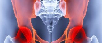
- A half-joint in humans is an intermediate form between synchondrosis and a discontinuous connection. Despite the presence of a gap with synovial fluid between the bones, the remaining parts of the joint have not yet been formed. There is only one such formation in the skeleton - the pubic symphysis. Its role is to “dampen” shocks during movements and help a woman during childbirth, slightly expanding the pelvis.
- The joints themselves create continuity in the human skeleton without compromising its integrity in terms of mobility. This feature is due to the similarity of structure - they are formed by three elements, regardless of shape. The articular surfaces of the bones are located inside the cavity, surrounded by a capsule and ligamentous apparatus. This entire complex is sealed and maintains the structure due to synovial fluid and negative pressure inside.
Let's look at the parts of the skeleton and see how diverse the compounds can be in the human body.
Scull
Almost all the bones of the human skull are connected by a continuous type - synostosis. In newborns, some areas of the joints have a soft tissue structure - they are called fontanelles. Their presence is due to the passage of the child through the birth canal and the “adjustment” of the baby’s head to it. In an adult, they are completely replaced by bone tissue.
The only joint of the head is the temporomandibular joint, which connects the lower jaw and the articular surface of the temporal bone. It is surrounded by many ligaments and covered by a powerful muscle - the chewing muscle. Thanks to its work, we can open our mouths and chew food.
Bones of the skull, shoulder, pelvic girdle and limbs
The skeleton of the head (skull) consists mainly of flat, immovably connected bones. The only movable bone of the skull is the lower jaw. The skull protects the brain and sensory organs from external damage and provides support for the facial muscles. The skull contains the initial sections of the digestive and respiratory systems.
The skull is divided into a large cerebral and smaller facial (visceral) sections.
The brain section is formed by bones:
- unpaired - frontal, occipital, sphenoid, ethmoid;
- paired - parietal and temporal.
The largest bones of the facial region:
- paired – zygomatic, maxillary, nasal, lacrimal;
- unpaired - the lower jaw and the hyoid bone located on the neck.
The skeleton of the body consists of the spine and rib cage.
The spine connects parts of the body, performs a protective function for the spinal cord and a supporting function for the head, arms and torso.
The spine is formed by 33–34 vertebrae.
Spinal sections:
- cervical (7 vertebrae);
- chest (12);
- lumbar (5);
- sacral (5);
- coccygeal (4–5).
In an adult, the sacral and coccygeal vertebrae fuse into the sacrum and coccyx.
The spine has 4 bends that act as shock absorbers: thanks to them, shocks when walking, running, and jumping are softened. The spine is formed by vertebrae. A typical vertebra consists of a body, from which an arch extends posteriorly, and processes extend from the arch. Between the posterior surface of the vertebral body and the arch is the vertebral foramen. Overlapping each other, the vertebral foramina form the spinal canal, which contains the spinal cord.
Thorax: Formed by 12 pairs of ribs, thoracic vertebrae and a flat chest bone - the sternum. The ribs are flat, curved bones, their posterior ends are movably connected to the thoracic vertebrae, and the anterior ends of the 10 upper ribs are connected to the sternum using flexible cartilage. This ensures the mobility of the chest during breathing. The two lower pairs of ribs are shorter than the others and end freely. The chest protects the heart, lungs, liver, stomach and large blood vessels from damage.
The skeleton of any limb consists of two sections:
- skeleton of a free limb;
- limb belts.
Upper limb belt:
- two shoulder blades;
- two collarbones.
The skeleton of the upper limb consists of three sections:
- brachial bone;
- forearm bones (radius and ulna);
- hand (3 sections - wrist, metacarpus, phalanges).
The humerus forms a movable joint with the scapula (shoulder joint), allowing various movements.
Lower limb girdle (pelvic girdle):
- three bones that are motionlessly connected to each other, fused with the sacrum, which allows them to withstand heavy physical loads and perform a protective function for internal organs.
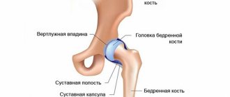
Each pelvic bone has a spherical socket into which the head of the bone of the free lower limb fits.
The skeleton of the lower extremities consists of three sections:
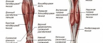
- femur;
- bones of the lower leg (tibia and fibula);
- foot (tarsal bones, metatarsals and phalanges).
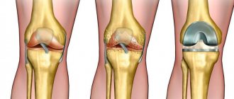
The femur and tibia with the small bone formation adjacent to them in front - the kneecap - form a very mobile knee joint.
Spine
Despite the fact that the vertebra is a fairly small bone, its connections are distinguished by the most complex structure in the human body. It contains a body and several processes that form separate joints with the upper and lower vertebrae. Therefore, the spine is one large joint formed from many small joints. The exceptions are its final sections - at the point of contact with the skull and pelvis.
- The vertebral bodies are connected by cartilaginous discs, which form a “soft cushion” for them. To prevent them from moving forward or backward, longitudinal ligaments run along the entire length of the spine.
- The lateral arches with processes from the inside strengthen the yellow ligaments.
- The spinous processes - lumps that protrude through the skin of the back - support the interspinous ligaments. Above the tops of the processes they grow together into a dense supraspinatus fascia, which rises to the neck and attaches to the back of the head. It, together with the neck muscles, holds the head in a straight position.
- The transverse processes are developed in the thoracic region and are strengthened by intertransverse ligaments.
- The vertebrae also have articular processes above and below, which, when connected to each other, form a joint with an axis of rotation.
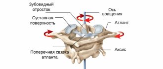
- The articulation of the first and second vertebrae in the neck is formed in the shape of a cylinder. It consists of a bone “tooth” and a corresponding notch, designed to turn the head to the side.
Connections to the skull and pelvis
The spine is connected to the head by the occipital joint - with the help of the first vertebra (atlas) and the condyles of the occipital bone. In general, it consists of about 5 separate formations. Movements in it can be performed in any direction - flexion, extension, head tilt, rotation.
On the atlas there are peculiar depressions resembling a spoon, which correspond to bony protrusions on the back of the head. Many small ligaments and simultaneous work with other joints provide such a large range of movements.
There are two joints in the pelvic area - the sacral and coccygeal. The last lumbar vertebra forms a connection with the sacrum through a disc - but thicker and stronger than in other sections. The coccyx is attached with the help of a cartilaginous plate and many ligaments that envelop it on all sides.
7.2.2. Skeletal features
The musculoskeletal system consists of the skeleton and muscles. It allows a person to perform various movements, and also protects internal organs from damage. The skeleton determines the shape of the body and muscles are attached to it. In the human body there are more than 220 bones that form the skeleton of the head, torso, upper and lower limbs and their girdles. In men, the mass of skeletal bones is 18% of body weight, and in women – 16%.
The connection of bones in the skeleton is divided into three types: fixed, semi-mobile and mobile. The fixed connection is represented by the bones of the skull, the semi-mobile connection is the connection of the vertebrae or ribs with the sternum, carried out with the help of cartilage and ligaments. Finally, the joints are movably connected. Each joint consists of articular surfaces, a bursa and fluid located in the articular cavity. Joint fluid reduces bone friction during movement. Joints are most often strengthened by ligaments, which limit the range of motion.
| 1 |
| Figure 7.2.2.1. Types of bone connections |
The human skeleton consists of bones. There are long (bones of the shoulder, forearm, thigh, lower leg), short (bones of the hand and foot) and flat (bones of the skull, scapula) bones. On top of the bones are covered with a dense shell - periosteum, through the small holes of which blood vessels pass that feed the bone. Thanks to the periosteum, the growth of bones in thickness and the fusion of bones during a fracture are ensured. The ends of the bone are covered with cartilage. Due to the division of cartilage cells, the bone grows in length. Behind the periosteum there is a compact, dense substance impregnated with calcium salts, and underneath it is spongy bone, which consists of many intersecting bone plates that give them strength. Long tubular bones have a cavity inside filled with bone marrow.
The skeleton consists of the bones of the head (skull), torso, upper and lower extremities.
| 2 |
| Figure 7.2.2.2. Types of joints |
The skull consists of the brain and facial sections. The brain section - the cranium - protects the brain from damage. The brain section is formed by the frontal, occipital, two parietal and two temporal bones. The facial part of the skull includes various large and small bones (for example, the upper and lower jaws, zygomatic and nasal bones). All of them are motionlessly connected to each other, except for the mandibular bone.
| 3 |
| Figure 7.2.2.3. Human skull |
The skeleton of the body is formed by the spine and rib cage. The spine includes 7 cervical, 12 thoracic, 5 lumbar, 5 sacral and 4–5 coccygeal vertebrae, according to which five sections of the spine are distinguished - cervical, thoracic, lumbar, sacral and coccygeal. The human spine, unlike the spine of animals, has four curves. Their appearance is associated with upright posture and helps soften shocks when walking, running, jumping, and protect internal organs and the spinal cord from concussions. Each vertebra consists of a body and an arch with several processes. Inside the spine is the spinal canal, which surrounds the spinal cord.
The thoracic vertebrae, ribs, and breastbone (sternum) form the rib cage, which is located in the upper part of the torso. The chest protects the heart and lungs located in it from damage. A person has 12 pairs of flat, arched ribs. The ribs are movably articulated with the vertebrae at the back, and at the front they (except for the two pairs of lower ribs) are connected to the sternum, located along the midline of the chest, using flexible cartilage. This allows the rib cage to expand or contract as you breathe.
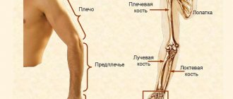
The skeleton of the upper limb (arm) consists of three sections: the shoulder, forearm and hand. The long humerus forms the shoulder. Two bones, the ulna and the radius, make up the forearm. Connected to the forearm is the hand, which consists of small bones of the wrist and metacarpus, forming the palm, and flexible movable fingers (humans have five of them, and the thumb, unlike animals, is opposed to the other four). With the help of the shoulder blades and collarbones, which form the shoulder girdle, the bones of the arm are attached to the bones of the torso.
The lower limb (leg) consists of the thigh, lower leg and foot. The hip is formed by the femur, which is the largest bone in our body. The lower leg consists of two tibia bones, and the foot consists of several bones, the largest of which is the heel bone. The lower limbs are attached to the body using the lower limb girdle (pelvic bones). In humans, the pelvic bones are wider and more massive than in animals. The bones of the limbs are movably connected to each other using joints.
| 4 |
| Figure 7.2.2.4. Human skeleton |
Incorrect body position for a long time (for example, sitting at a table with your head constantly bowed, incorrect posture, etc.), as well as some hereditary causes lead (especially in combination with poor nutrition and poor physical development) to poor posture. Poor posture can be prevented by developing the correct posture at the table, as well as by playing sports (swimming, special gymnastics complexes). Another common skeletal disorder is flatfoot - a foot deformity that occurs as a result of disease, fractures, or prolonged overload of the foot during the period of growth of the body. With flat feet, the foot touches the floor with the entire area of the sole. As preventive measures, it is recommended to select shoes more carefully and use a special set of exercises for the muscles of the lower leg and foot.
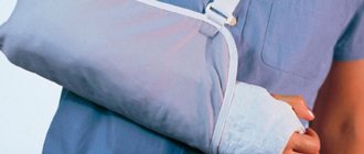
Excessive physical stress on a bone can cause it to fracture. Fractures are divided into open (that is, with the presence of a wound) and closed. Three quarters of all fractures occur in the arms and legs. Signs of a fracture are severe pain in the area of injury, deformation of the limb in the area of the fracture and impairment of its function. If a fracture is suspected, the injured person must be given first aid: stop the bleeding, cover the fracture site with a sterile bandage (in the case of an open fracture), ensure immobility of the injured area by applying a splint (any rigid object that is tied to the limb above and below the fracture site so , to immobilize both the damaged bone and both joints) and deliver the patient to a medical facility. There, using x-ray diagnostics, the fracture site is localized and it is determined whether the fragments are displaced. Then the bone fragments are combined (in no case should you do this yourself) and a plaster cast is applied, ensuring fusion of the bone. A less severe injury is a bruise (muscle damage from an impact, often accompanied by subcutaneous hemorrhage). Local application of cold (ice pack, cold water jet) can reduce pain in minor bruises.
A dislocation is a persistent displacement of the articular ends of bones, which causes dysfunction of the joint. Do not try to correct the dislocation yourself; this may cause additional injury. It is necessary to immobilize the damaged joint and apply cold to it; Warming compresses are contraindicated in this case. Then the victim must be urgently transferred to a doctor.
Rib cage
Its frame is formed by twelve pairs of ribs, thoracic vertebrae and sternum. Unlike animals, the human chest is much wider (laterally) and longer. This is due to the appearance of upright walking - relying only on the legs. Changing the position of the internal organs to vertical ensured straightening and increasing the volume of the chest.
The presence of a heart and lungs inside it obliges this formation to be strong and at the same time elastic. This is ensured by long and relatively soft ribs, as well as a large number of joints.
- At the back, the ribs are loosely fixed between two adjacent vertebrae, which allows them to rotate up and down when breathing.
- In front, they are connected to the sternum through synchondrosis, a sedentary cartilaginous connection.
Only the first seven pairs of ribs are attached directly to the sternum - the rest are fixed to the overlying connection.
Skeletal features
The musculoskeletal system consists of the skeleton and muscles. It allows a person to perform various movements, and also protects internal organs from damage. The skeleton determines the shape of the body and muscles are attached to it. In the human body there are more than 220 bones that form the skeleton of the head, torso, upper and lower limbs and their girdles. In men, the mass of skeletal bones is 18% of body weight, and in women – 16%.
The connection of bones in the skeleton is divided into three types: fixed, semi-mobile and mobile. The fixed connection is represented by the bones of the skull, the semi-mobile connection is the connection of the vertebrae or ribs with the sternum, carried out with the help of cartilage and ligaments. Finally, the joints are movably connected. Each joint consists of articular surfaces, a bursa and fluid located in the articular cavity. Joint fluid reduces bone friction during movement. Joints are most often strengthened by ligaments, which limit the range of motion.
|
| Types of bone connections |
The human skeleton consists of bones. There are long (bones of the shoulder, forearm, thigh, lower leg), short (bones of the hand and foot) and flat (bones of the skull, scapula) bones. On top of the bones are covered with a dense shell - periosteum, through the small holes of which blood vessels pass that feed the bone. Thanks to the periosteum, the growth of bones in thickness and the fusion of bones during a fracture are ensured. The ends of the bone are covered with cartilage. Due to the division of cartilage cells, the bone grows in length. Behind the periosteum there is a compact, dense substance impregnated with calcium salts, and underneath it is spongy bone, which consists of many intersecting bone plates that give them strength. Long tubular bones have a cavity inside filled with bone marrow.
The skeleton consists of the bones of the head (skull), torso, upper and lower extremities.
2 |
| Types of joints |
The skull consists of the brain and facial sections. The brain section - the cranium - protects the brain from damage. The brain section is formed by the frontal, occipital, two parietal and two temporal bones. The facial part of the skull includes various large and small bones (for example, the upper and lower jaws, zygomatic and nasal bones). All of them are motionlessly connected to each other, except for the mandibular bone.
3 |
| Human skull |
The skeleton of the body is formed by the spine and rib cage. The spine includes 7 cervical, 12 thoracic, 5 lumbar, 5 sacral and 4–5 coccygeal vertebrae, according to which five sections of the spine are distinguished - cervical, thoracic, lumbar, sacral and coccygeal. The human spine, unlike the spine of animals, has four curves. Their appearance is associated with upright posture and helps soften shocks when walking, running, jumping, and protect internal organs and the spinal cord from concussions. Each vertebra consists of a body and an arch with several processes. Inside the spine is the spinal canal, which surrounds the spinal cord.
The thoracic vertebrae, ribs, and breastbone (sternum) form the rib cage, which is located in the upper part of the torso. The chest protects the heart and lungs located in it from damage. A person has 12 pairs of flat, arched ribs. The ribs are movably articulated with the vertebrae at the back, and at the front they (except for the two pairs of lower ribs) are connected to the sternum, located along the midline of the chest, using flexible cartilage. This allows the rib cage to expand or contract as you breathe.
The skeleton of the upper limb (arm) consists of three sections: the shoulder, forearm and hand. The long humerus forms the shoulder. Two bones, the ulna and the radius, make up the forearm. Connected to the forearm is the hand, which consists of small bones of the wrist and metacarpus, forming the palm, and flexible movable fingers (humans have five of them, and the thumb, unlike animals, is opposed to the other four). With the help of the shoulder blades and collarbones, which form the shoulder girdle, the bones of the arm are attached to the bones of the torso.
The lower limb (leg) consists of the thigh, lower leg and foot. The hip is formed by the femur, which is the largest bone in our body. The lower leg consists of two tibia bones, and the foot consists of several bones, the largest of which is the heel bone. The lower limbs are attached to the body using the lower limb girdle (pelvic bones). In humans, the pelvic bones are wider and more massive than in animals. The bones of the limbs are movably connected to each other using joints.
4 |
| Human skeleton |
Incorrect body position for a long time (for example, sitting at a table with your head constantly bowed, incorrect posture, etc.), as well as some hereditary causes lead (especially in combination with poor nutrition and poor physical development) to poor posture. Poor posture can be prevented by developing the correct posture at the table, as well as by playing sports (swimming, special gymnastics complexes). Another common skeletal disorder is flatfoot - a foot deformity that occurs as a result of disease, fractures, or prolonged overload of the foot during the period of growth of the body. With flat feet, the foot touches the floor with the entire area of the sole. As preventive measures, it is recommended to select shoes more carefully and use a special set of exercises for the muscles of the lower leg and foot.
Excessive physical stress on a bone can cause it to fracture. Fractures are divided into open (that is, with the presence of a wound) and closed. Three quarters of all fractures occur in the arms and legs. Signs of a fracture are severe pain in the area of injury, deformation of the limb in the area of the fracture and impairment of its function. If a fracture is suspected, the injured person must be given first aid: stop the bleeding, cover the fracture site with a sterile bandage (in the case of an open fracture), ensure immobility of the injured area by applying a splint (any rigid object that is tied to the limb above and below the fracture site so , to immobilize both the damaged bone and both joints) and deliver the patient to a medical facility. There, using x-ray diagnostics, the fracture site is localized and it is determined whether the fragments are displaced. Then the bone fragments are combined (in no case should you do this yourself) and a plaster cast is applied, ensuring fusion of the bone. A less severe injury is a bruise (muscle damage from an impact, often accompanied by subcutaneous hemorrhage). Local application of cold (ice pack, cold water jet) can reduce pain in minor bruises.
A dislocation is a persistent displacement of the articular ends of bones, which causes dysfunction of the joint. Do not try to correct the dislocation yourself; this may cause additional injury. It is necessary to immobilize the damaged joint and apply cold to it; Warming compresses are contraindicated in this case. Then the victim must be urgently transferred to a doctor.
Shoulder girdle
It contains two low-moving joints that provide connection between the upper limb and the collarbone and scapula. The sternoclavicular joint is formed by formations of the same name, connecting the torso and arm. It provides additional mobility in the shoulder and protects it from excessive lateral movement.
The acromioclavicular joint is formed by the outer end of the clavicle and the process of the scapula (acromion). It can be felt at the very top and outer point of the shoulder, like a bony protrusion. It provides simultaneous movement of the shoulder girdle and arm.
Upper limb
Anatomically, it starts from the joints of the shoulder girdle and ends at the fingertips. Almost all compounds have a round shape and a simple structure.
The arm is divided into three main sections - the shoulder, forearm and hand. In humans, it is designed to perform small and precise movements, therefore the joints that form it are small and have great mobility.
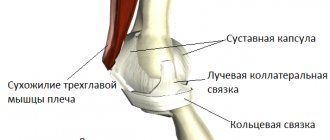
Shoulder joint
The largest joint of the arm, the shoulder, is located closest to the body. It is formed by the round head of the humerus and the corresponding articular cavity of the scapula. An interesting feature is the difference in their sizes - the head is twice as large as the recess. This deficiency is eliminated by a cartilaginous lip that covers the articular surface of the shoulder.
This connection is an example of skeletal changes caused by upright walking. In humans, it ceased to play a supporting function, and began to be intended for performing work. The shoulder paid for this with poor resistance to injury.
Elbow joint
It is a complex compound consisting of three separate formations. They are formed by the condylar end of the shoulder and two bones of the forearm - the radius and ulna. They are all connected to each other, but are located under a common capsule.
In the humerus-elbow and humerus-radial joints, the connection occurs in the shape of a cylinder - due to notches on the bones of the forearm and the protruding block of the shoulder. It ensures flexion of the arm at the elbow, and the radial-ulnar joint allows rotation of the hand.
Joints of the forearm and hand
The radius and ulna are connected to each other by cylindrical joints that are located at their ends. They provide turns with a brush, as when opening a door with a key. These movements are always combined with the inclusion of small joints of the hand.
The wrist joint has a rounded shape and is formed by four bones: the radius and carpals (scaphoid, lunate and triquetrum). Additionally, it contains cartilage discs that increase range of motion.
The most important are the metacarpophalangeal and interphalangeal joints. They are formed by small bones of the phalanges and wrist. All of them are characterized by a spherical shape, which ensures simultaneous and precise movements with the brush.
Human skeleton
Types of bones:
- Tubular
(have a tube) – shoulder, tibia; - Spongy
(no tube) – tarsus, vertebrae; - Flat
(do not have a tube, flattened) - frontal, scapula.
Bone connections:
- Fixed:
sutures of the skull, ossified cartilage between the pelvic bones - Semi-movable
(layers of cartilage, “half-joints”): between the vertebrae, between the ribs and the sternum - Movable
(joints): between the femur and pelvis, between the lower jaw and the skull, etc.
Scull
consists of two sections: the brain (frontal, occipital, two parietal, two temporal bones) and the facial (two maxillary, one mandibular, two zygomatic, two nasal, etc.) The occipital bone connects to the first vertebra. Functions of the skull: support and protection for the brain and sensory organs, chewing.
Spine
consists of vertebrae, between which there are cartilaginous discs (semi-movable joint). Each vertebra consists of a body (support), an arch (protecting the spinal cord) and processes (connecting the vertebrae to each other, ligaments and muscles). Sections of the spine: cervical (7 vertebrae), thoracic (12, articulate with the ribs), lumbar (5, the most massive), sacral (5, fused into the sacrum), coccygeal (3-5, rudimentary, fused into a single bone, the coccyx). The spine has 4 curves that spring when walking. Functions: support for organs, protection of the spinal cord.
Rib cage
consists of 12 thoracic vertebrae, 12 pairs of ribs and sternum. Ribs 1-7 reach the sternum with their cartilaginous ends, ribs 8-10 are attached to the cartilage of the 7th, ribs 11-12 end freely in the muscles. Functions: protection of internal organs (heart, lungs), participates in breathing.
Limb skeleton
consists of a belt (support for the limb) and a free limb (movement).
- Upper limb girdle (shoulder girdle): shoulder blades and collarbones.
- Free upper limb - 3 sections: shoulder (humerus), forearm (ulna and radius), hand (3 sections: wrist, metacarpus, phalanges of the fingers).
- The lower limb girdle is the pelvic bones. Until 12-14 years of age, each bone consists of 3 bones - the ilium, the pubis and the ischium.
- Free lower limb - 3 sections: thigh (femur), lower leg (tibia and fibula), foot (3 sections - tarsus, metatarsus, phalanges).
IF THE INFORMATION IS NOT IN THE MAIN SUMMARY, THEN IT IS IN THE DETAILED SUMMARY:
Skeletal tissue, Bone structure, Bone joints, Human skeleton, First aid for fractures
ASSIGNMENTS OF PART 2 OF THE USE ON THIS TOPIC
Part 1 tasks
TUBULAR Choose three correct answers out of six and write down the numbers under which they are indicated. Which bones of the human skeleton are tubular?
1) clavicle 2) tibia 3) cervical vertebra 4) terminal phalanx of the little finger 5) lunate bone of the wrist 6) radius
Answer
246
TUBULAR - FLAT Match the bones of the human skeleton and their types: 1) tubular, 2) flat. Write numbers 1 and 2 in the order corresponding to the letters.
A) radial B) frontal C) femoral D) tibia E) sternum E) occipital
Answer
121122
TUBULAR - SPONIOUS - FLAT Match the examples and types of human bones: 1) tubular, 2) spongy, 3) flat. Write the numbers 1, 2, 3 in the order corresponding to the letters.
A) humerus B) temporal bone C) carpal bones D) vertebral body E) frontal bone E) tibia bones
Answer
132231
FIXED - SEMI-MOBILE Establish a correspondence between the bones and the types of their connections in an adult: 1) semi-movable, 2) immobile. Write numbers 1 and 2 in the order corresponding to the letters.
A) occipital and parietal B) temporal and zygomatic C) parietal and frontal D) lumbar vertebrae E) thoracic vertebrae
Answer
22211
FIXED - MOBILE Establish a correspondence between an example of a bone connection and the type to which it belongs: 1) immobile, 2) movable
A) femur and tibia B) frontal and parietal bones C) occipital and temporal bones D) mandible and temporal bone E) sacral vertebrae
Answer
21121
FIXED - SEMI-MOBILE - MOBILE 1. Establish a correspondence between the example of the connection of the bones of the skeleton of an adult and the type of connection of these bones: 1) fixed, 2) mobile, 3) semi-mobile. Write the numbers 1, 2 and 3 in the correct order.
A) lumbar vertebrae B) mandibular bone and cranial bones C) frontal and parietal bones D) sacral vertebrae E) occipital and parietal bones E) bones of the hip joint
Answer
321112
2. Establish a correspondence between the type of bone connection (1-fixed, 2-semi-movable, 3-mobile) and the place in the human skeleton where such a connection exists. Write the numbers 1, 2 and 3 in the correct order.
A) sacral vertebrae B) shoulder and forearm C) thoracic vertebrae D) bones of the brain part of the skull E) lower leg and foot E) tailbone
Answer
132131
3. Establish a correspondence between the types of bone connections and examples: 1) fixed, 2) movable, 3) semi-movable. Write the numbers 1, 2, 3 in the order corresponding to the letters.
A) coccyx vertebrae B) hip joint C) knee joint D) skull bones E) spinal vertebrae E) elbow joint
Answer
122132
4. Establish a correspondence between the examples and types of bone connections: 1) movable, 2) semi-movable, 3) immobile. Write numbers 1-3 in the order corresponding to the letters.
A) bones of the skull B) lower jaw with other bones of the skull C) bones of the phalanges of the fingers D) vertebrae of the thoracic spine E) bones forming the pelvic bone E) bones of the upper free limb with the shoulder girdle
Answer
311231
5. Establish a correspondence between the bones and the types of their connection: 1) fixed, 2) mobile, 3) semi-mobile. Write numbers 1-3 in the order corresponding to the letters.
A) occipital and parietal B) 5th and 6th cervical vertebrae C) femoral and tibial D) rib and sternum E) pubic and ischial E) humerus and scapula
Answer
132312
WE COLLECT
A) temporal and parietal
B) temporal and mandibular
SEMI-MOBILE - MOBILE Establish a correspondence between the bones and the types of their connection: 1) joint, 2) semi-joint. Write numbers 1 and 2 in the order corresponding to the letters.
A) bones of the wrist and forearm B) vertebrae of the thoracic spine C) bones forming the phalanges of the fingers D) bones of the lower leg and tarsus E) pubic bones E) bones of the thigh and lower leg
Answer
121121
MOBILE Select three options. What bones are movably connected in the human skeleton?
1) temporal and parietal 2) thoracic vertebrae 3) lower jaw with skull 4) femoral and pelvic 5) frontal and parietal 6) thighs and legs
Answer
346
1. Select three correctly labeled captions for the drawing “Structure of the Human Skull.” Write down the numbers under which they are indicated.
1) frontal 2) occipital 3) lacrimal 4) ethmoidal 5) temporal 6) zygomatic
Answer
156
2. Select three correctly labeled captions for the drawing that depicts the structure of the skull. Write down the numbers under which they are indicated.
1) parietal 2) frontal 3) ethmoidal 4) auditory 5) occipital 6) lacrimal
Answer
125
FACIAL - BRAIN Establish a correspondence between the bone of the human skull and the section it belongs to: 1) facial, 2) brain
A) nasal B) occipital C) maxillary D) temporal D) zygomatic E) parietal
Answer
121212
BELT - LIMB Establish a correspondence between the skeletal bone of the human upper limb and the section to which it belongs: 1) limb girdle, 2) free limb. Write numbers 1 and 2 in the correct order.
A) clavicle B) humerus C) carpal bone D) radius E) scapula
Answer
12221
HANDS 1. Select three correctly labeled captions for the drawing “Structure of the upper limbs.” Write down the numbers under which they are indicated.
1) clavicle 2) scapula 3) ulna 4) radius 5) humerus 6) hand bones
Answer
126
2. Select three correctly labeled captions for the picture that depicts the structure of the skeleton of the human forelimb. Write down the numbers under which they are indicated.
1) humerus 2) talus 3) radius 4) ulna 5) metacarpal bone 6) phalanges
Answer
135
ARM SEQUENCE 1. Determine the correct sequence of arrangement of the bones of the upper limb, starting from the shoulder girdle. Write down the corresponding sequence of numbers.
1) Metacarpal bones 2) Humerus 3) Phalanges 4) Radius 5) Carpal bones
Answer
24513
2. Establish the sequence of connecting the bones of the skeleton of the upper limb, starting with the shoulder girdle. Write down the corresponding sequence of numbers.
1) radius and ulna 2) scapula and clavicle 3) phalanges 4) humerus 5) metacarpus 6) wrist
Answer
241653
LEGS Choose three correct answers out of six and write down the numbers under which they are indicated. The skeleton of the free lower limb includes bones
1) femoral 2) iliac 3) radial 4) pelvic 5) calcaneal 6) tibia
Answer
156
LEGS Select three correctly labeled captions for the drawing “Skeleton of the lower limb of a person” and write down the numbers under which they are indicated.
1) ischium 2) femur 3) radius 4) fibula 5) metatarsal bones 6) talus
Answer
245
Choose three correctly labeled captions for the drawing that depicts the structure of the human lower limb. Write down the numbers under which they are indicated.
1) ilium 2) femur 3) patella 4) tibia 5) radius 6) phalanges
Answer
234
LEGS SEQUENCE Arrange the bones of the lower limb in the correct order, starting from the pelvic girdle. Write down the corresponding sequence of numbers.
1) metatarsus 2) femur 3) tarsus 4) fibula 5) phalanges
Answer
24315
Analyze the table “Bone Connections”. Fill in the blank cells of the table using the terms in the list. Write down the selected numbers in the order corresponding to the letters.
1) trochlear 2) semi-movable 3) skull 4) chest 5) pelvis 6) wrist 7) upper limb upper limb belt
belt
Answer
527
Analyze the table “Types of bone connections.” Fill in the blank cells of the table using the terms and concepts given in the list. For each cell indicated by a letter, select the corresponding term from the list provided.
1) complete fusion of bones 2) between the bones there is a layer of dense connective tissue 3) maxillary and zygomatic 4) elbow and humerus 5) rib and sternum 6) semi-movable 7) hinged elastic
Answer
614
Analyze the table “Classification of Bones”. Fill in the blank cells of the table using the terms and concepts given in the list. For each cell indicated by a letter, select the corresponding term or concept from the list provided.
1) mixed 2) chest 3) free limb 4) flat 5) vertebra 6) humerus and radius 7) rib, occipital bone and sternum
Answer
436
SKELETON Choose three correctly labeled captions for the drawing “Human Skeleton”. Write down the numbers under which they are indicated.
1) ulna 2) radius 3) humerus 4) sacrum 5) femur 6) foot
Answer
356
Establish a correspondence between the characteristics and sections of the human skeleton: 1) the cerebral part of the skull, 2) the chest, 3) the upper limb. Write numbers 1-3 in the order corresponding to the letters.
A) the composition includes long tubular bones B) the fixed nature of the connection C) compressed in the anteroposterior direction D) the mobile nature of the connection E) the composition includes the occipital bone E) the semi-movable nature of the connection
Answer
312312
Establish a correspondence between examples of bones and sections of the skeleton: 1) skeleton of the head, 2) skeleton of the torso, 3) skeleton of the limbs. Write numbers 1-3 in the order corresponding to the letters.
A) pelvic B) sternum C) occipital D) sphenoid D) tibial E) radial
Answer
321133
© D.V. Pozdnyakov, 2009-2020
Lower limb
Starts from the groin folds and ends at the fingertips. The leg has three sections: the thigh, shin and foot. Their joints are the largest in the body.
All connections have a different structure, but perform one function - creating support for the body. In humans, the leg acquired a straighter shape and developed muscles as a result of evolution.
Hip joint
The basis for the leg is formed by the hip joint, which is formed by the large head of the femur and the acetabulum of the pelvis. The recess provides almost complete coverage of the head, which causes rare dislocations of this joint. Additionally, it is strengthened by powerful ligaments both outside and inside.
Despite its high strength, this is the most mobile joint of the leg. Full range of movements (even rotation) are possible in it.
Knee and ankle joints
The knee is formed by the condyles of the femur, the articular surface of the tibia and the patella. To create stability, there are two cartilage plates - menisci. Maintaining integrity is provided by many ligaments inside and outside the knee. It is supporting in function, and therefore is inactive - only flexion and extension are carried out.
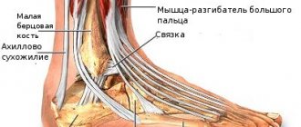
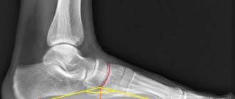
The ankle joint is shaped like a fork formed by the bones of the lower leg and foot. This structure is necessary for normal support of the leg - when standing, under body pressure, these formations firmly hold each other.
Joints (Anatomy)