If your joints are swollen and painful at night, your rheumatologist will suggest you check your rheumatology profile. This examination will help make an accurate diagnosis, monitor the dynamics of the disease and prescribe the correct treatment.
If rheumatic disease is suspected, the following tests are used:
- blood test for uric acid levels;
- blood test for antinuclear antibodies;
- blood test for rheumatoid factor;
- blood test for ACCP (antibodies to cyclic citrulline-containing peptide);
- blood test for C-reactive protein.
Detailed description of the study
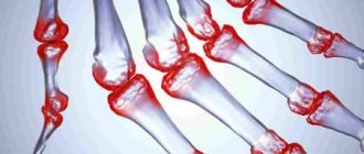
Rheumatoid arthritis (RA) is an autoimmune disease that is accompanied by the development of chronic damage to the joints—usually the hands and fingers—and internal organs. The course of RA can be rapidly progressive or slowly progressive. Along with damage to the joints, swelling and pain in them, stiffness of movement in the morning after sleep, the disease has such nonspecific manifestations as weakness, fatigue, and weight loss. The disease is characterized by the formation of specific rheumatoid nodules under the skin, as one of the most common extra-articular manifestations.
Delayed detection of RA has many adverse consequences. Long-term pathological changes in the joints cause impairment of their mobility and, without treatment, lead to disability. Deposits of pathological amyloid protein are found in organs and tissues, and amyloidosis develops. RA is also the cause of so-called tunnel syndromes, which are caused by compression of nerves and blood vessels in anatomical canals, or tunnels. The most common types are carpal tunnel syndrome and compression syndromes of the ulnar and tibial nerves.
Rheumatoid arthritis is differentiated from a number of other diseases that begin with joint damage. These include joint injuries, some acute infectious diseases such as influenza, rubella, measles, hepatitis, post-streptococcal arthritis, as well as gout, osteoarthritis and other rheumatic diseases.
This study is aimed at identifying RA and includes five important indicators. It is worth noting that RA has seropositive and seronegative forms, which are determined depending on the detection of rheumatoid factor in the blood.
Rheumatoid factor
Normally, immunoglobulins (antibodies, antibodies) protect a person from the harmful effects of microorganisms and foreign substances. They are proteins that differ in their structure and function. They are divided into five classes: IgG, IgM, IgA, IgD and IgE. Antibodies, united by the concept of “rheumatoid factor” (RF), most often belong to the IgM class, are produced against parts of one’s own IgG, altered in rheumatoid arthritis due to various not fully established reasons. Their determination is one of the key stages in establishing the diagnosis of RA.
SSR antibodies
Cyclic citrulline-containing peptide (CCP, CCP) is a protein in which the normally absent amino acid citrulline replaces the physiological amino acid arginine. Determination of antibodies to CCP makes it possible to determine the presence of the disease with a high probability, including in seronegative RA. For early arthritis, its sensitivity is 41-80% and specificity is 93-99%.
ASLO
Antistreptolysin-O (ASLO) are also a group of antibodies directed to bacterial molecules: group A beta-hemolytic streptococcus - streptolysin. This microorganism is one of the causative agents of sore throat, scarlet fever and some other diseases. Previous streptococcal infection is often associated with the subsequent occurrence of autoimmune diseases.
The study also contains two tests: determination of C-reactive protein and a clinical blood test. The concentration of the former may reflect the presence of an acute stage of RA and is associated with disease activity. Together, these two tests allow us to assess the presence of inflammatory processes and their intensity.
Thus, this complex study is widely used in clinical practice, since it can highly likely confirm or exclude RA; it is also used to monitor the effectiveness of treatment and assess the prognosis of the disease.
A detailed description of the studies and reference values are presented on the pages with descriptions of individual studies.
C-reactive protein test
C-reactive protein ( CRP ) is a very sensitive element of a blood test that quickly responds to even the slightest damage to body tissue. The presence of C-reactive protein in the blood is a harbinger of inflammation, injury, and the penetration of bacteria, fungi, and parasites into the body.
CRP more accurately shows the inflammatory process in the body than ESR (erythrocyte sedimentation rate). At the same time, C-reactive protein quickly appears and disappears - faster than the ESR changes.
Due to the ability of C-reactive protein to appear in the blood at the very peak of the disease, it is also called “acute phase protein.”
As the disease enters the chronic phase, C-reactive protein decreases in the blood, and when the process worsens, it increases again.
C-reactive protein is normal
C-reactive protein is produced by liver cells and is found in minimal amounts in the blood serum. The content of CRP in blood serum does not depend on hormones, pregnancy, gender, or age.
The norm of C-reactive protein in adults and children is the same - less than 5 mg/l (or 0.5 mg/dl).
A blood test for C-reactive protein is taken from a vein in the morning, on an empty stomach.
1 Blood test for uric acid levels
2 blood test for antinuclear antibodies
3 Blood test for rheumatoid factor
Causes of increased C-reactive protein
C-reactive protein may be elevated in the presence of the following diseases:
- rheumatism;
- acute bacterial, fungal, parasitic and viral infections;
- gastrointestinal diseases;
- focal infections (for example, chronic tonsillitis);
- sepsis;
- burns;
- postoperative complications;
- myocardial infarction;
- bronchial asthma with inflammation of the respiratory system;
- complicated acute pancreatitis;
- meningitis;
- tuberculosis;
- tumors with metastases;
- some autoimmune diseases (rheumatoid arthritis, systemic vasculitis, etc.).
With the slightest inflammation, in the first 6-8 hours the concentration of C-reactive protein in the blood increases tenfold. There is a direct relationship between the severity of the disease and changes in CRP levels. Those. The higher the concentration of C-reactive protein, the stronger the inflammatory process develops.
Therefore, changing the concentration of C-reactive protein is used to monitor and control the effectiveness of treatment of bacterial and viral infections.
Different reasons lead to different increases in C-reactive protein levels:
- The presence of chronic bacterial infections and some systemic rheumatic diseases increases C-reactive protein to 10-30 mg/l. With a viral infection (if there is no injury), the level of CRP increases slightly. Therefore, high values indicate the presence of a bacterial infection .
- If neonatal sepsis is suspected, a CRP level of 12 mg/l or more indicates the need for urgent antimicrobial therapy.
- In acute bacterial infections, exacerbation of some chronic diseases, acute myocardial infarction and after surgery, the highest level of CRP is from 40 to 100 mg/l. With proper treatment, the concentration of C-reactive protein decreases within the next few days, and if this does not happen, it is necessary to discuss other antibacterial treatment. If after 4-6 days of treatment the CRP value has not decreased, but remains the same and even increased, this indicates the occurrence of complications (pneumonia, thrombophlebitis, wound abscess, etc.). After surgery, the more severe the operation, the higher the CRP will be.
- During myocardial infarction, protein increases 18-36 hours after the onset of the disease, decreases after 18-20 days and returns to normal by 30-40 days. With angina pectoris, it remains normal.
- In a variety of tumors, elevated levels of C-reactive protein can serve as a test to assess tumor progression and disease recurrence.
- Severe general infections, burns, sepsis increase C-reactive protein to enormous values: up to 300 mg/l or more.
- With proper treatment, the level of C-reactive protein decreases already on days 6-10.
Preparation for rheumatological tests
In order for analyzes to show objective information, it is necessary to adhere to certain rules. You need to donate blood in the morning, on an empty stomach. Approximately 12 hours should pass between taking tests and eating. If you're thirsty, drink some water, but not juice, tea or coffee. It is necessary to exclude intense physical exercise and stress. You cannot smoke or drink alcohol.
The multidisciplinary clinic "MedicCity" provides diagnostics of the highest level, experienced, qualified rheumatologists and specialists in more than 30 specialties. We treat arthritis, arthrosis, vasculitis, lupus erythematosus, osteoporosis, gout, rheumatism and many other rheumatological diseases. Do not delay your visit to the doctor, contact us at the slightest symptoms. High-quality diagnosis is 90% of successful treatment!
References
- Rheumatoid arthritis. Clinical recommendations. Association of Rheumatologists of Russia, 2021. - 102 p.
- Rheumatology: National Guide / ed. E.L. Nasonova, V.A. Nasonova. - M.: GEOTAR-Media, 2008. - 720 p.
- Kishkun, A.A. Guide to laboratory diagnostic methods. - M.: GEOTAR-Media, 2007. - 800 p.
- Volkova, M.V., Kunder, E.V. Early arthritis: relevance, Immunopathology, diagnosis. Vestnik VSMU, 2013. - No. 3.
- Medical microbiology, virology and immunology: a textbook for medical students / ed. ed. A.A. Vorobyov. — 2nd ed., corrections and additions. - M.: Medical Information Agency, 2012. - 704 p.
Laboratory examination for joint pain
A comprehensive examination includes tests aimed at determining circulating autoantibodies and various biochemical markers of the acute phase of inflammation. Joint pain can be a sign of arthritis, including rheumatoid arthritis, arthrosis, osteoarthritis, gout, chondrocalcinosis, ankylosing spondylitis and other diseases. Allows you to identify a possible cause, as well as differentiate various forms of arthritis.
What tests are included in this complex:
· Clinical blood test (with leukocyte formula);
· Erythrocyte sedimentation rate (ESR);
· Fibrinogen;
· Antistreptolysin O;
· Uric acid in serum;
· C-reactive protein, quantitative (method with normal sensitivity);
· Rheumatoid factor (RF);
· Antinuclear factor on HEp-2 cells;
· Antibodies to extractable nuclear antigen (ENA screen).
What biomaterial can be used for research?
Deoxygenated blood.
Research method
· Flow cytometry: Clinical blood test (with leukocyte formula);
· Capillary photometry method: ESR;
· Clotting method (detection of side light scattering, determination of percentage by end point): Fibrinogen;
· Immunoturbidimetry: Antistreptolysin O, C-reactive protein, Rheumatoid factor;
· Enzymatic colorimetric method: Uric acid;
· Indirect immunofluorescence reaction: Antinuclear factor on HEp-2 cells;
· Enzyme immunoassay: Antibodies to extractable nuclear antigen (ENA screen).
How to properly prepare for research?
- During the day before the test, do not drink alcohol or take medications (as agreed with your doctor);
- Do not eat for 12 hours before the test;
- Avoid physical and emotional stress for 24 hours before the test;
- Do not smoke 3 hours before the test
General information about the study
Joint pain can be a sign of arthritis, including rheumatoid arthritis, arthrosis, osteoarthritis, gout, chondrocalcinosis, ankylosing spondylitis and other diseases.
Inflammation of the joints can also be caused by infectious or systemic diseases: influenza, scarlet fever, tuberculosis, gonorrhea, chlamydia, as well as a chronic focus of infection caused by staphylococci or streptococci. The basis of many joint diseases is the inflammatory process, which causes mobility impairments in the musculoskeletal system.
Inflammation is a biochemical protective reaction of the body in response to tissue damage; it can be both acute and chronic. For example, in rheumatoid arthritis, the process of systemic chronic inflammation is accompanied by an increase in erythrocyte sedimentation rate (ESR) and the concentration of acute-phase proteins such as fibrinogen and C-reactive protein. Fibrinogen is one of the factors known as “rheumatic tests”. The level of fibrinogen increases sharply in the blood when there is inflammation or tissue damage.
Antistreptolysin-O is one of the laboratory markers of rheumatism; it is used for the differential diagnosis of rheumatism and rheumatoid arthritis (in the case of RA, the level of antistreptolysin-O is much lower). An increase in this indicator indicates sensitization of the body to streptococcal antigens.
Elevated uric acid levels are one of the signs of gout, rheumatism, arthritis and other disorders. If the rate of synthesis of uric acid exceeds the rate of its elimination from the body, the process of purine metabolism is disrupted. The retention of this substance in the body affects the activity of the kidneys, renal failure develops, leading to inflammation of the joints, in which uric acid crystals are deposited in the joint (synovial) fluid.
Rheumatoid factor (RF) is one of the standard criteria for rheumatoid arthritis established by the American Association of Rheumatology (AAR). It is detected in 75-80% of patients with rheumatoid arthritis, but is not specific for rheumatoid arthritis, but indicates the presence of suspicious autoimmune activity. It is also found in Sjögren's syndrome, scleroderma, dermatomyositis, hyperglobulinemia, and B-cell lymphoproliferative diseases. About 30% of patients with systemic lupus erythematosus (SLE) who do not have signs of rheumatoid arthritis are RF-positive. The sensitivity of RF for rheumatoid arthritis is only 60-70%, and the specificity is 78%.
Rheumatoid factor is an antibody against immunoglobulin G (IgG) fragments. More often (up to 90% of cases) these antibodies belong to class M immunoglobulins (IgM); IgG, IgA, and IgE are rare. Despite its low specificity, the presence of RF is considered an important prognostic sign for the outcome of rheumatoid arthritis.
Antinuclear antibodies (another name is antinuclear factor) are a heterogeneous group of antibodies that react with various components of the cell nucleus. A healthy person with normal immunity should not have antinuclear antibodies in the blood or their level should not exceed the established reference values. The loss of a number of readily soluble components from the nucleus of HEp-2 cells (standardized cells used in the analysis) or their redistribution into the cytoplasm may be the reason for the detection of low titers of antinuclear factor in the HEp-2 cell line.
It is advisable to use, together with the determination of antinuclear factor, the determination of the specificity of antinuclear antibodies, which avoids false negative results in systemic rheumatic diseases. The definition of “specificity of antinuclear antibodies” refers to the determination of autoantibodies to specific antigens, for which the determination of extractable nuclear antigen (ENA screen) is used. ENA is a readily soluble component of the cell nucleus. This test includes the antigens RNP-70, RNP/Sm, SS-A, SS-B, Scl-70, centromeric protein B and Jo-1, and a number of them are produced recombinantly.
Due to its high sensitivity of 95-98%, the combined use of two tests allows for both early diagnosis of systemic diseases and clarification of the diagnosis of systemic diseases in the case of an unclear clinical picture.
The specificity of ENA screening is somewhat inferior to the specificity of testing for antibodies of the ENA group using the immunoblot method. This fact is of particular importance in the case of examination of persons suspected of having systemic lupus erythematosus, as well as mixed connective tissue disease. Taking this into account, if the result of ENA screening is positive, an additional confirmatory study is performed - immunoblot.
Detection of antibodies indicates the presence of an autoimmune disorder, but does not indicate a specific disease because the test is a screening test. The goal of any screening is to identify people at increased risk of a particular disease.
What is the research used for?
- Differential diagnosis of arthritis;
- Diagnosis of systemic autoimmune diseases;
- To diagnose rheumatoid arthritis and Sjögren's syndrome, and to distinguish them from other forms of arthritis and diseases with similar symptoms.
When is the study scheduled?
- For symptoms of an autoimmune disease (prolonged fever, joint pain, fatigue, weight loss, skin changes);
- When identifying changes characteristic of systemic connective tissue diseases (increased ESR, level of C-reactive protein, circulating immune complexes);
- For rheumatoid arthritis (determining the activity of the process, prognosis and control of treatment of the disease);
- If you suspect gout (the main symptom is pain in the joints, most often in the big toe);
- If you have morning stiffness or joint stiffness.
What do the study results mean?
To make a diagnosis, it is important to use a comprehensive examination, which includes laboratory diagnostics, clinical data and modern methods of instrumental examination of joints: CT, MRI, ultrasound.
Clinical blood test (with leukocyte formula)
Reference values: decoding of the general blood test (see detailed description)
The leukocyte formula is usually interpreted depending on the total number of leukocytes. If it deviates from the norm, then focusing on the percentage of cells in the leukocyte formula can lead to erroneous conclusions. In these situations, the assessment is made based on the absolute number of each type of cell (in a liter - 1012 / l - or microliter - 109 / l).
More details about the results: https://www.helix.ru/kb/item/02-005#subj12
Erythrocyte sedimentation rate (ESR)
Reference values:
| Floor | Age | Reference values |
| Male | Up to 15 years | 2 - 20 mm/h |
| From 15 to 50 years | 2 – 15 mm/h | |
| Over 50 years old | 2 - 20 mm/h | |
| Female | Up to 50 years | 2 - 20 mm/h |
| Over 50 years old | 2 – 30 mm/h |
In arthritis, the inflammatory picture of the blood is clearly expressed with a sharp increase in ESR to 40-80 mm.
More details about the results: https://www.helix.ru/kb/item/02-007#subj12
Fibrinogen
Reference values: 1.8 - 3.5 g/l
More details about the results: https://www.helix.ru/kb/item/03-011#subj12
Antistreptolysin O
Reference values:
| Age | Reference values |
| Up to 14 years old | 0 – 150 IU/ml |
| Over 14 years old | 0 – 200 IU/ml |
More details about the results: https://www.helix.ru/kb/item/06-007#subj12
Serum uric acid
Reference values:
| Floor | Reference values |
| Male | 202.3 - 416.5 µmol/l |
| Female | 142.8 - 339.2 µmol/l |
More details about the results: https://www.helix.ru/kb/item/06-033#subj12
C-reactive protein, quantitative (method with normal sensitivity)
Reference values: 0 - 5 mg/l
More details about the results: https://www.helix.ru/kb/item/06-182#subj12
CRP more than 10 mg/l indicates acute inflammation, chronic disease, injury, etc. For viral infections, metastases, sluggish chronic and some systemic rheumatic diseases, the concentration of CRP is 10-30 mg/l; for bacterial infections, exacerbation of some chronic inflammatory diseases (for example, rheumatoid arthritis) and tissue damage (surgery, acute myocardial infarction) - 40-100 mg/l (sometimes 200 mg/l), for severe generalized infections, burns and sepsis - up to 300 mg/l or more.
Rheumatoid factor
Reference values:
More details about the results: https://www.helix.ru/kb/item/13-020#subj12
Antinuclear factor on HEp-2 cells
Reference values: antibody titer not exceeding 1:160 is considered negative
Positive result: antibody titer 1:320 or more
- A negative result in a patient with signs of an autoimmune process does not exclude the presence of an autoimmune disease;
- ANAs are detected in 3-5% of healthy people (10-37% over the age of 65 years);
- If the test for antinuclear antibodies is positive, it is necessary to perform an immunoblot of antinuclear antibodies to clarify the type of autoimmune disease and make a diagnosis.
More details about the results: https://www.helix.ru/kb/item/13-045#subj12
Antibodies to extractable nuclear antigen (ENA screen)
Reference values: negative
When immunosuppressive therapy is prescribed, the test result may be negative.
More details about the results: https://www.helix.ru/kb/item/13-046#subj12
What can influence the result?
- Uremia may result in a false-negative ANA test result;
- Many drugs are associated with the development of drug-induced lupus and the appearance of ANA in the blood;
- Some drugs lower fibrinogen levels: anabolic steroids, phenobarbital, streptokinase, urokinase, and valproic acid;
- False-negative results in the analysis of antistreptolysin O can be observed in nephrotic syndrome, as well as treatment with corticosteroids and some antibiotics; hypercholesterolemia and liver diseases lead to overestimated indicators;
- Stress, intense physical activity, a diet rich in purines, anabolic steroids, niacin, epinephrine, thiazide diuretics, beta blockers, furosemide and some other drugs can lead to falsely elevated uric acid levels;
- The rate of false-positive rheumatoid factor test results increases with patient age.
Who orders the study?
Rheumatologist, immunologist, orthopedist, neurologist, therapist.
Also recommended
[13-007] Anti-double-stranded DNA antibodies (anti-dsDNA), IgG
[13-014] Antibodies to cyclic citrulline-containing peptide, IgG
[13-026] Anti-citrullinated vimentin (anti-MCV)
[13-063] Antinuclear antibodies, IgG (anti-Sm, RNP/Sm, SS-A, SS-B, Scl-70, PM-Scl, PCNA, dsDNA, CENT-B, Jo-1, anti-histone, anti nucleosomes, Ribo P, AMA-M2), immunoblot
[20-024] Circulating immune complexes (CIC)
Literature:
1. Nasonov E.L., Aleksandrova E.N. Modern standards of laboratory diagnosis of rheumatic diseases. Clinical recommendations / BKhM, M. - 2006.
2. Rheumatology: National Guide / ed. E.L. Nasonova, V.A. Nasonova. – M.: GEOTAR-Media, 2008. – 720 p.
3. Clinical rheumatology (a guide for practitioners) / ed. Corresponding Member of the Russian Academy of Medical Sciences, Professor V.I. Mazurova. – St. Petersburg: Foliant Publishing House LLC, 2001. – 416 p.
Complications
Any form of arthritis has serious complications, so you should not delay treatment.
See how easily the disease can be cured in 10-12 sessions.
If a sick person is not prescribed comprehensive treatment in a timely manner, the course of the disease may become more complicated:
- necrosis of the femoral head, which will lead to disability;
- severe damage to the cardiopulmonary system;
- amyloidosis, in which the functions of internal organs are affected.
To reduce the risk of developing severe complications with seronegative arthritis, you must follow all your doctor’s recommendations.
Changes in the blood with gouty arthritis
Characteristic of the pathology is damage to the small joints of the foot, less commonly the knee joint.
A characteristic sign of gouty inflammation is an increase in uric acid levels. Normally, in men the biochemical indicator does not exceed 460 µM/l, for women it is lower – 330 µM/l.
There is a category of healthy people whose plasma uric acid levels are elevated, but they do not suffer from gout. A number of cases of clinical manifestations of inflammation of the knee joint with normal laboratory parameters have been noted. It is worth additionally conducting a study of the amount of uric acid in daily urine.
Biochemical indicators for gout are determined by an increase in the following components:
- Haptoglobin is a protein whose main purpose is to bind free hemoglobin. In a healthy person, the concentration does not go beyond 450 – 1600 mg per 1 liter of blood
- Seromucoid is a complex fraction of plasma proteins that have carbohydrate chains in their chemical composition. The norm for this indicator should not exceed 1.6 mmol per liter.
With gouty inflammation, a general nonspecific indicator is determined - an increase in ESR, a change in the amount of fibrin.
Rheumatoid arthritis is a difficult disease to diagnose, and as part of its diagnosis, many tests are prescribed to determine the form of the disease. The article describes in detail all the most effective tests.
Diagnostic criteria
Rheumatoid arthritis is characterized by erosive and destructive lesions of predominantly small peripheral joints. The disease is dangerous due to the gradual destruction of joints and disorders of the internal organs. A decrease in their functional activity can be tracked by changes in the composition of biological fluids - blood, urine, synovium. Often, biochemical studies help to definitively make a diagnosis of rheumatoid arthritis, confirming the results of radiography, MRI, CT, and arthroscopy. If you suspect the development of pathology, the following tests are necessary:
- general urinalysis;
- general blood test, including calculation of ESR (erythrocyte sedimentation rate);
- biochemical blood test to determine the levels of liver enzymes, creatinine, C-reactive protein;
- detection of rheumatoid factor, anticitrullinated and antinuclear antibodies;
- identifying markers of hepatitis development as a differential diagnosis of reactive arthritis.
These are the most common methods for clinical detection of joint disease. Diagnosis of rheumatoid arthritis using laboratory tests simultaneously allows you to assess your overall health.
Biopsy of the synovium of the joint
The rheumatoid type of arthritis is accompanied by inflammation of the synovial membrane of the joint. Research of materials in this area allows us to more accurately decipher the stages of the pathogenesis of the disease and develop more progressive treatment methods.
Immunohistochemical features of the synovium are excellent indicators of disease activity, and the use of biomarkers for laboratory purposes helps to identify new potentially effective agents in the fight against rheumatoid arthritis. An important quantitative indicator here is the content of macrophages in the subsynovial layer, which characterizes how treatable the disease is.
Bibliography:
- https://otnogi.ru/bolezn/artrit/pokazateli-krovi-pri-artrite-i-ix-normy-pri-analize.html
- https://revmatolog.net/diagnostika/analizy-na-revmatoidnyy-artrit
Stages of the disease
There are 4 stages of development of seronegative rheumatoid arthritis:
- Initial.
During the first one and a half to two months, signs of intoxication and initial damage to one large joint develop. - Early.
This is the first 12 months of the disease. Articular degenerative changes progress, the lesions become symmetrical. Signs of systemic damage appear. - Progressive.
Develops from one to two years. Irreversible damage increases in the affected areas, and damage to the small joints of the hand and foot occurs. - Launched.
After two years. Due to ankylosis, joint function is impaired and the limb becomes immobile.
Stages of seronegative rheumatoid arthritis
How the disease develops
The main reason for the development of seronegative rheumatoid arthritis is a genetic factor - a family predisposition to the disease.
Therefore, if one of your close relatives suffers from this articular pathology, you need to take care and avoid exposure to various factors that can trigger the autoimmune process.
These factors include:
- stress, heavy physical and mental stress;
- any acute and chronic infections; Especially often, various types of herpes viruses are “accused” of involvement in CHRA, including the Epstein-Barr virus, which causes mononucleosis;
- traumatic lesions of the limbs;
- hypothermia;
- prolonged exposure to the sun;
- living in cold, damp areas.
Chondroprotectors: what are they, how to choose, how effective are they?
Joint pain at rest
Seronegative rheumatoid arthritis begins when, under the influence of these factors, an autoimmune process is launched in the body, which is based on an imbalance between cytokines that support and suppress inflammation. Cytokines are information molecules that regulate physiological processes in cells. Inflammatory cytokines predominate, which is the basis for an imbalance of the immune system and a long-term inflammatory process that develops in the connective tissue.
First of all, the connective tissue of the joint cavity is affected: the synovial membrane becomes inflamed and grows, cartilage and bone tissue suffer, and joint function is impaired.
There is connective tissue in the internal organs, so they can also be damaged. With a long-term pathological process, protein metabolism is disrupted and protein-polysaccharide complexes - amyloid - are deposited in the internal organs. Amyloidosis is a serious complication that causes damage to the function of internal organs - heart, liver, kidneys, lungs.
Symptoms
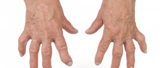
The symptoms and course of seronegative rheumatoid arthritis differ from the typical manifestations of seropositive RA. The main manifestation of the latter is the gradual damage to the small joints of the hands and feet. Seronegative arthritis presents differently.
Course of seronegative rheumatoid arthritis
First signs
The onset of the disease is usually not gradual, but acute. Suddenly there is a fever. The temperature can be either low-grade or very high, with changes and the appearance of heavy sweating throughout the day. The general condition is disturbed: weakness, malaise, headache, dizziness appear, which are characteristic of the initial stage of the disease.
Stiffness of movement in seronegative arthritis is almost never observed, but the pain syndrome is quite severe. Swelling and pain appear in the area of one large joint. Most often these are the knees, elbows or ankles. At the onset of the disease, there is a characteristic absence of small articular lesions of the hand and foot - this is a rare clinical symptom in this form of the disease.
In the lymph nodes with seronegative arthritis, the following changes occur: they become dense and painless.
Sometimes signs of damage to internal organs appear from the very beginning. If the heart is affected, this is shortness of breath and palpitations.
Obvious symptoms
Gradually, the fever gives way to normal or subfebrile temperature. After six months, the inflammatory process in seronegative arthritis becomes symmetrical due to the appearance of inflammation in several more joints. After some time, the process spreads further, inflammation may occur in the small joints of the fingers and toes - signs of polyarthritis develop. The hands and fingers take on a characteristic shape.
Joint pain increases, destructive changes quickly develop, pronounced signs of ankylosis (fusion of cartilage and bones), and impaired limb function appear. Quite often, with seronegative rheumatoid arthritis, the hip joint is affected. Erosion of the articular surfaces is insignificant, but there is a rapid proliferation of connective tissue, closure of the joint space and the formation of ankylosis.
Systemic manifestations in seronegative RA are also not long in coming: signs of damage to the heart (endocarditis), lungs (pleuritis), kidneys (nephritis), and intestines appear. The liver and spleen enlarge. By prescribing timely treatment, it is possible to achieve long-term remission during this pathology and suppress the progression of the disease. If this is not done, the disease will progress.
The most dangerous symptoms of seronegative arthritis
The most dangerous signs are:
- fever for a long time;
- multiple lesions of large and small joints;
- the appearance of severe shortness of breath at rest.
If these symptoms appear, you should immediately seek medical help.