Arthritis is a general term that means inflammation in a joint. Joint inflammation is characterized by redness, swelling and pain in the joint area. Rheumatoid arthritis is a type of chronic arthritis that typically affects joints on both sides of the body (such as the hands, wrists, or knees). The symmetry of the lesion is one of the criteria for differentiating rheumatoid arthritis from other types of arthritis. In addition to affecting the joints, rheumatoid arthritis can cause abnormal changes in the eyes, skin, lungs, heart or nerves. Rheumatoid arthritis affects people differently. In some cases, joint damage occurs gradually over several years. In other cases, rheumatoid arthritis progresses rapidly, without long periods of remission. On average, rheumatoid arthritis occurs in 1% of the population. And although it occurs 2-3 times more often in women than in men, the manifestations of rheumatoid arthritis in men are much more pronounced than in women. Rheumatoid arthritis typically occurs in middle age, but is not uncommon in the elderly and children.
The exact cause of rheumatoid arthritis is unknown, but the disease is thought to occur due to a combination of genetic, environmental, and hormonal factors. In rheumatoid arthritis, certain factors serve as triggers that cause an autoimmune reaction in the body and an effect on joint tissue. A certain role of smoking as a factor provoking the immune system cannot be ruled out.
Research has not yet been able to identify the role of genetic factors in the development of rheumatoid arthritis. But, nevertheless, in some patients it is possible to identify a genetic predisposition to this disease.
Once the immune system begins to work against its own tissues, lymphocytes migrate from the blood into the joints and the tissue lining the joints, called the synovium. There, lymphocytes produce substances that cause inflammation. An increased number of lymphocytes and inflammatory factors causes irritation of the joint and wear of cartilage tissue, inflammation of the joint capsule. Inflammation of the joint capsule causes excessive secretion of synovial fluid. As the cartilage thins, the joint space decreases, and conditions arise for bone surfaces to rub against each other.
Since the inflammatory process involves all tissues of the joint, inflammation can involve adjacent bone surfaces, which can manifest itself as damage to bone tissue (erosion). All these disorders lead to pain, swelling and a local increase in temperature in the joint area. Rheumatoid arthritis is characterized by a positive reaction to rheumatoid factor. But this test can be positive for other diseases.
People with rheumatoid arthritis may have mild anemia. Blood tests may also reveal an elevated erythrocyte sedimentation rate (ESR) or increased level. With reactive protein, which are markers of inflammation.
Some people with rheumatoid arthritis may also test positive for antinuclear antibodies.
There are 3 main factors as the cause of the disease (rheumatological triad)
- Genetic predisposition
- Hereditary tendency to autoimmune reactions
- More common in carriers of a certain MHC II class antigen: HLA - DR1, DR4
- Infectious factor
- paramyxoviruses - mumps, measles, respiratory syncytial infection viruses
- hepatoviruses - hepatitis B virus
- herpes viruses - herpes simplex viruses, herpes zoster, Cytomegalovirus, Epstein-Barr virus
- retroviruses - T-lymphotropic virus
- Triggering factor (hypothermia, hyperinsolation, intoxication, mutagenic medications, endocrinopathies, stress, etc.). For women, breastfeeding for a longer period of time reduces the likelihood of developing RA. Breastfeeding for 24 months or longer reduces the risk of developing RA by half.
Bone erosion in pathology
Erosion of bone structures is divided into three types:
- Marginal or superficial - detected already at the second stage in joints where there is no cartilaginous structure and friction is more severe.
- Compression - occurs due to aggravation of the process and failure of the bone at the site of injury.
- Deforming - formed due to the destruction of the bone plate, visible bone deformation is visually observed, characteristic of stage 4 of pathology.
Single erosions of the bones of the wrist
When bone erosions occur, it is practically impossible to cure the disease. Long-term multicomponent therapy only helps to stop the progressive process.
Diagnosis of the disease
Based on a biochemical blood test , changes in the joints visible on x-rays , and the use of basic clinical markers . A blood test examines ESR, rheumatoid factor, platelet count, etc. The most progressive analysis is the titer of antibodies to cyclic citrulline-containing peptide - ACCP, anti-CCP, anti-CCP. The specificity of this indicator is about 90%, while it is present in 79% of sera from RA patients.
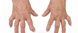
Diagnostically important clinical features are the absence of changes in skin color over the inflamed joints, the development of tenosynovitis of the flexors or extensors of the fingers and the formation of amyotrophies, typical deformities of the hands, the so-called “rheumatoid hand”.
The criteria for an unfavorable prognosis are:
- early damage to large joints and the appearance of rheumatoid nodules;
- swollen lymph nodes;
- involvement of new joints during subsequent exacerbation;
- systemic nature of the disease;
- persistent increase in ESR;
- early appearance (during the first year) and high titers of rheumatoid factor;
- early (up to four months) radiological changes in the affected joints - rapid progression of destructive changes;
- detection of antinuclear antibodies and LE cells;
- carriage of HLA-DR4 antigens; poor tolerability of basic drugs.
In the treatment of rheumatoid arthritis, systemic drug therapy with various groups of drugs is used.
Symptoms
Rheumatoid arthritis (RA) is a chronic disease. Symptoms of rheumatoid arthritis can come and go and are different for each patient. Some patients experience long periods of remission. During remission, rheumatoid arthritis is not active and symptoms may be minimal or absent. In other patients, symptoms may be constant for months.
Although rheumatoid arthritis can involve various organs, the joints are primarily affected. At the onset or exacerbation of the disease, inflammation of the joints occurs. Inflammation is the body's natural response to infection or other factors, but in rheumatoid arthritis, inflammation occurs spontaneously and without obvious cause. Unlike osteoarthritis, the symptoms of rheumatoid arthritis are more severe (severe pain, fatigue, stiffness, fever, loss of appetite).
With rheumatoid arthritis, there may be pain and stiffness in the arms, knees, ankles, legs, and neck. Pain usually occurs on both sides, but sometimes occurs on one side. Thus, there is a symmetrical involvement of the joints in the inflammatory process. Inflammation in the joints interferes with normal physical activity (driving, walking and movements necessary for daily activities).
Stiffness in movement in the morning can quickly disappear or bother you for several hours. Feeling tired is sometimes debilitating. The inflammatory process can cause loss of appetite and weight loss. In addition, it is possible that the temperature of the skin rash may increase or the lungs or heart may be involved in the inflammatory process. These symptoms appear when the area of the autoimmune process expands beyond the joints.
The most common symptoms of joint inflammation:
- Stiffness (stiffness). The joint is difficult to move and the range of motion is limited. Unlike other types of arthritis, where there is also stiffness in the joints, the duration of stiffness in rheumatoid arthritis is longer (more than an hour) until the range of motion normalizes.
- Swelling. Fluid accumulates in the joint and it becomes swollen; it also contributes to stiffness.
- Pain. Inflammation in the joint makes it more sensitive and painful. Prolonged inflammation also contributes to pain.
- Hyperemia and increased local temperature. The joints may be slightly warmer to the touch and redder than the surrounding skin.
- Various joints are affected (hands are almost always affected) and the process is symmetrical.
- Symptoms of secondary organ damage in RA.
- Rheumatoid arthritis can involve various organs, resulting in certain symptoms:
- Fatigue (excessive fatigue)
- General weakness
- Loss of appetite, which sometimes leads to weight loss
- Muscle pain
These symptoms are similar to those of an acute viral infection, but are more intense and last longer.
Rheumatoid nodules are small growths under the skin that most often appear on the elbows. Sometimes they are painful.
Damage to the lungs or pleura is usually asymptomatic. When shortness of breath develops, medications that reduce the inflammatory process are used
Rheumatoid arthritis can affect the structures of the larynx and cause dysphonia.
Rheumatoid arthritis can cause inflammation of the mucous membranes around the heart, usually without symptoms. As symptoms develop, this may include shortness of breath and chest pain. In addition, damage and narrowing of the coronary arteries occurs.
Eye damage occurs in less than 5% of patients with rheumatoid arthritis. This manifests itself as redness of the eyes, dryness, and soreness in the eyes.
Treatment and prevention of rheumatoid arthritis
The pathology is treated by a rheumatologist. Treatment of the disease is complex and multicomponent:
- Diet therapy. The diet should be dominated by foods rich in calcium (milk, cottage cheese, fresh vegetables and herbs); if you are overweight, there is a need to lose weight to reduce the load on the joints of the lower extremities. Products that help maintain cartilaginous structures are also useful: jelly, agar-agar, jellied meat, jellied fish.
- Drug treatment.
- Pathogenetic therapy. Non-steroidal anti-inflammatory drugs (Diclofenac, Nimesil) and steroids (Prednisolone, Dexamethasone) are prescribed; they help relieve pain and reduce inflammation. To prevent the autoimmune process, cytostatics (Methotrexate, Cyclosporine) and biological drugs (Rituximab, Enbrel) are prescribed.
- Etiotropic therapy. Antibacterial agents (Bicillin) are used to prevent relapse of streptococcal infection.
- Symptomatic therapy. Includes the use of chondroitin preparations and hyaluronic acid preparations to maintain cartilage structures, calcium with vitamin D3.
- Physiotherapy and therapeutic exercises. Allows, with the help of physical exercises, electrophoresis, magnetic therapy, to improve hemodynamics in the affected area and activate regeneration processes.
- Operative methods. They are used in extreme radiological stages (third and fourth) in the presence of dislocations, contractures and ankylosis, and pathological fractures.
Prevention of rheumatoid arthritis is the timely and correct treatment of infectious diseases and the prevention of the appearance of chronic lesions. In the initial stages (1-2), it is necessary to develop fine motor skills of the hands and arms - this helps to avoid permanent changes due to normal hemodynamics.
Such patients are recommended to start embroidering, knitting, and drawing. It is also necessary to eat properly to prevent degenerative pathologies of cartilage and bone structures.
Rheumatoid arthritis is a serious pathology that, at extreme stages of development, leads to disability of the patient. The disease can develop both in adulthood and in childhood. Progression leads to a deterioration in overall health and the appearance of persistent irreversible changes in the joint area.
- X-ray of the wrist and hand: what is it and what does it show?
Are the radiological signs of RA different from other types of arthritis?
Using an x-ray, the doctor is able to make a differential diagnosis between different types of inflammatory joint disease. For example, with gouty arthritis, urate crystals (accumulations of uric acid) are visualized on x-rays.
The places where crystals are localized are periarticular tissues and joint space.
At the end parts of the epiphyses (edge of the tubular bone), one can notice the presence of a sclerotic border, and in the cortical layers - cystic defects. The soft tissues along the affected joints are enlarged, and the joint space is widened.
Radiological signs of psoriatic arthritis are different. Osteoporosis, which is an integral companion of RA, in this case occurs only in mutilating forms of the disease. Psoriatic arthritis is accompanied by the appearance of erosions in the distal interphalangeal joints. Such lesions form in the marginal zones of the joints, and then spread to the center.
The tops of the terminal and middle phalanges are ground off, the articular surface is concave, resulting in the visual effect of a “cup and saucer” or “pencils in a glass.”
X-ray signs of glenohumeral periarthritis (inflammation of the soft tissues around the shoulder joint) are the presence of deposits of small calcium crystals (calculous bursitis), osteoporosis of the head of the humerus (this symptom is especially pronounced in the ankylosing form of the disease associated with fusion of the joint and, as a result, its complete immobility).
Detailed x-ray description of the hands at different stages of rheumatoid arthritis.
There are 4 radiological stages of rheumatoid arthritis.
An example of a description of radiographs of the hands:
1st stage. An X-ray of the hands in a direct projection shows deformation of the proximal interphalangeal joints of the 2-3 fingers in the form of moderate thickening. Regional (periarticular) osteoporosis of the phalanges of the fingers of both hands is noted, the compact layer of the phalanges is moderately thinned.
2nd (A) stage. An X-ray of the hands in a direct projection shows deformation of the proximal interphalangeal joints of the 2-4 fingers due to thickening of the para-articular tissues. Regional periarticular osteoporosis of the phalanges of the fingers of both hands is noted, the compact layer of the phalanges is thinned. In the head of the middle phalanx of the 2nd finger there is a cyst-like lucency and a moderate narrowing of the joint space of the proximal interphalangeal joint of the same finger.
2nd (B) stage. An X-ray of the hands in a direct projection shows deformation of the proximal interphalangeal joints of the 1st-5th fingers due to thickening of the paraarticular tissues. There is pronounced regional periarticular osteoporosis of the phalanges of the fingers of both hands, the compact layer of the phalanges is thinned. In the heads of the middle phalanges of the 2nd to 4th fingers there is a cystic subchondral restructuring and narrowing of the joint space of the proximal interphalangeal joints of the 2nd to 5th fingers. In the heads of the middle phalanx of 2-3 fingers, marginal erosions measuring 2-2.5 mm in diameter with clear contours are detected (symmetrical changes).
3rd stage. An X-ray of the hands in a direct projection shows deformation of the proximal interphalangeal joints of the 2-5 fingers due to significant thickening of the paraarticular tissues, narrowing of the joint spaces and deformation of the subchondral plates of the interphalangeal joints. There is pronounced regional periarticular osteoporosis of the phalanges of the fingers of both hands, the compact layer of the phalanges is thinned. In the heads of the middle phalanges of the fingers there are multiple cyst-like lucencies and marginal usurations. Multiple ulnar subluxations of the metacarpophalangeal joints (ulnar deviation) are detected.
4th stage. An X-ray of the hands in a direct projection shows a pronounced deformation of the interphalangeal and metacarpophalangeal joints of the hand, the joint spaces are deformed and sharply narrowed with the presence of multiple subchondral cysts and marginal metaphyseal erosions. Severe widespread osteoporosis is noted. Severe, multiple ulnar subluxations of the metacarpophalangeal joints (ulnar deviation), partial osteolysis of the heads of the 2-5 main phalanges and 3-4 metacarpal bones, ankylosis of the 2nd metacarpophalangeal joint (symmetrical changes) are detected.
According to the classification, rheumatoid arthritis belongs to the group of autoimmune pathologies with a chronic course, which develops when a person’s immune status is weakened. The exact reasons why this pathology develops have not been identified to this day.
Predisposing factors include genetic predisposition, previous infectious processes (measles, mumps, hepatitis B), the effect of toxic substances on the human body, menopause and other autoimmune pathologies.
With the active progression of RA, symmetrical damage to the joints of the legs and arms (elbows, shoulders, hips, knees, hands and feet) develops. Small joint structures located on the upper extremities are usually affected first. First, the synovial membrane is involved in the inflammatory process, then this process affects the cartilage tissue, and as a result, the formation of erosions and irreversible deformation of the joints.
Arthritis can affect more than just joint elements. There are examples when it involves other organs and systems in the process. And the following changes begin to develop in the body: atrophy of the skeletal muscles, enlarged lymph nodes, the liver does not fully perform its functions, the gastrointestinal tract, lung tissue and heart muscle are affected, and possible damage to the skin.