Osteomyelitis is the medical term for infection of bone tissue. Infections can enter bone tissue through the bloodstream or spread from nearby tissues. Osteomyelitis can begin in the bone itself if an infection occurs during an injury.
In children, osteomyelitis most often affects the long bones of the legs and upper arms, while in adults, osteomyelitis of the spinal bones (vertebrae) is more common. People with diabetes may develop osteomyelitis in the lower extremities if there are trophic ulcers on the legs. If previously osteomyelitis was considered an incurable disease, now this disease can be successfully treated. Most patients with osteomyelitis require surgery to remove dead bone tissue along with intensive antibiotic therapy (strong antibiotics given parenterally for at least 6 weeks). With osteomyelitis of the spine, infection of the vertebrae occurs. It is a fairly rare cause of back pain, especially in young, healthy adults.
As a rule, the infection enters the vertebral body hematogenously (through the bloodstream). The veins in the lower part of the spine also drain the pelvis (Watson's venous plexus) and thus infections easily enter the spine. Therefore, in most cases, the cause of vertebral osteomyelitis is infections that occur after urological procedures (for example, cystoscopy).
Classification
Osteomyelitis can be classified according to location, route of penetration, and severity of the process.
If we talk about the course of the disease, then there is an acute or chronic type.
If we are talking about how the bone tissue became infected, then it is worth noting three types of osteomyelitis - post-traumatic, hematogenous, odontogenic.
If we take into account the localization of the purulent process, then it is worth distinguishing between the disease of the lower jaw and the upper jaw.
Acute osteomyelitis in children
This form of the disease is most often observed in children. Here the clinical picture has pronounced symptoms that are characteristic of general intoxication. Inflammation and destruction also develop. This happens not only in the bone, but also in those soft tissues that are around.
Osteomyelitis in children often occurs as a result of certain characteristics of the body (functional, anatomical):
- there is hypersensitivity to infections, a high rate of reactivity;
- bone tissue is actively growing;
- when baby teeth erupt or change, the jaw structures are rebuilt;
- The Haversian canals are wider, and the bone trabeculae are quite thin;
- abundant network of blood vessels.
Risk factors
Bone tissue is generally resistant to infection. For osteomyelitis to occur, conditions must be present that increase the bones' vulnerability to infection.
Recent injuries or surgeries. In case of severe bone fractures or deep punctures, infection penetrates into the bone or nearby tissue. Surgical repositioning of bone fragments or endoprosthetic surgery may also inadvertently introduce infection into the bone.
Poor circulation. When blood vessels are damaged or the blood flow through them is disrupted, conditions arise for a deficiency of immune cells responsible for counteracting microbes and preventing the small number of microbes that accidentally enter the tissue from multiplying. What begins as a small cut can progress to a deep ulcer that can involve deeper tissue and even bone. Diseases that impair blood circulation include:
- Diabetes
- Peripheral artery disease often associated with smoking
- Sickle cell anemia
Medical catheters
A medical catheter connects the outside world with the internal organs. And although these catheters are necessary for some conditions, they can serve as conduits for infections into the body. Therefore, catheters increase the risk of infection, including osteomyelitis. Examples include:
- Catheters for dialysis
- Bladder catheters
- Intravenous catheters, which are necessary for long-term administration of medications (months or even years)
Intravenous drug administration
People who inject drugs are at greater risk of developing osteomyelitis because they typically use unsterile needles and do not clean the skin before injections.
Common causes of osteomyelitis.
The following groups of people are most susceptible to developing osteomyelitis:
- Elderly patients
- Intravenous drug addicts
- Patients with a weakened immune system
The immune system may be weakened as a result of the following conditions:
- Long-term use of corticosteroids to treat systemic diseases such as rheumatoid arthritis.
- Insulin-dependent diabetes mellitus
- Patients with transplanted organs
- Acquired immunodeficiency syndrome (AIDS)
- Malnutrition
- Cancer
Intravenous drug use is causing an increase in the number of patients with spinal infections. As a rule, the microorganism most often affecting the spine is Staphylococcus aureus, and among drug addicts who use intravenous drugs, Pseudomonas aeruginosa is often the causative agent of a spinal infection. Treatment of these two pathogens requires different antibiotic treatments.
In the recent past, Mycobacterium tuberculosis has been a common cause of spinal infections. At present, spinal tuberculosis practically does not occur in developed countries and is common only in poor, backward countries with a low standard of living. But drug addicts may have a tuberculous infection.
Most vertebral body infections occur in the lumbar spine due to the nature of the venous blood flow. With tuberculous lesions of the vertebrae, the cervical and thoracic spine are most often affected.
Causes
- Acute odontogenic osteomyelitis occurs from teeth that have been affected by caries or from pathogenic microorganisms that have penetrated. Typically, a similar form is diagnosed on the lower jaw. At the same time, the age period of children is 7-12 years;
- acute hematogenous osteomyelitis is the result of an infection that appeared from other foci. For example, it could be omphalitis (when the umbilical ring becomes inflamed), purulent mastitis, which the baby’s mother suffers from. If we talk about older children, they can become infected as a result of skin infections, tonsillitis, and purulent otitis media. This form occurs in the upper jaw and is accompanied by infection of the internal organs;
- Acute traumatic osteomyelitis is rarely diagnosed. It can be caused by infection of the line where the jaw fracture is noted. This occurs in case of injury, also after operations in this area. Often this disease occurs if the bone fragments are not properly treated, if bone fragments are immobilized, and also if carious teeth are caught in the fracture line and the skin and oral mucous membranes are damaged.
Treatment of chronic odontogenic osteomyelitis of the jaw
Treatment of chronic odontogenic osteomyelitis of the jaw is complex and includes surgical intervention and drug treatment.
I. During exacerbation of chronic osteomyelitis, the symptoms of acute inflammation are first relieved. If the causative tooth has not been removed previously, then it must be removed this time. Adjacent mobile teeth are trepanned and splinted, if not removed according to indications (after assessing their viability and x-ray examination). Sanitation of the oral cavity is mandatory and all chronic sources of infection are removed to prevent complications during subsequent procedures.
To facilitate the outflow of pus, fistulas or wounds are expanded, and primary surgical treatment of subosseous and perimandibular purulent foci is performed.
An important part of the surgical stage of treatment is sequestrectomy. After evaluating the radiograph, the formed sequesters are removed. Removal is performed through an intraoral or extraoral incision. Large sequestra in the area of the body and ramus of the lower jaw, as well as in the area of the infraorbital margin and zygomatic bone, are removed extraorally. Sometimes large necrotic areas of bone are broken into several parts for ease of removal. Incisions are made along the natural folds of the face for better aesthetics.
After removal of sequesters, attention is paid to granulations and sequestral capsule. Using a curettage spoon or even a milling cutter, pathological tissue is removed until the signs of healthy bone are revealed: alveolar bleeding, white bone, hard bone tissue.
The free space is filled with a biosynthetic osteotropic drug: kolapol, kolapan, etc. The wound is tightly sutured, drainage is left. The stitches are removed after 7-10 days.
II. Next, let's move on to drug treatment. As with other purulent diseases, etiotropic, pathogenetic and symptomatic treatment is carried out.
To eliminate the cause of the disease, the surgeon removes the causative teeth. But the infection remains in the blood, so the patient is prescribed antibacterial drugs: macrolides, cephalosporins. It is also worth prescribing the patient antifungal agents (Deflucan 150 mg once a week).
Since the patient’s immunity is reduced, it is recommended to prescribe immune drugs such as thymalin, T-activin, levomisol, staphylococcal toxoid.
In case of extensive damage to bone tissue, the patient is recommended to follow a gentle diet to prevent a pathological fracture of the jaw.
To reduce the symptoms of inflammation, detoxification and anti-inflammatory therapy is carried out. Physical therapy exercises and physiotherapy are individually selected for the patient to restore function.
Chronic osteomyelitis of the jaw. Outcomes and complications
Outcomes:
- Favorable - if the patient contacts a maxillofacial surgeon in a timely manner and receives adequate treatment, the patient’s full recovery is possible.
- Unfavorable – if treatment is insufficient and the patient contacts a doctor late, the following may occur:
- Exacerbation of the disease
- Jaw deformity,
- Jaw fracture - occurs due to minor physical impact, from which a healthy jaw would not be damaged,
- Complications of osteomyelitis: Abscesses and phlegmon of soft tissues of the face,
- Thrombosis of facial vessels and cavernous sinus,
- Mediastinitis,
- Death.
Symptoms
Acute osteomyelitis appears very suddenly. If children are small, then they are noted to be moody and lethargic. They don’t want to eat, they don’t sleep well, and their temperature rises. If the child is older, then he may feel unwell, general weakness, and may complain of toothache.
When the disease is still in its early stages, the following manifestations may be present:
- bone pain. Initially it has a local character, and then diffused;
- the oral mucosa swells, hyperemia is noted;
- on the side that is affected, the soft tissues become swollen;
- the face becomes asymmetrical;
- chewing muscles may spasm.
If an injury occurs, signs of the disease appear after 3-5 days. The child feels much worse, the temperature rises, and pus may also be released if the mucous membrane has been damaged.
Is it possible to prevent the disease?
To protect your child from various types of illnesses, you need to carefully monitor his actions, diet and follow the drinking regime. It is important to protect your baby from falls, bruises and skin injuries. If an injury does occur, you need to carefully treat the wound and apply a sterile bandage or plaster. Strict adherence to sanitary and hygienic standards for caring for an infant plays an important role. The main thing is that if any negative manifestations concerning the baby’s health occur, immediately seek help from a pediatrician.
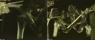
Diagnostics
It is based on X-ray examination. When the disease is just beginning, the images may show symptoms of osteoporosis (in the area where the inflammatory process occurs). And after a few days, radiographs already show foci of destruction. After another 2-3 weeks, bone necrosis is already visible, as well as the places where sequestra have formed.
Treatment
Here it is necessary to carry out certain measures that will help eliminate foci of inflammation and restore those functions that have been impaired.
Every case of childhood osteomyelitis must be treated urgently. To do this, you should call a doctor and admit the child to the hospital. If surgical intervention is performed on time, then we can talk about a positive prognosis and outcome.
In the case of odontogenic osteomyelitis, the tooth that provoked the inflammatory process should be quickly removed. When it comes to children, teeth can be removed, both baby and permanent teeth. After tooth extraction. the hole acts as natural drainage. However, sometimes the bone marrow spaces are opened to improve the drainage of pus.
After the tooth has been removed, the subperiosteal abscesses are opened or a periostotomy is performed.
During sanitation, they not only open, but also thoroughly clean the destruction cavity and remove necrotic masses from there. Antibiotics and antiseptics are used for washing, and drainage is also done. As for conservative therapy, special drugs from the groups of immunostimulants, anti-inflammatory and antibacterial are used.
Secondary chronic
In children, rapid chronic inflammation is noted. After 3 weeks, acute symptoms disappear, but the child does not become healthier.
When it comes to chronic inflammation, there is an active process of destruction, as well as melting of the elements of bone substance. After which areas of necrosis form. In addition, thanks to the intraosseous structures and periosteum, bone tissue is actively restored. The peculiarity of this type of osteomyelitis is the presence of the rudiments of permanent teeth. If they are involved in pathology, they may then die, and their behavior will resemble sequestration.
Symptoms of chronic osteomyelitis
When the inflammatory process becomes chronic, acute symptoms decrease. 10 days after the disease began, the child feels better (appetite and sleep return to normal, fever and symptoms of intoxication disappear). But children still feel weak, they get tired quickly, they sweat profusely, and their skin is pale. Sometimes children say that their jaw hurts slightly on the side that is affected.
During the examination of such a patient, you can notice certain manifestations of the inflammatory process:
- over the place where the focus of osteomyelitis was, there are soft tissue infiltrates;
- pain is felt when the child's jaw is probed;
- There are fistulas in which pus is released. They can be multiple or single;
- lymph nodes in the neck and jaw increase in size and hurt;
- The sockets of the tooth that were removed do not heal well. It may also leak pus;
- teeth are very loose.
When the disease worsens, the symptoms resemble acute osteomyelitis.
Diagnosis of chronic osteomyelitis
An x-ray should be taken to confirm the diagnosis. It will allow you to see the foci of destruction, those rudiments of teeth that have died, sequestration. If the lesion is extensive, then jaw fractures may even be detected.
Treatment of chronic osteomyelitis
When it comes to secondary osteomyelitis of a chronic form, a conservative treatment method is used:
- antibiotic therapy. Which drugs are suitable? All this is determined after the patient undergoes a culture of the pus discharged from the fistulas. Thus, the type of microorganism that provoked the pathology and its sensitivity to different drugs will be determined;
- desensitizing therapy. This includes antihistamines, which will remove the allergic reaction and also increase the body's resistance;
- therapy that can be used to stimulate the immune system and strengthen the body as a whole;
- physiotherapy. For this purpose, UHF therapy and laser irradiation are performed.
To perform an osteotomy or sequestrectomy, you must have the following indications:
- sequesters that are large in size and do not undergo spontaneous lysis for a long time;
- there are rudiments of permanent teeth that have died. They support inflammation;
- there was a risk of amyloidosis of internal organs.
If the disease worsens, the treatment will be identical to that in the case of acute osteomyelitis. But the main method is surgical intervention, during which lesions with pus are opened and drainage is performed.
Therapeutic measures
Part of the child's treatment may take place in the hospital.
Therapy for osteomyelitis in newborns consists of several stages, and is therefore carried out in the hospital and at home. The main thing is to promptly identify the signs of the disease, conduct a full examination and select effective treatment based on the nature of the main pathogen. Great responsibility when treating a baby falls on the parents.
Independent actions
First of all, you need to monitor the baby’s condition and note any visual or behavioral changes. If you suspect the development of the disease, you should immediately contact your pediatrician. Next, it is important that parents follow all the doctor’s recommendations and provide the child with proper care. Spa treatment is of great importance, so it is recommended to worry about organizing your trip in advance. Every six months, parents are required to provide a routine examination and, if necessary, re-treatment.
Doctor's actions
Osteomyelitis requires immediate medical intervention. Therefore, the baby is hospitalized for a number of therapeutic measures. Initially, the doctor prescribes medications aimed at activating protective functions and strengthening the immune system. A course of antibiotics is used to combat a specific type of bacteria. If the infection is caused by a violation of the integrity of the skin, then the affected area is covered with an antiseptic plaster. If complications develop in the form of an abscess with the accumulation of purulent exudate, surgery is performed under general anesthesia.
Primary chronic
It's rare to see a form like this. Typically, this disease is caused by weakly active microorganisms. They penetrate from teeth affected by caries. They are the ones who maintain inflammation for a long time.
This form of osteomyelitis is characterized by a productive nature, since the processes of destruction observed in bone tissue are weakly expressed. However, during this process new bone is actively formed.
If we study the clinical picture of primary chronic osteomyelitis, then the acute period is omitted. The child's condition generally remains the same, only fatigue, pale skin, and weakness appear. As for local signs, the area of the causative tooth hurts a little, the jaw bones and their thickness increase, and also symptoms of lymphadenitis appear in the lymph nodes. There are no fistulas on the skin or mucous membranes.
At an early stage, the productive form of the disease can be easily treated with conservative methods. If you choose the right means, you can achieve positive results quite quickly and without consequences. If the case is advanced, then surgical intervention will be required. It is used to remove too much bone tissue, as well as dead tooth germs. After that, they model the correct jaw contour.
Possible complications
In the future, the pathology can lead to arthritis.
Osteomyelitis provokes a number of negative consequences in a newborn child. As a consequence of the disease, severe pathologies of the musculoskeletal system appear. The structure of bone and cartilage often suffers, which leads to their deformation and impaired joint mobility. Prolonged inflammation provokes the development of degenerative changes in osteochondral tissue, which causes the formation of arthritis.
Purulent lesions of the bones of the head lead to the development of meningitis, which poses a danger to the health and life of the baby.
Consequences and rehabilitation
Osteomyelitis can have quite serious consequences in childhood:
- defects associated with bone tissue;
- jaw fractures that cause abnormal joints to form;
- the jaw is deformed;
- absence of permanent teeth, edentia;
- arthrosis, ankylosis, arthritis that relate to the joints of the temples and lower jaw;
- the jaw grows much more slowly, microgeny is noted;
- soft tissues are deformed and look like scars.
Due to the above complications, cosmetic defects may occur, but they do not have any effect on the masticatory apparatus or its functioning.
In order to restore the anatomy of the face and jaw to the maximum and ensure its normal functioning, it is necessary to take certain rehabilitation measures:
- sometimes plastic surgery is used. But this is done only after the facial bone stops growing;
- If adentia develops, then temporary dental prosthetics can be done. And as soon as the skeleton is finally formed, then permanent prosthetics are allowed;
- To improve the temporomandibular joint and its functioning, it is worth applying physiotherapeutic procedures.
All children who have suffered osteomyelitis that affected the jaw area are registered at the dispensary. They are required to visit the dentist at least 2 times a year.