Learn more about diseases starting with the letter “A”: Abdominal migraine, Absence, Brain abscess, Abuse headache, Agnosia, Pituitary adenoma, Adrenoleukodystrophy, Acalculia, Akathisia, Alternating syndromes, Amaurotic idiocy, Werdnig-Hoffmann amyotrophy, Kugelberg-Welander amyotrophy, Amnesia, Angioneurosis, Cerebral aneurysms, Anomalies of brain development, Chiari anomaly, Kimerli anomaly, Apallic syndrome.
Kimerli's anomaly is a pathology that can be either acquired or congenital. This disease occurs in 12-30% of people on Earth. The disease consists of the presence of an additional bone arch in the structure of the first cervical vertebra. The free movement of the vertebral artery with the help of this arch is limited, and artery compression syndrome also appears.
The anomaly manifests itself in the form of tinnitus, uneven gait, dizziness, muscle weakness and lack of coordination. In the presence of pathology, ischemic stroke and TIA may occur. To detect this pathology, duplex scanning of the vessels of the head and neck is used, magnetic resonance angiography and radiographic examination of the craniovertebral junction are performed. Vascular disorders must be treated conservatively and comprehensively.
Kimerli's pathology is a craniovertebral malformation, that is, congenital disorders in the structure of the articulation of the skull and the first cervical vertebrae. The anomaly causes both compression of the vertebral artery and chronic ischemia in the posterior regions of the brain. This ailment in itself does not belong to a number of diseases and its presence in the body does not indicate that this is the cause of all disorders in the area of the vertebral artery.
When examining patients with this anomaly, only one out of 4 people shows a relationship between the anomaly itself and the development of the syndrome.
Pathogenesis
Both vertebral arteries (left and right) have their origin in the corresponding subclavian arteries. Each of these vertebral arteries is located in a canal formed by the foramina of the transverse processes of the vertebrae located in the area of the cervical spine. Passing through the entire cervical region, the vertebral artery enters the skull through the foramen magnum. The cerebellum is supplied with blood through the vertebrobasilar system, which is formed by the left and right vertebral arteries with their branches.
The vertebral artery can move freely in the wide bony groove in which it lies horizontally during head movement. It enters this groove straight from the cervical canal, while bending around the cervical vertebra on its way.
Kimerly's disease is characterized by the presence of a bone arch, which is located above the bone groove, and this arch can completely limit the movements of the vertebral artery passing through this place.
Atherosclerosis, lesions of the vascular wall in the presence of vasculitis, osteochondrosis and spondyloarthrosis of the cervical spine - all of these diseases can arise from pathology.
Classification
In modern neurology, there are only 2 types of pathology:
- the first is the presence of a bone arch connecting the process of the atlas with its posterior arch;
- in the second variant, the disease appears in the form of a bone arch between the transverse process of the atlas with its articular process.
Kimerli's disease can manifest itself on both sides of the first cervical vertebra, or with a unilateral character (located on one side of the first cervical vertebra). Also added to all this is the division of the anomaly into incomplete and complete. In the presence of a complete form of the disease, the bone arch is in the form of a semicircle, while in an incomplete anomaly it is expressed in the form of an arched outgrowth.
Types of pathology and their symptoms
Neurologists distinguish two main types of damage:
- A complete anomaly
. It suggests the presence of an atlanto-occipital ligament, in which, over time, hardening and closure of the ring in the posterior section is observed. This process is characterized by noticeable compression of the artery and serious disturbances. - Incomplete
. There is no closure, only one bone arch is observed. In some cases, it is not detected due to the absence of certain symptoms. When diagnosing, it requires observation by the attending physician.
By type of formation there are:
Advertising:
- Congenital form
. The pathology begins in the womb, so it is impossible to prevent its occurrence. The lesion is not genetically transmitted and manifests itself in children of school age, especially during active physical activity. Requires constant medical supervision, physiotherapeutic procedures and maintenance medications. - Acquired
. It becomes a complication of diseases of the spine, cartilage and bone tissue. Often occurs with the progression of osteochondrosis. Therapy requires an integrated approach, since damage can cause other neurological disorders.
By localization:
- One-sided
. It involves damage to only one artery and progresses only on one side of the spine. - Double-sided
. Accordingly, it affects both vertebrae and compresses both arteries.
In all varieties, the symptoms are similar, so anamnesis is not the main diagnostic measure in determining the defect.
Before starting treatment, it is necessary to determine the causes and location.
The lesion is not inherited, so the pathology can be diagnosed in a child with absolutely healthy parents.
Most often, radiculitis becomes a consequence of spinal injuries or the active progression of osteochondrosis. Usually the lesion is diagnosed in old age, but in recent years more and more cases of the disease have been reported in people over 30-35 years of age. Read more in the article: “how to treat lumbar radiculitis at home.”
Symptoms
The disease is accompanied by clinical manifestations that appear due to a decrease in blood flow to the posterior parts of the brain. Due to these manifestations, patients develop symptoms such as tinnitus, manifested in the form of hissing, whistling or ringing, and also flickering “stars” and darkening in the eyes.
All these factors begin to act more intensely during head turns. Due to the fact that with Kimerli's pathology, blood flow to the cerebellum is significantly reduced, symptoms such as dizziness and instability while walking may appear. All this also intensifies when turning the head in any direction.
If people with Kimerli's anomaly have overstrained neck muscles or the head is in an uncomfortable position, this may even result in loss of consciousness.
With a more severe course of the disease, tremor of the arms and legs, loss of coordination, hypoesthesia, muscle weakness of parts of the face or body, and headache may occur. In addition, there may be the presence of transient ischemic attacks in the vertebrobasilar region, but the most dangerous of all of the above is the manifestation of ischemic stroke.
Causes of the anomaly
At the moment, the causes of the Kimmerle anomaly and the factors that provoke the development of the malformation are not clear. Experts divide this pathology into several types, which sometimes explains their origin, for example, congenital and acquired. According to statistics, 10% of newborns have such a bridge. The development of an acquired anomaly is associated with diseases of the spine.
Diagnostic methods
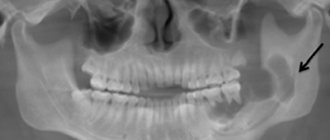
If the patient has a lack of blood circulation in the vertebrobasilar system, an X-ray of the skull and spine in the cervical region is first performed. Typically, such a disease is clearly visible on lateral radiographs of the craniovertebral junction area.
It may also be necessary to consult with an otolaryngologist if the patient has tinnitus to subsequently rule out ENT pathology. Audiometry and other hearing tests are performed. In addition, a vestibular analyzer test is prescribed.
The neurologist needs to eliminate other possible causes of vertebrobasilar insufficiency, since the previously identified Kimerli anomaly may not necessarily be the cause of vertebral artery syndrome. In this case, it is necessary to perform contrast angiography, which will help detect diseases such as thrombosis. Also, angiography will reveal compression of the vessel by any space-occupying formation, presented in the form of a tumor, cyst or brain abscess.
A number of hemodynamic studies will help determine how clinically significant Kimerli's anomaly is. With the help of these studies, in this disease, it is possible to identify the place where compression of the vertebral artery occurs, as well as its dependence on the position of the head and neck.
Treatment
Treatment is only necessary if the patient has clinical and hemodynamic signs of circulatory disorders in the vertebrobasilar region, which are associated with the presence of Kimerli's anomaly in the cervical spine.
People with this pathology should beware of strong physical exertion, sudden turns of the head and many sports, such as wrestling, gymnastics, etc. When visiting massage and manual therapy, it is necessary to warn the doctor about the presence of the disease. If the patient’s condition worsens, this is a reason to immediately seek help from a specialist.
Basically, Kimerli's disease, which can lead to clinical manifestations of vascular insufficiency, is subject to conservative treatment. Vascular therapy is performed, which is mainly aimed at improving blood flow to the brain.
Also, in the presence of Kimerli’s anomaly, special drugs are used under the strict supervision of a coagulogram, which improve the rheological properties of the blood. At the same time, complex therapy includes nootropics, neuroprotectors, as well as metabolic medications such as Piracetam, Aminobutyric acid, Meldonium.
Today, if a patient has a Kimerli anomaly, there is no urgent need for surgical treatment. Surgery may be required only if decompensated vertebral artery syndrome occurs, which leads to a clear lack of blood flow to the vertebrobasilar.
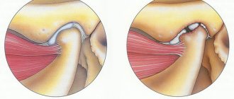
If, nevertheless, the operation occurs for Kimerli’s pathology, then it consists of resection of the abnormal arch and mobilization of the vertebral artery. After the operation, the patient needs to walk for about 2-4 weeks with a special Shants collar. But most often, it is enough for the patient to be observed periodically by a qualified neurologist to prevent the development of vertebral artery syndrome.
In addition, many people are interested in the question of a deferment from the army if they are diagnosed with Kimerli’s anomaly. This anomaly in itself does not allow one to receive a deferment from the army, since it is not a disease. A deferment can be given only in case of any complications that have arisen due to this disease.
Additional measures
Patients diagnosed with a mild form of Kimerly's anomaly should definitely adhere to certain precautions that will help them avoid serious complications. Namely, they need to avoid in every possible way sharp turns of the head, excessive physical exertion, performing physical exercises while standing on their heads, as well as sports games and activities that involve hitting their heads. Such activities include football, gymnastics, basketball and others.
Before a session of manual therapy or massage, the patient must warn the attending physician about the presence of Kimerli's anomaly. He must remember that in the event of a sharp deterioration in his health, he needs to urgently consult a neurologist for advice so as not to miss the early stage of the disease and begin treatment on time.
Preventive measures
If you exercise, you should consult with a qualified physician about the safety of these activities. Together, choose the most suitable therapeutic exercises to improve blood flow. You can contact an experienced chiropractor who, with his “magic” hands, can improve blood circulation in the neck area where the disease is located.
In addition to all of the above, sometimes it’s enough to walk in the fresh air more often. Such walks stimulate the immune system and stabilize a person’s general condition, help improve blood pressure and limit the production of the stress hormone, which is found at almost every step of modern people.
Kimmerle anomaly in children
As a rule, this pathology is diagnosed at an early age by chance. Kimmerle anomaly is formed in a child at the stage of intrauterine development, but a specific cause for this process has not been identified. A child can live with this disorder for a long time and not suspect it. An aggravating factor may be:
- scoliosis;
- osteochondrosis;
- strong physical activity;
- prolonged sitting position (for example, at a computer).
Due to compression of the blood supply channel to the brain, children may study worse, and endurance may be reduced. The following symptoms actively manifest themselves as Kimmerle disease progresses:
- nausea;
- prostration;
- weakness;
- vegetative-vascular dystonia;
- dizziness;
- unstable emotional background.