This article describes the anatomy of the human hand. You will learn everything about joints, muscles, tendons and skin.
Children study the structure of the human body at school, and students may also need such information for specialized education. The structure of each part of the body is complex. It can be difficult to learn their names.
Read all about the human skeleton in another article on our website. This is educational and interesting information.
This article describes the structure of the human hand, with the name of the basic parts, features, and so on. Read on.
Internal structure of the human body: names of the basic parts of the right and left hands, features, photos
The name of the basic parts of the right and left hand
The internal structure of the human body is studied by such a science as anatomy. Hands are the upper limb of the human body, which allows you to take objects, touch them and evaluate them. Below you will find the name of the basic parts of the right and left hands and their features. The musculoskeletal limb consists of several tissues:
- Bones are a solid organ that performs a musculoskeletal function. Serves as a frame for all other elements of the hand.
- Muscle is an organ that consists of muscle tissue. They are involved in the musculoskeletal system and the transmission of nerve impulses.
- Ligaments are an organ representing the formation of connective tissue. They hold the human skeleton and internal organs together.
- Cartilage is elastic connective tissue. There are no blood vessels or nerves inside the cartilaginous junction.
- Tendons are formations of connective tissue.
- Blood capillaries are thin vessels that participate in the blood circulation process.
- Nerve fibers are extensions of nerve cells. Their main role is to distribute nerve impulses.
Like any complex structure in the human body, the right and left hands consist of basic sections. See the photo above for more details. Sections of the human hand:
- Shoulder girdle
- Shoulder
- Forearm
- Brush
Each zone is connected to another department through a joint. This ensures mobility of the upper limbs. There are 32 bones in one human hand .
Functional role
Speaking about the anatomy of the hand, one cannot fail to mention the functional and clinical role of its structural features.
The first lies in the features of the function performed by the limb. Thanks to the complex structure of the hand, the following is achieved:
- A strong belt of the upper limbs holds the free part of the arm and allows you to perform enormous loads.
- The moving part of the arm has complex but important joints. Large joints have a large range of movements that are important for the operation of the hand.
- The fine articulations and work of the muscle structures of the hand and forearm are necessary for the formation of precise movements. This is necessary to carry out daily and professional activities of a person.
- The supporting function of fixed structures is complemented by muscle movements, the number of which is especially large in the hand.
- Large vessels and nerve bundles provide blood supply and innervation to these complex structures.
The functional role of the anatomy of the hand is important for both the doctor and the patient to know.
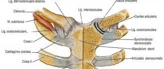
The structure of the bones of the shoulder girdle of the human arm with names in pictures: skeleton, photo
Bones of the shoulder girdle of the human arm
The skeleton of the bones of the shoulder girdle of the human arm represents: two pairs of shoulder blades and clavicles, which provide support and motor activity of the upper limbs.
The structure of the bones of the shoulder girdle of the human arm
Below you will find the buildings with names. Above in the picture everything is visible and described in detail. The right and left shoulder blades resemble a flat triangular bone located on the side of the back. It is slightly curved outward in the direction from the costal arches. The blade consists of several elements:
- Top corner
- Top edge
- Shoulder cutting
- Neck of the scapula
- Medial edge
- Subscapular fossa
- Subarticular tubercle
- Lateral edge
- bottom corner
The structure of the bones of the shoulder girdle of the human arm.
The lateral edge has a thickening for connection with the head of the humerus. The lower angle of the scapula ends at the level of the eighth rib. Along its axis is the key bone, which is connected to muscle fibers. The subarticular tubercle on the scapula allows you to make circular movements with your hands.
Collarbone
Another tubular bone belonging to the group of the shoulder joint is the clavicle. It is located in a horizontal position in the chest at the border with the neck. The bone serves as a connecting link between the sternum and the shoulder blades. The collarbone supports the entire muscular frame of the shoulder girdle.
Clinical role
In order to properly treat diseases, understand the symptoms and diagnosis of diseases of the upper limb, you need to know the anatomy of the hand. Structural features have a significant clinical role:
- The large number of small bones leads to a high incidence of bone fractures.
- Movable joints have their own vulnerabilities, which is associated with a large number of dislocations and arthrosis of the hand joints.
- Abundant blood supply to the hand and a large number of joints leads to the development of autoimmune processes in this area. Among them, arthritis of the small joints of the hand is relevant.
- The wrist ligaments, which tightly cover the neurovascular bundles, can compress these formations. Tunnel syndromes occur, requiring consultation with a neurologist and surgeon.
A large number of small branches of nerve trunks is associated with the phenomena of polyneuropathy during various intoxications and autoimmune processes. Knowing the anatomy of the upper limb, one can assume the clinical features, diagnosis and principles of treatment of any disease.
It can be useful:
- Anatomy of the human neck
- Anatomy of the elbow joint
- Kyphosis of the thoracic spine
- Anatomy of the spine, structural features of the vertebrae
- Spinal cord and spinal nerves
The structure of the muscles of the shoulder girdle of the arm, the functions of the shoulder: description
The structure of the muscles of the shoulder girdle of the arm
The muscle tissue of the shoulder girdle of the arm includes the following muscles:
- Deltoid
- Supraspinatus
- Infraspinatus
- Subscapularis
- Big round
- Small round
Here is the detailed structure and function of the muscles of the shoulder and arm:
Deltoid:
- These are superficial muscle fibers that are located above the shoulder joint.
- In shape it resembles an inverted Latin letter “Delta”, which is where its name comes from.
- The structure of the deltoid muscle consists of three groups: scapular, acromial and clavicular.
- Each component ensures the movement of the hand in different directions.
Supraspinatus muscle:
- Resembles the shape of a triangle, which is located in the supraspinatus fossa of the scapula.
- She is responsible for abducting the shoulder to the sides.
Infraspinatus muscle:
- It resembles the shape of a flat triangle located in the infraspinatus fossa of the scapula.
- Its main function is to extend the shoulder in the shoulder joint.
Subscapularis muscle:
- Located in the central area, between the muscles of the chest and shoulder.
- It is responsible for lifting heavy objects and extending the shoulder.
Teres major muscle:
- It is located from the lower angle of the scapula to the tubercle of the humerus.
- In its structure it resembles a square shape, but when contracted it takes on a rounded shape.
- Its role is to break the shoulder and rotate along circular axes.
Teres minor:
- It is a continuation of the teres major muscle with a similar structure and functionality.
- Its location begins in the area of the scapula and reaches the greater tubercle of the humerus.
A more detailed description of the structure of the muscles of the human arm is described in the picture below:
The structure of the muscles of the shoulder girdle of the arm
Anatomical structure of the human forearm: skeleton, drawing
Anatomical structure of the human forearm
The human forearm belongs to the category of long bones. Its anatomical structure is simple. The skeleton has two sections:
- Elbow bone
- Radius
They are connected to each other by interosseous membranes. This is clearly visible in the picture above. More details:
The ulna is a paired organ of the forearm of a triangular shape with a thickened structure at the top. The ulna becomes thinner towards the bottom. It has three departments:
- Upper section of the tubular bone . In this part there is a trochlear notch, which has two processes: anterior and posterior, as well as a radial notch connecting the processes with the radius.
- Base (body) . The section has a curve at the front.
- The lower part of the tubular bone. This part contains the head, styloid process and articular circumference.
Along its entire length it is covered with muscle fibers, with the exception of the posterior edge.
The radius is a paired organ of the forearm with a triangular shape. She has:
- The head is the widest and thickest place at the upper end of the bone.
- The neck is a narrowing that is located under the head.
- The tuberosity is the junction of the tendon of the main muscle of the shoulder.
- Styloid process located on the lateral side.
- The dorsal tubercle is located on the posterior surface of the rounded section of the tubular bone.
- The carpal articular surface is the point of connection with the bones of the wrist.
The main function of bones is a framework for the muscle layer, joints and cartilage, which provide motor activity to the arm.
Diagnostics
The diagnosis of arthritis of small joints of the hands is established on the basis of examination and questioning of the patient and is confirmed by laboratory and instrumental studies:
- Laboratory tests
- reveal the content of components confirming the inflammatory process, the presence of infectious pathogens and antibodies to them, as well as rheumatoid factor. - Instrumental
:- Ultrasound
– reveals the presence of excess fluid in the joint; - radiography
- the presence of bone changes (narrowing of the joint space, bone ankylosis); - MRI
– presence of changes in soft articular and periarticular tissues; - arthroscopy
- endoscopic examination, allows you to see the inner lining of the joint and all changes in it; During diagnostic arthroscopy, you can take fluid or a piece of tissue for examination.
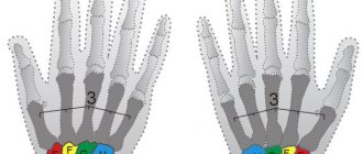
Structure of the human wrist: description
The structure of the human wrist
The human wrist is a section located between the bones of the forearm and the metacarpal bones. It has eight small bones, which are divided into two types: proximal and distal. Here is a description of the building:
The proximal view has four types of bones:
- Scaphoid - located in the first row of the wrist.
- Lunar - located in the second row on the radial side. The shape of the bone resembles a crescent, which is why it got its name.
- Triangular - located in the first row of the wrist. Has a convex surface.
- Pea-shaped - resembles an egg or oval in shape. It is located in the thickness of the tendons.
The distal section has four types of bones:
- The trapezoid bone has a concave structure and is located next to the triquetral bone.
- The trapezoid bone connects the trapezoid bone with five short tubular bones.
- The head bone is the largest of the carpal bones. It has a spherical shape.
- The hamate bone connects the capitate bone and the second row of carpal bones.
The main function of the wrist is the circular movements of the hand and its correct position.
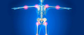
Common pathologies
Human hands are susceptible to various inflammatory and degenerative processes . At the same time, a person can encounter diseases in the hand area even at a young age.
Tenosynovitis
Inflammatory process in the tendons , accompanied by pain, limited hand movements, and the inability to perform simple everyday tasks. Develops due to excessive muscle overload, injuries to the wrist, fingers, and shoulder. It can also occur against the background of chronic diseases (diabetes mellitus, sexually transmitted infections, rheumatoid arthritis), sore throat, flu, hormonal imbalance.
Tunnel (carpal) syndrome
Characterized by compression of the median nerve in the carpal tunnel . Occurs against the background of compression or traumatic injury to the hand. The pathology can be provoked by arthritis, arthrosis, tenosynovitis, tumor formations in burrow tissues, and diabetes mellitus. Accompanied by numbness, tingling, burning of fingers, pain in the hands, impaired grasping function, muscle atrophy, and discoloration of the skin.
Osteoarthritis
A pathological process occurring in the cartilage tissues of the joints, leading to degenerative changes and their destruction . The risk of developing the disease increases with age. However, even in young people, the health of the joints of the hands can deteriorate due to obesity, poor nutrition, hormonal imbalance, frequent hypothermia, injuries, and chronic illnesses. The patient experiences pain in the fingers and wrists during exercise, cannot perform full movements, and feels stiffness in the hands in the morning.
Gouty arthritis
inflammatory disease that affects the joints of the hands and feet. Accompanied by pain in the fingers, swelling, redness of the skin, and local fever. It can develop due to poor nutrition, the presence of concomitant pathologies, and the use of certain medications.
Raynaud's syndrome
A disease characterized by impaired blood supply to the blood vessels of the hands. In most cases, it is a symptom of other pathologies, such as rheumatoid arthritis. Causes occasional pain, pallor, cyanosis of the skin of the hands, a feeling of numbness and tingling in the fingers.
Coordinated work of all elements of the hand is possible if all its main parts function normally. Only then does a person remain able to work and can perform even the most complex tasks, for example, those associated with embroidery, typing, writing, driving, etc.
Anatomy of the structure of the human hand: skeleton, bones, muscles
Anatomy of the structure of the human hand
The skeleton of the human hand has the most complex structure. The composition includes 27 bones , which are divided into groups:
- Wrist
- Metacarpus
- Fingers
The bones are connected to each other by cartilage tissue. More detailed anatomy of the structure:
Anatomy of the structure of the human hand
The metacarpus is five tubular bones that have no special names. They are simply numbered with Roman numerals I – V from thumb to little finger. The structure of each bone is divided into three sections: the head, the body and the base. The head is connected to the bones of the fingers, and the base to the bones of the wrist.
The bones of the metacarpus are similar in joints to each other. The only difference is the third finger, which has a styloid process. All bones of the metacarpus are connected to each other by phalanges. The metacarpus performs a motor function and helps to hold objects in the hands.
fingers , except the thumb, have three phalanges:
- Proximal
- Average
- Distal
The longest phalanx is the proximal one, and the shortest one is the distal one. The middle phalanx connects the proximal and distal parts.
Anatomy of the structure of the human hand
Sesamoid bones - they are located in the thickness of the tendons. Sesamoid bones are located on the palmar surface, but in some exceptions they can be found on the dorsal surface. Their main function is to increase the strength of the shoulder muscles.
Anatomy of the structure of the human hand
Muscles and ligaments are responsible for power loads and lifting objects. Hand mobility and fine motor skills of fingers depend on muscle tissue. Tendons and ligaments securely hold the bones in a stationary state.
Innervation and blood supply
All parts of the hand are penetrated by tiny capillaries , so the lower part of the upper limb receives nutrients and oxygen in sufficient volume. With normal systemic blood circulation, due to the intensive blood supply to the tissues, the motor functions of the hand are performed clearly, and damaged tissues heal quickly.
go from the forearm to the hand area : ulnar and radial . Running along a special channel along the wrist joint, they are located between the muscles and bone structures of the hand. An anastosmic plexus is formed between them, which has the appearance of a deepened arc. Small blood vessels pass from the arch to each finger , intertwining and forming a network. This choroid plexus helps prevent cuts and wounds from losing a lot of blood.
The innervation of the hand, tactile, temperature, pain sensitivity of the hand up to the fingertips is provided by the ulnar, radial, median nerve .
Structure of the human thumb: bones and muscles with names
The structure of the human thumb
The structure of the human thumb: bones and muscles with names.
The structure of the thumb consists of two phalanges:
- Proximal
- Distal
At the end of the phalanx there is a bony plane that connects the phalanges to the joints. The thumb has a wide variety of muscles compared to other fingers:
Structure of the human thumb
- Abductor pollicis brevis muscle
- Opponus pollicis muscle
- Flexor pollicis brevis
- Adductor pollicis muscle
There are no muscles in the fingers themselves at all. Flexion and extension movements are carried out by the muscles of the palm and forearm.
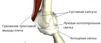
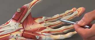
The structure of the joints of the human hand with drawings: elbow, shoulder, wrist, fingers
The structure of the joints of the human hand with drawings.
Normal functioning of the musculoskeletal system is impossible without articular tissue, which is covered with a synovial membrane and an articular capsule. Here is the structure of the joints of the human hand with drawings - elbow, shoulder, wrist, fingers:
The structure of the joints of the human hand with drawings
Elbow joint:
- It is divided into three sections: radial, humeral and ulnar.
- The wrist joint is a movable connecting link between the bones of the hand and forearm.
- It resembles an ellipse in shape.
- Performs a very important motor function - flexion and extension of the hand.
- The joint is strengthened by a large number of ligaments.
The structure of the joints of the human arm
Shoulder joint:
- It connects the bones of the shoulder to the shoulder blades.
- The shoulder joint is the most mobile joint in the human body, allowing movement without stiffness.
- The shoulder joint allows for circular movements, as well as flexion and extension of the arm.
The structure of the shoulder joint looks like this:
- Articular process of the scapula
- Head of humerus
- Joint space
- Acromion - acromioclavicular joint
There are many carpal joints, but they are smaller in size than those described above. Therefore, to make it easier to remember, they should be divided into several different groups. The classification of hand joints looks like this:
The structure of the joints of the human hand
- The midcarpal joint is the connection between the first and second lines of bones at the base of the wrist.
- The carpometacarpal joints are the connection of two rows of bones at the wrist with the bones that lead to the fingers themselves.
- Metacarpophalangeal joints - the connection between the phalanges of the fingers and the metacarpal bones leading to them.
- Interphalangeal joints - there are 2 of them on all fingers (except for the big one, since it has 1 such joint).
The structure of the tendons of the human hand is described below. Read on.
Treatment for hand pain
Treatment methods for arm pain directly depend on the causes that caused them:
- Treatment of cervical osteochondrosis and intervertebral hernia is aimed at eliminating the root cause of the disease and includes: various therapeutic and diagnostic blockades;
- intravenous infusions of drugs;
- radiofrequency ablation of individual nerves or joints.
- Treatment of arthritis requires eliminating inflammation, which helps minimize pain.
- Treatment of arthrosis consists of improving blood supply to joint tissues and stimulating collagen synthesis, which ultimately reduces or completely eliminates pain.
- Treatment of bursitis is primarily aimed at fixing the joint in one position. It includes medication, massage and physiotherapy.
Experienced specialists at the multidisciplinary CELT clinic have modern treatment technologies that will eliminate the problem causing hand pain, restore functionality and improve the quality of life.
Make an appointment through the application or by calling +7 +7 We work every day:
- Monday—Friday: 8.00—20.00
- Saturday: 8.00–18.00
- Sunday is a day off
The nearest metro and MCC stations to the clinic:
- Highway of Enthusiasts or Perovo
- Partisan
- Enthusiast Highway
Driving directions
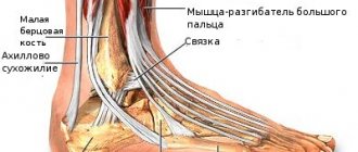
Anatomy of the structure of the human hand: tendons of the shoulder, forearm, wrist, hand, finger
Anatomy of the human arm: tendons
Tendons are connective tissue that allows the full transmission of muscle load. Anatomy of the structure of the human hand - tendons of the shoulder, forearm, wrist, hand, finger:
Tendons are divided into two layers:
- Deep
- Surface
More details:
- Each connection has its own bed, which is located between the soft tissues.
- Tendons provide soft gliding without friction and wear of the joints.
- The ability of the hand to perform its direct functions depends on their condition.
- The palmar part contains the largest part of the tendons.
- Superficial ones go to each finger of the hand.
- The deep tendons end at the level of the nail phalanx.
- The extensor tendons are located on the back of the hand under a small layer of fat.
The connection of tendons with muscle tissue occurs due to collagen structures that fuse with muscle fibers.
Possible injuries
The most common hand injuries include injuries to the hands in the area of the wrist and wrist joint , since it is not covered by muscle tissue. Athletes often experience dislocations and sprains in this area of the body.
Bone fractures in the hand area are not common. They are detected using radiography. The damaged area is cooled, anesthetized and immobilized with a plaster or fixing bandage. When a bruise occurs, the upper limb swells, so it is advisable to take decongestant medications. To shorten the recovery period, warming is used.
A fall on the arm can cause a dislocation . In this case, the blood vessels and nerve fibers running along the wrist joint are stretched or compressed. As a result, the victim experiences severe pain, numbness, and subsequently loss of sensitivity.
Valeria
General doctor
Ask a Question
You should not try to correct a dislocation in the wrist area on your own, as there is a risk of a fracture and worsening the condition. It is necessary to immobilize the hand, apply ice and take the person to a medical facility.
In addition to the bone elements in the hand area, the ligamentous apparatus is often injured . When a ligament is sprained/torn, the fingers may not lose mobility, but may move in unusual places, for example, bend in the opposite direction. A ligament rupture is an extremely painful injury, comparable to a broken bone. It leads to joint dislocation, requiring long-term treatment and recovery.
The structure of the skin of human hands: photo with description
The structure of the skin of human hands
Skin is the longest organ in the human body. Its main function is to protect against external negative factors. You see the photo with description above. Here is the structure of the skin of a person’s hands; it has three layers:
The epidermis is a thin stratum corneum that reaches a thickness of no more than 0.05 millimeters . Epidermal cells produce keratin. There are no blood vessels in the epidermis.
The structure of the epidermis includes:
- Stratum corneum
- Shiny layer
- Granular layer
- Spiked layer
- Basal layer
The basal layer contains substances responsible for the production of melanin. This substance protects the skin from aggressive sun rays and ultraviolet radiation. The cells of the basal layer are constantly dividing, which promotes renewal processes. Old cells change their shape and undergo the process of keratinization. They gradually peel off from the skin throughout a person's life.
The granular layer has a diamond shape, which is elongated parallel to the surface of the skin.
Dermis - by it we mean the inner layer of the skin, in which the sweat and sebaceous glands are located, which act as cleansers of the body from excess moisture and salts.
The hypodermis is a deep fatty layer that protects against cold and serves as the basic basis for the remaining layers.
It is worth noting:
The skin of the palm has distinctive features from all other parts of the body:
- Increased wear resistance
- There are no hair follicles or sebaceous glands in the palm
- There are many sweat glands on the skin of the palms
The skin of the hands is the main protector of our body, so it should always be given special attention.
The structure of nails on human hands: description
The structure of nails on human hands
Human nails are the most unique part of the human body. The anatomical structure is complex, but by studying it, you can learn a lot of interesting things. The body of the nail is located in the nail bed. Growth rate up to 4 mm per month. The nail is a dense, shiny and elastic coating that has a pink tint if the person is not sick. Read more about the structure of the nail in another article on our website at this link .