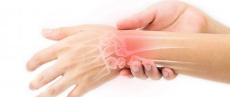
| X-ray of the hand at the German Family Clinic : research and interpretation of the image. Make an appointment for an x-ray by phone, (812) 337-12-12. Hand imaging is the very first study of the internal structure of the human body that was made by projecting the degree of attenuation of X-rays as an image onto film. At that time, William Roentgen did not yet know that even two centuries later, this diagnostic method would be the most informative in the case of injuries and pathological changes in joints and bones. In addition, it was discovered that x-rays of the hand make it possible to fairly accurately determine the anatomical age of any person, as well as his growth reserves. |
The condition of the joints of the hand can tell a specialist a lot, depending on which joints have undergone changes, the nature of the destructive changes, and the location of the pathologies. X-ray results can serve as an important indicator for diagnosis.
X-ray of the hand: when is it prescribed and what does it show?
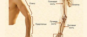
The bones of the hand are represented by the wrist, metacarpus and fingers. The wrist joint, formed by the radius bone, acts as the ligamentous apparatus of the hand.
X-ray of the hands allows you to:
- check the integrity of joints and bones;
- identify areas of inflammation in the bones (for example, arthritis);
- see osteophytes;
- detect necrotic processes in bone tissue, joint erosion;
- identify soft tissue calcification
- determine bone age
Thus, based on the X-ray images obtained, the diagnostician can make the following diagnoses:
- fracture, crack;
- dislocation;
- rheumatoid arthritis;
- osteoarthritis;
- arthritis (inflammation of the joints);
- systemic lupus erythematosus;
- calcification
In addition, using an X-ray of the hand, you can determine the structure of the skeleton. The fact is that in the event of the development of a systemic inflammatory (rheumatic) disease, it is the joints of the hands that will be the first to react. In this case, an x-ray of the hand will help to establish the stage of the inflammatory process based on a number of accompanying phenomena (thickening of the joints, calcifications, bone necrosis, osteolysis).
The most common diagnosis is a broken finger or fracture of the hand - more than 30% of all fractures. Moreover, this problem often occurs in children, on whose bodies radiation exposure is extremely undesirable. Therefore, parents doubt whether it is necessary to carry out an x-ray procedure if there are sufficient clinical signs of a fracture: swelling, cyanosis of the tissue, deformation of the natural shape, significant pain that intensifies during movement, deterioration in finger mobility. According to experts, an x-ray is still necessary in order to visualize the relative position of bone fragments and joints, and the condition of nearby tissues. Moreover, the symptoms of finger fractures are very similar to those of a dislocation or bruise. Therefore, in order not to take risks, but to prescribe adequate and timely therapy, an x-ray is necessary. The dose for this type of X-ray diagnostics will be only 0.001 mSv, which corresponds to the dose of natural radiation received in less than one day.
Classification of hand bone fractures
Depending on the presence or absence of skin damage over the fracture, the following are distinguished:
- closed fractures - the integrity of the skin is not compromised;
- open fractures - in the area of damage there is a wound in which bone fragments can be identified.
According to the position of bone fragments:
- without displacement - the broken bone maintains its position, the fragments exactly touch along the fracture line;
- with displacement - bone fragments diverge to the sides and, as a result, cannot heal along the fracture line without their comparison - reposition.
According to the involvement of articular structures in the fracture:
- extra-articular fractures - the fracture line passes outside the joint cavity;
- intra-articular fractures - the fracture line is located inside the joint cavity;
- fracture-dislocations - a violation of the integrity of the bone in combination with a dislocation in the adjacent joint.
According to the location of the fracture:
- wrist bone fractures;
- metacarpal bone fractures;
- fractures of the phalanges of the fingers.
You can also classify hand fractures depending on the number of fragments, degree of displacement, and infection. The etiology of the fracture is also important - whether it was traumatic or pathological - arising against the background of bone disease. All these factors influence the choice of treatment tactics for fractures and, ultimately, the possibility of completely restoring the function of the damaged hand.
Contraindications for X-ray diagnostics of hands
This radiation study can be performed for emergency indications even for pregnant and lactating women. Difficulties will arise when X-raying the hands of patients suffering from nervous disorders due to uncontrollability of hand movements (absolute immobility is required for an informative image).
As already noted, children are more affected by ionizing radiation. However, when it comes to diagnosing fractures or dislocations, the doctor may prescribe this procedure. Considering that the degree of radiation exposure is insignificant, parents should not worry. However, if possible, it is best to reduce your radiation dose by going to a clinic that has equipment that allows you to take digital X-rays.
Metacarpal fractures
The long, thin metacarpal bones are often broken by a punch or direct trauma. Muscle traction and movements in the hand until the fracture is immobilized often lead to displacement of bone fragments. There are epiphyseal fractures, when the fracture line is localized in the area of the bone heads, and diaphyseal fractures, when the fracture line is located in the bone body.
First metacarpal fracture
The cause is a blow with a bent first finger, less often a direct blow to the first metacarpal bone.
Fracture of the base of the first metacarpal bone . A typical injury for boxers and MMA fighters. A Bennett fracture is a separation of the base of the first metacarpal bone, which is held by ligaments, with simultaneous dislocation of most of it in the carpometacarpal joint. Rolando's fracture is a comminuted fracture-dislocation of the first metacarpal bone. Both injuries present with pain, deformation and swelling in the “anatomical snuffbox” area - the area under the base of the first finger - with increased pain when moving or trying to make a fist. Diagnosis is carried out taking into account complaints, trauma history, examination of the area of injury and x-ray of the hand. Bennett and Rolando fractures are treated surgically using osteosynthesis - restoring bone integrity by fixing fragments with metal knitting needles, pins or plates.
Fracture of the middle part of the first metacarpal bone . More often it occurs due to a direct blow to the bone. It manifests itself as pain, swelling and deformation in the area of the first metacarpal bone. The diagnosis is established taking into account the patient's complaints, information about the mechanism of injury, examination of the area of the first metacarpal bone and x-ray examination of the bones of the hand. Treatment is plaster immobilization for a period of 4-5 weeks; if the fragments are displaced, preliminary closed reposition is required. If conservative reduction is ineffective, an operation - pin osteosynthesis - is performed to compare the fragments.
An example of Dr. Valeev’s operation to restore after a fracture of the first metacarpal bone:
Before surgery:
After operation:
Fracture of II, III, IV, V metacarpal bones
The cause is a blow with a fist or a fall on fingers clenched into a fist. They can be single, but more often several metacarpal bones are broken, usually the fourth and fifth. It manifests itself as pain, swelling and deformation of the hand, and a hematoma often occurs. Diagnosed on the basis of complaints, history of injury, objective examination and X-ray results of the bones of the hand. To treat a non-displaced fracture, immobilization is performed for a period of 4-5 weeks. If fragments are displaced, closed reduction is indicated, and if it is ineffective, skeletal traction or pin osteosynthesis is indicated.
Advantages of X-ray diagnostics using digital equipment
The use of low-dose digital equipment has the following advantages, due to which a significant reduction in radiation exposure is achieved in comparison with the traditional film method:
- the process does not involve X-ray film or chemicals;
- the area under study is clearly limited - the beam flow is directed exclusively at the area being examined;
- the exposure time to X-rays required to obtain a high-quality image has been reduced;
- the clarity of the resulting images eliminates the need for repeated radiography due to the lack of information in the resulting image
In the German clinic, thanks to the use of modern equipment manufactured by GE, a reduction in radiation exposure is achieved in comparison with the traditional method of obtaining x-rays by almost 40%. Even the study of infants is permissible using such equipment. The price of an X-ray of the hand using digital equipment is quite affordable.
Preparing for an X-ray
To take an x-ray of the right and left hands, no preliminary preparation is needed. Before the procedure, you need to remove rings, bracelets, watches and place your hands (or arm) on the work table of the X-ray machine as directed by a radiologist.
To protect from the influence of ionizing radiation those parts of the patient’s body that are not involved in the study, special lead aprons or vests are used.
Determining a person's biological age using radiography
Based on the degree of ossification (bone formation), one can determine the biological age of a person. The name “bone age” is also used. The wrist joint and hand are made up of a large number of small bones that undergo changes with age. Experts, knowing a certain sequence of the ossification process, can draw a conclusion about the biological age of a person.
The ability to determine “bone age” is significant for endocrinology in order to diagnose growth pathologies in children (stunted growth, or vice versa), as well as a number of chromosomal abnormalities, ovarian and adrenal tumors and other diseases. If there is a suspicion of dysfunction of the parathyroid glands, expressed in a disorder of calcium and phosphorus metabolism, X-ray diagnostics of the hand will also help determine the cause and make a diagnosis.
The health of a child is an invaluable asset for loving parents. And if something goes wrong during the child’s growth, then you need to contact qualified doctors. For the fifth year in a row, the German clinic has been ranked first in the ranking of young clinics in the Pediatrics category. The clinic employs highly qualified doctors, and the diagnostic equipment is made by a world-famous manufacturer - GE. By contacting here, you will undoubtedly receive advice from competent specialists.
Wrist fractures
The bones of the wrist, due to their shape, structure and position, are broken quite rarely. The most susceptible bone to fracture is the scaphoid, the large bone at the base of the big toe. Injuries to the lunate and pisiform bones of the wrist also occur. The triquetral bone, as well as the bones of the distal row - polygonal, trapezoid, capitate and hamate - are subject to fractures extremely rarely; usually their fractures are combined with dislocations in the corresponding joints.
Scaphoid fractures
The cause is a fall on a bent hand, a blow with a fist, or a direct injury to the wrist. The following options are possible:
- intra-articular fracture of the scaphoid - the fracture line is located inside the cavity of the wrist joint;
- extra-articular fracture - separation of the tubercle of the scaphoid;
- de Quervain's fracture-dislocation - a simultaneous fracture of the scaphoid and dislocation of its proximal fragment and the lunate from the wrist joint.
Symptoms are pain and swelling at the base of the thumb, inability to move the hand at the wrist joint, or clench the hand into a fist. The diagnosis is established based on the patient’s complaints, data on the nature of the injury, examination and radiography of the hand bones. Sometimes, in the absence of displacement of fragments, the fracture line with all its signs is not determined. In this case, immobilization is still carried out with repeated radiography after 7-10 days, when, due to the activation of regenerative processes, the fracture line becomes clearly visible.
Treatment is immobilization with a plaster cast for a period of 4 weeks, followed by monitoring and prolongation of immobilization in case of insufficient consolidation of the fracture. In case of displacement of fragments and fracture dislocation, closed reduction is ineffective; fixation of fragments of the scaphoid bone with a wire is indicated. Fractures of the scaphoid are often complicated by the development of a false joint or lysis of bone fragments due to damage to the blood vessels supplying them during injury. Therefore, it is important to follow all the doctor’s recommendations and take control photographs in a timely manner to avoid complications and deterioration in the function of the wrist joint. After restoring the integrity of the scaphoid bone, physiotherapeutic treatment and exercise therapy are indicated to restore hand function.
Lunate fractures
The cause is a fall on a bent hand or direct injury, a blow to the wrist. It manifests itself as pain and swelling, intensifying with movements in the third, fourth and fifth fingers and with extension of the hand. The diagnosis is established taking into account complaints, the mechanism of injury, an objective examination of the area of damage and the results of an x-ray examination. To treat a lunate fracture, a plaster cast is applied for 4-8 weeks. Recovery usually proceeds without complications.
Fractures of the pisiform bone
The cause is a blow with the edge of the palm or direct injury. It manifests itself as pain and swelling of the wrist on the little finger side, increasing pain when it moves. The diagnosis is made taking into account complaints, anamnesis of injury, examination of the area of damage and radiography of the bones of the hand. For complete consolidation of a fracture of the pisiform bone, 4-5 weeks of immobilization are sufficient. The injury is rarely complicated.
How is the X-ray procedure performed?
An X-ray of the hand is obtained as follows:
- the patient sits down at the table;
- places one or both hands palms down;
- straightens the fingers as far as possible and brings them together;
- remains motionless for several seconds while the radiography process takes place.
X-rays of the fingers are done in the same way, but the fingers do not close. An X-ray of the hand can be taken in one projection (more often) or in two; fingers should always be examined in two projections.
Fracture of the phalanges of the fingers
The cause is a blow with the fingers, an injury while fixing the fingers, or a direct blow to the phalanges. Fractures of the phalanges of the fingers can be:
- intra-articular;
- extra-articular;
- single;
- multiple - within one finger or several;
- combined with dislocations in the metacarpophalangeal or interphalangeal joints.
Symptoms: pain, swelling, hematoma, deformity. The pain intensifies when you try to move your fingers. The diagnosis is established on the basis of complaints, trauma history, objective examination and X-ray results. To treat a fracture of the phalanges of the fingers without displacement, fixation is performed with a plaster cast for 3-4 weeks. In case of fracture-dislocations, joint reduction is performed; in case of displacement of fragments, closed reduction is performed. If it is not possible to compare the fragments using a closed method, skeletal traction or pin osteosynthesis is indicated.
