A false joint of the femoral neck is formed due to improper or incomplete fusion of the bone due to injury. The reason may be inaccurate comparison of fragments (in many cases it is almost impossible to do this using a bloodless method) and fixation in this form, disruption of metabolic processes, insufficient blood supply to the bone area, pinching of the joint capsule by fragments, early activation of the patient. The problem most often occurs after a femoral neck fracture in older people.
The process of the appearance of a false joint is quite long, it begins with smoothing and sclerosis of the ends of the fragments, sometimes a cartilaginous layer and even a certain volume of joint fluid is formed on the surfaces of nearby bone fragments. In other words, the resulting formation is called “pseudoarthrosis.” The femur, left without proper load, undergoes pathological changes: the normal structure of the tissue is disrupted, demineralization occurs, and signs of osteoporosis are visible on the pictures. The surrounding muscles atrophy, metabolic processes slow down even more.
Minimally invasive endoprosthetics in the Czech Republic: doctors, rehabilitation, terms and prices.
Find out more
Short description
False joint (pseudoarthrosis) is a violation of the integrity of the diaphysis of a tubular bone with the presence of pathological mobility.
Code according to the international classification of diseases ICD-10:
- M84.1 Fracture nonunion [pseudarthrosis]
- M96.0 Pseudarthrosis after fusion or arthrodesis
Frequency - acquired (post-traumatic) account for 2–3% of all fractures. Most often they occur on the tibia, forearm bones, and less often on the femur and humerus. Congenital ones are localized on the lower leg and account for 0.5% of all congenital pathologies of the musculoskeletal system.
Classification • By etiology •• Congenital •• Acquired • By type •• Fibrous pseudarthrosis without loss of bone substance •• Fibrous - synovial pseudarthrosis (true pseudarthrosis) •• Pseudarthrosis with loss of bone substance (bone defect).
3. Symptoms and diagnosis of false joints
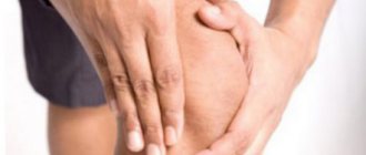
The site of bone nonunion is always clearly visualized. It is characterized by:
- swelling;
- mobility;
- muscle atrophy;
- scarring and proliferation of connective tissue.
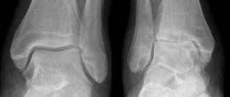
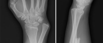
The clearest picture can be obtained from an x-ray, which clearly shows: the existing gap between the processes of the bone, the closure of the bone marrow canals, and the sclerosis of the bone endings.
An essential criterion for diagnosing a “false joint” is the time that has passed since the fracture. Such injuries have certain time standards for the rehabilitation period. If the period has expired and fusion has not occurred, then the fracture is assigned the status of ununited or slowly healing. If the period is exceeded twice, a diagnosis of “false joint” is made.
About our clinic Chistye Prudy metro station Medintercom page!
Causes
Pathogenesis. A false joint (post-traumatic) is one of the forms (or stages) of imperfect osteoregeneration. Formally, delayed consolidation is distinguished - a condition when, after a fracture, the average period necessary for healing of a fracture of a given localization has passed, but consolidation is not clinically and radiologically determined. A false joint is a condition when, after a fracture, twice the average time required for its fusion has passed, but there are no signs of consolidation. This division, despite a certain convention, is of great clinical importance. Slow consolidation allows hope for fusion. The diagnosis of pseudarthrosis excludes fusion without additional stimulation and is actually an indication for surgery.
The development of congenital pseudarthrosis is based on neurotrophic disorders. In fibrous dysplasia, the cause of the development of a pseudarthrosis is the presence of foci of poorly differentiated bone tissue, which is unable to perform an adequate supporting function.
Risk factors. General and local factors can be identified that predispose to the formation of a pseudarthrosis.
• General •• Endocrine diseases and pathology of metabolism, especially electrolyte •• Infectious diseases •• General circulatory disorders (shock, blood loss) •• Innervation disorders •• Combined trauma and multiple fractures •• Combined lesions
• Local •• Extensive tissue destruction •• The presence of diastasis between fragments and tissue interposition •• Unstable osteosynthesis •• Inadequate exercise therapy regimen •• Local infectious process •• Local circulatory disorders (for example, femoral neck).
2. Reasons for the formation of false joints
There are two groups of reasons causing the formation of a pseudarthrosis: local and general.
Local disturbances are usually associated with mechanical damage. Moreover, these disorders can occur at different stages after injury. A number of local disorders that provoke the formation of a pseudarthrosis are associated solely with the severity of the injury. These are open fractures with exposure of bone tissue, injuries with loss of a significant part of the bone, crushing of a muscle over a large area, osteomyelitis, purulent complications, trophic disorders as a result of damage to blood vessels and conductive nerves.
Another group of local disorders covers causes associated with improper management of patients after a fracture. These include errors and shortcomings in the treatment and regimen, insufficiently careful comparison of bone fragments, immobilization disorders, incorrect selection of a fixator, and excessive motor activity of the patient himself after injury.
A special group includes local causes due to the peculiarities of the anatomical structure of the areas where the bone fracture occurred.
General (trophic) causes are a combination of internal factors and background conditions that impede the process of proper bone fusion. These include:
- presence of chronic infections;
- metabolic disorders, especially those related to calcium-phosphorus metabolism;
- endocrine diseases;
- vitamin deficiency and general weakness;
- reduced immunity;
- vascular insufficiency;
- trophic complications at the site of injury.
Visit our Traumatology and Orthopedics page
Treatment
Treatment • Conservative (stimulation of repair, osteogenesis) is ineffective • Surgical - preference is given to low-traumatic treatment methods - compression - distraction osteosynthesis • If ineffective - resection of the ends of fragments with subsequent lengthening (bilocal compression - distraction osteosynthesis); bone grafting with a graft on a feeding pedicle using microsurgical techniques • When treating congenital pseudarthrosis, the treatment is complex - medication and physiotherapeutic treatment aimed at improving neural trophism in combination with low-traumatic types of immobilization or bone grafting.
ICD-10 • M84.1 Fracture nonunion [pseudoarthrosis] • M96.0 Pseudoarthrosis after union or arthrodesis
Pseudoarthrosis or false joint
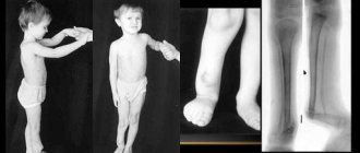
The percentage of victims of this pathology is quite high - 10% of the total number of limb fractures. Often, the formation of pseudarthrosis occurs in the lower leg, but it happens that it appears on the hips or shoulders.
Pseudarthrosis formation
As a rule, pseudarthrosis is the result of faulty bone healing after a fracture. But such a pathology can also be congenital. If the fetus did not receive enough nutrition to form bone, the result could be the formation of a pseudarthrosis.
Diagnostics
But how to diagnose a false joint? It is enough to take a picture with an X-ray machine - it will show whether there is a hole between the bone fragments and whether there is connective tissue in it. An x-ray is usually taken if the bone has not recovered after two periods of time for healing, and tissue fusion has not occurred. Pseudoarthrosis can be easily distinguished from a simple fracture that has not healed by the presence of a plate of bone blocking the medullary canal. As a result, cartilage tissue is formed, which, in turn, ensures the mobility of the false joint. This joint does not function because the ligaments and muscles do not support it.
Classification of pseudarthrosis
There are several classifications of pseudarthrosis, but the most common is based on its formation:
- the place where the bones grow together hurts and its mobility is abnormal;
- a feature of fibrous pseudarthrosis is the proliferation of fibrous tissue;
- non-ctoric is characterized by circulatory disorders;
- true. It is very similar to the real one (hence its name), most often affects long bones and is formed due to the appearance of a synovial capsule;
- hypertrophic (joint mobility is low, bone tissue grows at the ends of the fragments).
Causes
The main cause of pseudarthrosis is a violation of the process of bone fusion after injury. As a rule, bones do not heal properly due to incorrect treatment. The following factors add to the problems and have a strong influence on the formation of a pseudarthrosis:
- infected fracture;
- blockage of the bone marrow canal with fragments;
- the cast was not worn enough;
- violations of immobilization;
- lack of a clear logic of treatment or a combination of these methods;
- destruction and disruption of bone during surgery;
- penetration of soft tissue or foreign body between fragments;
- circulatory disorders.
It also happens that damaged bones do not heal properly due to the fact that the body has a certain physiological feature.
Pseudoarthrosis is a disorder that also has some risk factors for its development. For example, metabolic disorders can significantly impair the process of bone fusion, which will lead to the formation of a false joint. In addition, the presence of chronic bone diseases such as osteoporosis or rickets, an unbalanced diet, problems with the endocrine system, chronic infections and cancer can contribute to the development of pseudarthrosis.
As a rule, the most common site of occurrence of a pseudarthrosis is the femoral neck. There are many factors involved, such as old age and osteoporosis, that make this disease common. In addition, this feature can occur in the tibia, radius or shoulder.
How to determine the disease
Any pathology is characterized and described by a number of signs. They are not detected immediately, but after the plaster is removed. These include:
- shortening or deformation of the injured limb;
- physical activity causes pain, while pain is not felt when touched;
- bone mobility is observed in usually immobile places;
- muscles are weakened;
- muscle atrophy due to lack of movement.
Methods for diagnosing pseudarthrosis
After palpation, examination and conversation with the patient, the doctor, as a rule, prescribes an X-ray examination. The picture is taken in two projections; it usually shows the main signs showing pseudarthrosis. These include the absence of callus, the ends of the bones are smooth and sometimes rounded, there is a distance between the bones that is noticeable, and the presence of a depression. Sometimes X-rays are not enough, in which case a computed tomography scan or examination using isotopes is performed.
Treatment of false joints
When treating pseudarthrosis, an individual approach and a method suitable for a particular case are important. The main goal is to restore the bone and correct its deformity. As a rule, surgery comes to the rescue. There are several methods for treating pseudarthrosis, which also depend on the pathology. Atrophic pseudarthrosis is treated with bone grafting - cartilage tissue is removed, due to which doctors achieve maximum convergence of the bone fragments. However, it is only carried out if there is no infection. Hypertrophic pseudarthrosis is treated with stable osteosynthesis. Surgery is not necessary here. There is also a treatment method called compression-distraction osteosynthesis.
An important step is immobilization. The limb is immobilized for more than six months. Of course, this is not enough to restore bone health, so other treatment methods are also used together.
To avoid the occurrence of this disease, you need to follow all the doctor’s recommendations, because it is better to prevent the disease. However, if there is a predisposition and pseudarthrosis does appear, it is important to start treating the joints
.
Author: K.M.N., Academician of the Russian Academy of Medical Sciences M.A. Bobyr
False joint of the femoral neck
The continuous development of the disease, the deterioration of the patient’s quality of life and his condition is a reason to begin treatment. Without therapy, the patient will experience disability. This is not a congenital pseudarthrosis, treatment recommendations for which may be conservative. Such pseudarthrosis occurs in the lower leg and affects the tubular bones. Problems with the femoral neck are more serious and require urgent treatment.
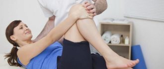
For false joints, clinical recommendations depend on the patient’s condition, his age, and the complexity of the situation. In some cases, surgery is completely ruled out. Among conservative methods, the patient can be offered hardware treatment, physiotherapy, and gentle massage. If the patient is young, then taking special medications can still start the processes of osteosynthesis.
Orthopedic treatment methods for false joints involve the selection and installation of special endoprostheses. This method of treatment is effective in most cases and helps even those patients who have long suffered from pseudarthrosis. An endoprosthesis is a replacement for a damaged joint, which is made of modern materials. The risk of rejection of such materials is eliminated.
After installation of the endoprosthesis, the patient requires rehabilitation measures so that he can normally restore motor functions of the leg. The new prosthesis works correctly, it loads the muscles and tendons. The correct position of the leg for support is created.
Other treatments
For pseudarthrosis, surgery can offer other treatment methods: installation of autografts, open reduction, reconstructive surgery. The autograft is obtained from the tibia, the material is formed into a rod, which is passed through the femoral neck. The fragments are connected using a rod. The method is suitable for minor damage and preservation of the head of the femoral neck.
The method of intra-articular osteosynthesis by reduction in an open manner involves fixation of the fragment using a pin. Fibrous tissue is removed and the surgical site is completely cleaned. If the head of the femoral neck is completely destroyed, then reconstructive surgery is recommended. During this treatment, reconstruction of the neck and head is performed.
If the described methods do not help, then endoprosthetics is performed. This method has no alternative and is considered the most effective even with serious complications.