directions
The ankle joint is one of the most complex parts of the human musculoskeletal system. Anatomically, this joint consists of vessels, muscles, nerves, tendons and ligaments. The ankle joint is responsible for stability of the body as well as mechanical movement. It is involved in activities such as walking, running, squatting, jumping, etc. This is a plastic and very mobile joint, but at the same time vulnerable to various types of injuries and diseases. Inflammation, pain and other ailments most often affect the ankle joint, especially during intensive professional sports, where the risk of injury is high.
Our clinics in St. Petersburg
Structural subdivision of Polikarpov Alley Polikarpov 6k2 Primorsky district
- Pionerskaya
- Specific
- Commandant's
Structural subdivision of Zhukov Marshal Zhukov Ave. 28k2 Kirovsky district
- Avtovo
- Avenue of Veterans
- Leninsky Prospekt
Structural subdivision Devyatkino Okhtinskaya alley 18 Vsevolozhsk district
- Devyatkino
- Civil Prospect
- Academic
For detailed information and to make an appointment, you can call +7 (812) 640-55-25
Make an appointment
We draw your attention to the schedule of technological breaks in the CT and X-ray rooms.
In this case, radiography of the ankle joint helps to make or clarify the diagnosis, see pathological processes and changes.
Today, despite the large number of diagnostic methods, radiography is still not losing ground and remains one of the simplest, most accessible, and informative ways to diagnose diseases of the musculoskeletal system, in particular the ankle joint.
Getting an X-ray examination of the ankle joint in St. Petersburg is not particularly difficult. X-ray diagnostic machines are available in almost all medical institutions. But if you want to take a high-quality x-ray with good and safe equipment, then get advice from a highly qualified specialist, and avoid sitting in long lines, come to a multidisciplinary medical center. The trauma department of the Medical Center accepts patients with various injuries and diseases any day of the week. The clinic's emergency room is equipped with a modern digital X-ray machine Clinomat, which allows you to take full X-ray images.
Dislocations, bruises, ankle fractures, pain, swelling and other unpleasant symptoms and ailments require a careful examination by a specialist and a diagnostic study in the form of x-rays.
Where to get tested
Examination by a traumatologist
If you receive an injury, you should immediately contact a traumatologist. He orders an on-site examination. Typically, emergency rooms are equipped with such devices. If you wish, you can go to a private clinic, where it is possible to conduct a paid examination and take an X-ray of the joint for a fee without a doctor’s referral.
The price of such an examination ranges from 300 to 600 rubles. This is a fairly low price for a hardware examination, considering how much an ultrasound or computed tomography costs.
The image is given to the patient or doctor immediately after the examination. No description is required; the doctor gives his conclusion based on the contours and features of the location of the bones. A traumatologist or orthopedist interprets and analyzes the photo of the joint.
Indications for ankle radiography
Most often, an X-ray examination of the ankle joint is prescribed in the following cases:
- fractures of the tibia, cracks;
- ankle fractures;
- fractures of the base of the fifth metatarsal;
- fractures of the finger phalanges;
- bruises;
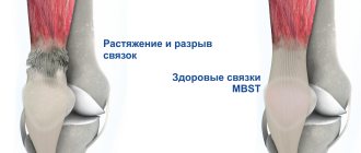
- damage to cartilage, ligaments;
- tendon ruptures;
- dislocations and subluxations;
- flat feet;
- hallux valgus;
- arthritis, arthrosis;
- rheumatoid arthritis;
- osteoporosis;
- osteoarthritis;
- osteomyelitis;
- gout;
- tumor processes;
- necrosis of the phalanges of the fingers, etc.
Often, pain in the ankle joint can appear several years after the injury and make itself felt in bad weather, during temperature changes and poison a person’s life, hindering movement, causing him to suffer from aching pain, etc.
The above ailments arise due to injuries, autoimmune diseases, immune failures, genetic predisposition, hormonal changes, past infectious diseases, excess weight and other negative factors.
Main symptoms for an x-ray
X-ray of the ankle joint
needed when a number of signs indicating the presence of pathological processes appear. X-rays provide clear images of bone tissue and reveal the cause of the disease and its severity.
Location, shape and size of bones
The shape and placement of bones relative to each other can be disrupted due to congenital pathologies or acquired injuries and damage. Thickening called “hyperostosis” may occur in areas of the bones. They are a consequence of pathological growth of the periosteum, inflammation or poisoning of the body. Also, an x-ray of the lower leg in different projections, including the lateral one, can show that the bone is underdeveloped or atrophied.
Conditions of bone surfaces
An inflammatory or tumor process can cause destruction of the outer part of the bones. The reverse process is not uncommon; ossification of areas of the periosteum and their calcification periodically occur. This process is called "periostosis" or "periostitis", depending on what caused the disease. Also, sometimes the periosteum peels off from the bone, which is clearly visible on an x-ray of the ankle joint.
Actually, bone structures
The procedure allows you to detect violations of bone integrity. Often the cause of structural changes is osteoporosis. In this case, there are much fewer bone beams per unit volume of bones. The tissue in the images is more transparent; an enlarged bone marrow canal is revealed. Osteosclerosis is characterized by an increase in the number of bone beams. The x-ray of the lower leg will differ from the norm; osteolysis or necrosis will also be visible in the images.
Joint space
This process is a consequence of injury or dystrophy and is characterized by a narrowing of the joint gap. Such a change, noticeable in a photo of an x-ray of the lower leg in frontal or lateral projection, may indicate the development of arthritis or arthrosis. The consequence of untimely treatment can be ankylosis - fusion of joints. The disease can be of fibrous and bone type.
Apical fracture of the outer ankle
This is an injury in which only the fibula bone is broken, while the tibia remains functional. The person can walk and only sometimes feels pain. There is not too noticeable swelling. It happens that people do not even seek help from a specialist, believing that the injury is not dangerous.
But that's not true. Then, during additional studies, negative changes in the nerve may be discovered. It is better to immediately complete the entire course of the study and receive adequate treatment.
Information content of radiography
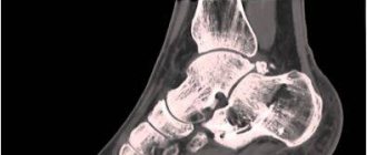
The essence of the method is that the diseased organ is exposed to x-rays. As a result, the internal structure of objects is projected onto X-ray sensitive film. In some modern devices, X-rays are projected onto an electronic matrix. Such devices are much more expensive than analog ones, and accordingly, the fee for the procedure increases.
The image on the radiograph is two-dimensional. The denser the structure of an object, the more clearly it is visualized. Thus, information about the condition of bone tissue is characterized by a high degree of reliability and accuracy. Pathological changes can affect all elements included in the structure of the ankle joint. These include:
- Supracalcaneal (talus) bone. It forms the lower part of the ankle. The surface of the bone is surrounded by cartilage, and its task is to distribute the weight of the human body throughout the foot.
- Fibula. In the lower part it has articulation with the supracalcaneal bone. The distal end is the outer malleolus of the ankle.
- Tibial. In its lower part, this bone forms the medial malleolus, enclosing the supracalcaneus.
There is a group of elements that are not so clearly displayed on an x-ray. These are internal and external ligaments, distal tibiofibular syndesmosis, Achilles and other tendons, blood vessels, flexor and extensor muscles.
In case of joint damage, it is important to diagnose the problem in time. Otherwise, the mobility of the foot will be limited, and the quality of life will significantly deteriorate.
Ability to work after surgery
Due to swelling and severe pain, the victim has the right not to go to his main place of work for at least 10 days. Then a medical commission meets and decides to close or extend the sick leave. But the patient cannot stand on his leg for at least 3-4 weeks. The timing of removal of plaster or splint depends on the speed of fusion and on the individual characteristics of the body.
So, the splint, when the fracture heals well, will be removed only a month after the ankle fracture without displacement. But even in this case, one cannot speak of good performance if a person earns his living by physical labor. However, if he is busy with intellectual work, he can return to work in three weeks, but he must not skip physical therapy classes.
Fracture with displacement and without displacement
The clinical picture depends on the severity of the injury. A unimalleolar fracture, even without displacement, looks like a minor ligament tear.
How to identify a non-displaced ankle fracture?
- The hemorrhage may be local or may not exist at all.
- Leaning on your leg is often possible, but painful.
- A severe fracture sometimes causes swelling, which can spread greatly.
- There is a symptom of pain irradiation.
The pain can be sharp or cutting. But always intense. Although each patient feels pain differently.
What is a symptom of irradiation? This symptom occurs when the traumatologist presses his fingers on the leg a few centimeters above the fracture, the patient feels acute pain in this very place.
If the fracture is displaced, the joint is deformed. It is obvious to the eye that an angle has formed between the foot and the lower leg. A person cannot move independently. When closed with displacement, the bone can penetrate into the muscle. When soft tissue is damaged, a large hematoma is discovered. If the fracture is open, there will be bleeding that needs to be stopped. Heavy bleeding leads to hypotension and loss of consciousness.
Anesthesia for ankle surgery
During planned trauma surgery, regional anesthesia is usually used. However, the age of the victim, the presence of chronic diseases, and whether alcohol is present in the blood are taken into account. Doctors know that the condition of a patient with severe blood loss worsens with regional anesthesia, and therefore it is better to use general anesthesia with mechanical ventilation.
How is medicated sleep ensured? The person is administered one of the following drugs: Diazepam or Phenazepam from benzodiazepines, possibly Propofol. But it is contraindicated to use Ketamine, since this substance is a hallucinogen. The anesthesiologist performs a femoral and sciatic nerve block. Sometimes it is also necessary to relax the patient’s muscles; muscle relaxants are used for this purpose.
The patient should be monitored for the first time after recovery from local or general anesthesia. The victim may exhibit some residual effects after the administration of chemotherapy.
Massage for injuries and fractures
When and how many times can you massage your leg? Massage on the ankle can be done from the first days of applying the plaster. Warming up soft tissues is certainly beneficial, as it improves blood flow and significantly improves skin tone. Soft tissues suffer during a fracture and need care. This massage can be performed by both the victim’s relatives and himself, if others do not have the time. However, this must be done carefully and correctly so as not to cause harm.
In general, all physical procedures, proper diet and massage help speed up the time after an ankle fracture when you can step on your foot.
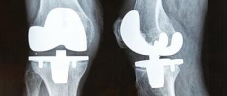
Surgical intervention. Indications. Recovery time
From the moment the victim is admitted to the hospital, within 3-4 days he should undergo surgery if he needs it. During this time, the doctor must study all the data and plan how the bone will be collected.
In general, there are the following types of operations:
- Osteosynthesis of the medial malleolus. The indication for surgery is supination fractures. The ankle is installed using a special nail at a right angle.
- Osteosynthesis of fragments of the tibia.
- Fixation of the tibiofibular joint. When there is a fracture of both the medial ankle and fibula.
- Osteosynthesis of the lateral malleolus. This is how pronation fractures are operated on.
Why are plates used? Sometimes, in an accident, for example, the bones are shattered, and the traumatologist has to collect the fragments with screws and give the ankle its anatomical shape again. Then the ligaments have to be sutured.
When an x-ray reveals an avulsion from the inner malleolus to compare the fragments, a titanium plate is placed. It holds the bones well and helps for a while. But it is still a foreign object in the leg, and therefore it needs to be removed as quickly as possible. Usually the metal is removed sometime after 3-5 months after surgery.
Surgery is also indicated when the ankle has not healed properly and the patient feels constant pain at the fracture site. If treatment is delayed, the patient will feel worse and his recovery period will be significantly delayed.
After the operation to restore the bone is completed, the doctor adjusts the plaster so that the wound can then be washed. After all, the wound is disinfected every day for a long time.
It happens that bones heal poorly, especially in old age. Then the bone grows together in six months or a year. This means that you need to go for a consultation with an endocrinologist. Poor thyroid condition leads to problems with bone healing. This means that you need to take additional iodized medications.
Ankle fracture. Rehabilitation
After the plaster or splint is removed, it takes a long time to take care of recovery. The person will attend rehabilitation courses and perform certain exercises to train the muscles.
First, light exercises are performed to flex and extend the foot. At first, approximately 3 weeks after the injury, small movements of the feet can already be done, but by placing the limb in a basin with warm water. Warm water will make it less painful.
Then you need to gradually complicate the tasks. If the rehabilitation course takes place at home, you can use different objects for exercises - tapes with which you gently stretch the toe of the foot or a sewing machine with a foot pedal.
Doctors advise doing exercises with a ball. For exercise therapy, you need a chair and a ball or half-deflated ball, which the victim must roll on the floor with his sore leg. Then you need to try to grab the ball with both feet and lift it. This is a more difficult level.
In general, it is better for exercise therapy to be conducted by a special instructor. During the training process, it is important not to harm the leg by overdoing it. You need to take these classes to restore normal ankle mobility and normal gait.
In addition to therapeutic exercises, the victim is also prescribed electrical stimulation of muscles, paraffin baths, hydrotherapy, massage and more. Why is rehabilitation needed for an ankle fracture? All these procedures relieve pain, help resolve hematomas and improve the overall metabolic process in tissues.
Also, during the recovery period, the victim needs to eat more foods containing potassium, calcium and phosphorus. But you need to be careful with the amount of phosphorus. Since its excess does not lead to improvement, but to deterioration in calcium absorption.
The need for diagnostics
For various reasons, specialists from different fields of medicine may refer you for an ankle x-ray. Most often, radiographs are taken for traumatologist patients. In such cases, the procedure allows us to identify the following problems:
- inflammation of the synovial membrane;
- dislocation;
- fracture;
- cracks;
- congenital pathologies;
- heel spurs;
- degenerative changes;
- arthrosis;
- osteophyte;
- arthritis;
- flat feet.
- inflammation of the synovial membrane;
- dislocation;
- fracture;
- cracks;
- congenital pathologies;
- heel spurs;
- degenerative changes;
- arthrosis;
- osteophyte;
- arthritis;
- flat feet.
The surgeon writes out a referral for an x-ray when it is necessary to determine the extent of the pathological process that has developed for various reasons, for example, due to diabetes mellitus. The oncologist refers patients with cancer to x-rays of the ankle. The goal is the same: to assess the current condition of the joint. Patients of an orthopedic surgeon undergo an X-ray examination in cases where there is a suspicion of flat feet or another type of foot deformity.
Impaired flexion and rotation of the ankle
It happens that after the plaster is removed, it is discovered that the fusion did not occur correctly. In this case, some adverse consequences are possible. For example, developing arthrosis of the ankle. Over time, this leads to difficulty flexing and straightening the foot, and difficulty walking.
To ensure that no problems occur, the doctor must check several times to ensure that all the bones fit together accurately after reducing a displaced ankle fracture. To do this, X-ray images of the limb are taken each time. Only if everything is correct, then the plaster is securely fixed and the patient is sent home to rest.