directions
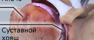
Shoulder pain is not always associated with injuries caused by falls or accidents. The shoulder joint experiences a huge daily load, which, due to its anatomical structure, leads to injuries such as dislocations, subluxations, sprains, etc. The shoulder joint itself is a rather complex structure of the human body, consisting of fibers not strengthened by muscles, which allows the arms to be mobile and perform the functions of movement, rotation, tilt, extension, and flexion. At the same time, such a design leads to frequent injuries and diseases of the joint.
Currently, work is underway on the website to change the price list; for current information, please call: 640-55-25 or leave a request, and an operator will contact you.
Our clinics in St. Petersburg
Structural subdivision of Polikarpov Alley Polikarpov 6k2 Primorsky district
- Pionerskaya
- Specific
- Commandant's
Structural subdivision of Zhukov Marshal Zhukov Ave. 28k2 Kirovsky district
- Avtovo
- Avenue of Veterans
- Leninsky Prospekt
Structural subdivision Devyatkino Okhtinskaya alley 18 Vsevolozhsk district
- Devyatkino
- Civil Prospect
- Academic
For detailed information and to make an appointment, you can call +7 (812) 640-55-25
Make an appointment
We draw your attention to the schedule of technological breaks in the CT and X-ray rooms.
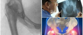
The nature and extent of injury, the presence of disease and pathology can be determined by such an informative diagnostic study as radiography of the shoulder.
In St. Petersburg, you can have an X-ray of the shoulder joint done in almost any medical institution - from a clinic to a hospital and a multidisciplinary medical center. But if you want to save time without wasting it in queues, or you need urgent examination and emergency medical care due to an injury, if you want to have an X-ray of the shoulder joint done with good and safe equipment, then sign up and come to the medical office. The trauma department of the clinic is equipped with the latest digital X-ray device Clinomat (made in Italy), which allows us to quickly, efficiently and fully provide all types of X-ray examinations.
Is it possible to do CT scans for children?
In most clinics, a CT scan of the shoulder joint in St. Petersburg is possible only if the child has reached 5 years of age, and if contrast is used - 12 years. A child's body is susceptible to radiation 5 times more than an adult's, so compelling reasons are needed to conduct the study. The absolute vital indication is a suspicion of a malignant tumor with metastatic lesions of the skeletal bones. In all other cases, the expected benefit should be higher than the potential harm, so it is better to undergo diagnostics without the use of radiation exposure. A CT scan of the shoulder joint can be performed on a child only with the direction of a doctor or consultation.
Two important conditions are immobility during the examination and preserved nasal breathing. If a child has a runny nose, it is better to postpone diagnosis or pre-drip vasoconstrictor drops into the nose.
Before the study, you need to have an explanatory conversation about the safety of the procedure and the need to strictly follow the specialist’s commands.
In what cases is an x-ray examination of the shoulder prescribed?
An X-ray of the shoulder joint is prescribed by a doctor if there is a suspicion of a fracture of the neck (surgical and anatomical) of the humerus, dislocation of the head of the humerus, damage to the clavicle, the presence of joint diseases such as arthritis, osteoarthritis, etc. X-ray of the shoulder joint is also done in case of restriction of movement of the upper limb, pain during movement, swelling, crunching, injury (suspicion of dislocation or fracture) of the shoulder, to exclude tumor processes, foreign bodies, malformations and congenital pathologies, etc.
Preparing for X-rays
When planning a procedure, the patient is recommended to wear loose clothing that can be easily removed. At the same time, there should be no metal fasteners or decorative elements on it. It is better to leave all jewelry at home - any metal object can distort the results of the study.
If radiography with the introduction of a contrast agent is prescribed, then there are some restrictions on the consumption of food and liquids - a “starvation” diet is recommended for 4-6 hours. Additionally, it is necessary to conduct an analysis of the body's sensitivity to a certain type of contrast agents.
What does an x-ray of the shoulder joint show?
On an X-ray of the shoulder joint, the radiologist will be able to see various damage caused by injuries or diseases, confirming or refuting the diagnosis. For example, X-ray signs of diseases such as arthrosis, arthritis, etc. clearly visible on an x-ray of the shoulder. The doctor will be able to examine in detail and evaluate the condition of the humerus, scapula, the distance between the bones that form the joint, the contours and condition of the joint space, etc. Based on the X-ray findings, the attending physician makes a diagnosis and develops a treatment regimen.
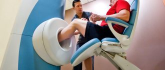
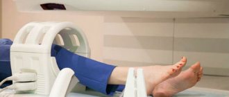
Features of CT scan of the shoulder
A CT scan of the shoulder joint does not require special preparation, but it is better to come to the diagnostic center in loose clothing without metal fasteners, since metal on tomograms gives a false shadow, and it will be uncomfortable to lie in tight clothing. If you plan to administer contrast, it is necessary to clarify the level of creatinine in the blood: kidney disease with impaired function or an acute process is a contraindication to the use of the drug. The amount of contrast agent is calculated based on body weight. At a diagnostic clinic in St. Petersburg, before performing a CT scan of the shoulder joint with contrast, a screening test for creatinine can be done. The result will be ready in 10 minutes. Patients with diabetes mellitus, after approval from the endocrinologist, should adjust their therapy 48 hours before undergoing an enhanced CT scan of the shoulder joint. When performing a CT scan of the shoulder joint, iodine-containing substances are used. The doctor must be warned in advance about allergic reactions to contrast: urticaria-type rash with skin itching, Quincke's edema, anaphylactic shock (swelling, difficulty breathing), in this case, a CT scan of the shoulder joint is performed without administering the drug. Hunger can provoke autonomic reactions: dizziness, palpitations, sweating, etc., so you should come to the procedure after a light snack. Take the results of previous studies (ultrasound, radiography, CT/MRI) with you, this will allow the doctor to get an idea of the dynamics of the process.
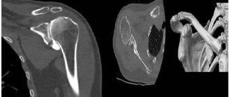
Photo of CT examination of the shoulder joint
The procedure for performing an X-ray examination of the shoulder joint
When performing an X-ray of the shoulder joint, the patient must undress to the waist. Depending on the type of projection (direct or lateral), the patient lies on a special table, or the picture is taken in a standing position. The patient is covered with a lead apron, which protects the person from the negative effects of X-rays. The duration of the procedure is several minutes. After this, the image is processed, a radiologist describes the image and issues a conclusion, the patient is sent to the attending physician or to recommended specialists.
Operating principle
X-ray of the shoulder joint is a research technique characterized by fairly high accuracy and accessibility. Provided that the image is taken correctly and is deciphered by a qualified specialist, there is no reason to doubt the correctness of the results obtained.
The method is based on the use of X-rays of a given range aimed at the area under study. Since soft tissues are able to transmit X-rays and hard tissues are able to absorb them, a clear image is obtained. In this case, all soft tissues in the image will be dark, and hard tissues will be light. This is a key factor why radiography is used when diagnosing pathologies of bones and joints. If it is necessary to study the condition of soft tissues or the vascular system, another diagnostic method is prescribed - these objects are not visible on an x-ray.
Procedure
The shoulder X-ray procedure takes on average 15 minutes, although the actual scanning process takes less than a second. During the procedure, the patient is wearing a lead apron to protect the reproductive organs.
An X-ray of the shoulder can be performed either in an X-ray room or using portable X-ray machines, which are used in operating rooms or intensive care units.
The technician or radiographer positions the patient (lying or standing) and then goes into the adjacent equipment room. As a rule, two photographs are taken - in frontal and lateral projection. When taking pictures, the patient needs to hold his breath for a short time. Stillness is necessary to avoid blurring of the image.
Indications and restrictions
The main conditions under which an X-ray of the shoulder joint is indicated:
- pain in the shoulder area;
- partial loss of motor activity in the shoulder joint;
- dislocations or subluxations;
- arthritis or arthrosis (change in skin color in the area of the shoulder joint);
- the need to assess the dynamics of the chosen treatment method;
- formed neoplasms;
- the need to monitor congenital pathologies in the structure of the shoulder joint.
Often x-rays are performed despite the absence of direct indications. This category of people includes athletes and people whose work involves high physical stress on the shoulder joint. To prevent the development of pathological processes in the connective and bone tissue of this category of citizens, it is recommended to undergo x-rays at least once every six months.
The need for radiography is determined by the attending physician, taking into account the likely harm that can be caused to the body by the radiation dose.
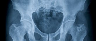
Prosthesis (direct projection)
Relative contraindications to the procedure include age under 15 years. During pregnancy and lactation, examination is prohibited and can only be carried out in extremely serious cases.
During the procedure
The patient will not feel anything during the test. The x-ray room may be cool due to the air conditioning used to operate the equipment.
The positions required to take x-rays may be uncomfortable for the patient, but the patient will only have to remain in this position for a few seconds.
After the x-rays are taken, the patient will be asked to wait a few minutes because if the images are blurry, the x-rays will have to be repeated.
Why do you need an x-ray?
Shoulder (image is normal)
The image visualizes manifestations of pathologies: violation of the integrity of the bone, growth of cartilaginous elements along the edges, areas of salt deposits, etc.
Through the procedure, complications and secondary pathologies associated with diseases of other organ systems are identified. Radiography helps plan the therapeutic course and monitor its course.
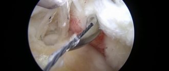
Titanium plate for a clavicle fracture
The technique does not demonstrate a pathological condition in all cases. Probably, during the examination, changes are observed in the soft tissue; they are not visible on the negative.
Common joint pathologies
The cause of pain in the shoulder can be both injuries and various diseases (mainly rheumatoid-inflammatory). The table below lists a number of the most commonly diagnosed pathological conditions for which it is recommended to take an X-ray of the shoulder joint:
| Pathology | Characteristic |
| Dislocation | A common occurrence is anterior dislocation. As a result of the injury, the bone head pulls out the articular lip from the articular scapular “fossa”. Dislocation occurs due to injury or awkward movement |
| Fracture | It is observed when the integrity of bone tissue is violated. Possible injury to the neck or tubercle of the joint, fracture (crack) of the bone. Occurs under the influence of external force |
| Arthrosis | Dystrophy of bone tissue and cartilaginous inclusion of the joint. There is further deformation of the structural elements, impairment of the functional ability of the hand |
| Arthritis | Development of the inflammatory process in musculoskeletal structures. Cartilage tissue wears out - the limb loses its functional ability |
Pain can also be caused by: sprained ligaments, osteochondrosis, neuritis, etc.
Decoding the results
Tissues in the human body are distinguishable by density. This fact is manifested in the differentiated shades of the structures in the image. The highlighted areas are called shadows, and the dark areas are called highlights. The presence of all these structures in the resulting image is considered the norm.
The radiologist begins to describe the examination results. The doctor evaluates the shoulder joint using an x-ray: describes areas of darkening, structural elements, identifies neoplasms and foci of inflammation.
At this moment, it is better for the patient to be near the doctor’s office for the following reasons:
- the specialist may need additional data, for example, a description of the circumstances of the injury or characteristics of the symptoms;
- a visual inspection of the area being surveyed may be required;
- it is possible that there may be a need for functional tests (when the patient makes a series of hand movements, testing is carried out for sensory impairment).
After assessing the results, the radiologist creates a report that will be shown to the attending doctor.