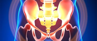
The pelvis is a part of the skeleton that is located at the base of the spinal column. It not only provides support for the internal organs, but also provides fixation to the legs to the body. That is why his health plays an important role, and x-rays of the pelvic bones are an integral component of diagnosis for diseases and injuries in this area.
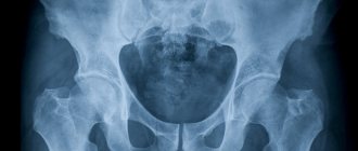
It is a highly informative diagnostic method that allows you to obtain data on the condition of the bone structures and joints of the pelvis, determine damage and pathologies of soft tissues and internal organs. Radiography is widely used in clinics, so it is an affordable method, including in terms of price. You can take an x-ray of the bones of the small and large pelvis at the diagnostic center of the multidisciplinary clinic CELT. We have modern equipment, and our specialists practice an individual approach, which invariably allows us to achieve high quality radiographic images.
X-ray of the pelvic bones - RUB 2,200.
15 - 20 minutes
(duration of procedure)
Indications
Diagnosis is carried out exclusively as prescribed by a doctor in the presence of clinical manifestations that indicate the following pathological changes in the pelvic bones:
- Cracks, hematomas, bruises, fractures, dislocations;
- Reduced density of bone structures (osteoporosis);
- Developmental anomalies;
- Subluxations and dislocations of the hips;
- Lesions of the joints of an infectious nature, as well as due to disruption of the body’s metabolic processes;
- Neoplasms of benign or malignant nature;
- Metastatic foci with neoplasms located in other places;
- Assessing the effectiveness of treatment tactics and the need for its correction.
Contraindications
Modern medicine identifies a number of conditions in which radiographic examinations are not recommended. These include:
- Pregnancy and breastfeeding in women;
- A large dose of radiation received over the past year;
- The patient's age is up to 15 years;
- The patient's serious condition.
Despite the safety of modern techniques, it is generally accepted that even a minimal dose of radiation can have a negative effect on patients in the above groups. At the same time, if absolutely necessary (if it is vital), radiography is still performed.
Advantages of X-ray of the pelvic bones over other methods
Modern diagnostic medicine has a fairly large number of highly informative methods for studying the pelvis, but radiography has not lost its relevance until now and has a number of advantages over other radiation techniques.
Advantages of pelvic bone x-ray:
- accessibility of the method - it requires only an X-ray machine;
- relatively low cost of the diagnostic procedure;
- absence of even minor discomfort or discomfort for the patient;
- the usual duration of radiography of the pelvic bones does not exceed 7-10 minutes;
- quick receipt of research results;
- high information content of the technique and the possibility of using it for dynamic assessment of the condition of the bone and joint structures of the pelvis;
- reduction of radiation exposure to the body through the use of modern high-precision digital X-ray machines.
Preparatory stage
The preparation is not particularly difficult. The patient is simply required to adhere to a certain diet two days before the scheduled examination date. It involves avoiding foods that stimulate increased gas formation:
- cabbage;
- legumes;
- black bread;
- dairy products.
Because of them, the visualization of the structures of the pelvic area is reduced by an order of magnitude, which may cause the need for a repeat study, once again exposing the body to radiation. And, if the disease or pathology is at an early stage of development, then it is unlikely to be examined at all due to increased gas formation.
An exception to the preparation rule is for patients who are urgently admitted to the hospital department. The doctor makes a decision on the spot, in case of receiving a blurry and uninformative image, about the choice of further diagnostics.
If the examination is routine, you will need to stop eating twelve hours before the procedure. It doesn’t hurt to do a cleansing enema the day before to better prepare the area to be examined for visualization during the examination.
An alternative is to use special pharmaceutical products that are freely available. Their cleansing effect is identical.
Another important point of preparation will be to warn the doctor about whether the patient has built-in electronic mechanisms in the body. We are talking about built-in hearing aids, pacemakers and other similar devices that may fail during this study. Moreover, their location does not play a significant role.
But the presence of metal inserts, such as supporting pins, pins or staples after previous fractures, does not pose a serious threat, including in terms of the information content of the study. It is enough to simply warn the radiologist and the attending physician about this feature in advance.
Also, immediately before the examination, you will need to remove metal jewelry and other removable metal elements that can limit the information content or clarity of the resulting image.
Having figured out how to prepare, all that remains is not to forget about two important contraindications:
- pregnancy;
- childhood.
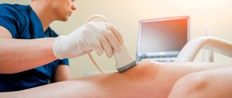
The first point applies to women at any stage of gestation. Despite the fact that for adults the device provides radiation exposure within the limits of the officially permissible dose, for the fetus it can become an impetus for the appearance of developmental anomalies. We are talking about disorders in mental or physical development. For pregnant women, they usually choose to study the condition of the pelvis using a more gentle examination format - ultrasound or MRI diagnostics, starting from the second trimester of pregnancy.
Almost the same applies to the situation with children under fifteen years of age. But here the contraindication is classified as relative. In practice, this means that if the benefits of the study predominate over the possible harm, X-rays are still indispensable.
To reduce radiation exposure to other parts of the body, special lead aprons are used. They are designed to protect the thyroid gland from excessive load, because it is this organ that is considered the most sensitive to the effects of radioactive radiation.
The standard radiation level for a pelvic examination ranges from 1.57-2.23 mSv. The exact indicator depends on the number of projections required to establish a diagnosis, as well as the release date of the device itself. The older it is, the more unnecessary radiation it will emit.
Indications
Like any other diagnostic procedure, an x-ray of the pelvic bones is performed for clear medical reasons and only on the direction of a doctor. This study is indicated if the patient has symptoms indicating changes in the pelvic bones, such as:
- fractures and injuries;
- developmental anomalies;
- damage to articular joints;
- benign or malignant neoplasms;
- metastases from tumors of other localizations;
- foci of rarefaction of bone structures in osteoporosis.
Also, this technique can be used to establish indications for other diagnostic procedures, to clarify treatment tactics in case of a diagnosis, and to dynamically assess the effectiveness of therapeutic measures.
Advantages of X-ray diagnostics using digital equipment
The main advantage, of course, is the minimization of the resulting radiation. This effect is achieved due to a number of factors:
- no X-ray film or chemicals are used;
- the device limits the area under study as much as possible, and thus reduces the load on adjacent organs that do not require X-ray diagnostics;
- The procedure time required to obtain a high-quality image is reduced.
In addition, the advantage of the digital radiography method is the ability to obtain clear, informative images, eliminating the need to repeat the procedure due to poor image quality. With the digital image format, it is easier and more functional for specialists - thanks to special programs, it is possible to “glue”, for example, long tubular bones or parts of the spine. It is convenient to store digital images for both doctors and patients in an electronic archive or on any digital media.
The German clinic has X-ray diagnostic equipment that can be used even for infants, and experienced specialists with extensive experience in pediatrics will help make an accurate diagnosis.
Attention: the prices presented on the website are not a public offer. Please check the cost of services by calling +7 (812) 432 32 32.
Preparing for X-ray of the pelvic bones
In emergency cases, when an immediate diagnostic procedure is required, an x-ray of the pelvic bones is performed without prior preparation. If the patient undergoes the examination as planned, then it is recommended that, for two days before the x-ray, he temporarily exclude from his diet foods that can cause activation of fermentation and gas formation processes in the intestines: fresh baked goods, legumes, apples, grapes. In the evening before the test, it is advisable to do a cleansing enema with a weak saline solution and drink a drug that has an adsorbing effect. These preparatory procedures reduce the influence of intestinal contents on the quality of the radiographic image and increase the information content of the resulting image of the pelvic bones.
Contrast X-ray of the pelvic organs - effective diagnosis of female infertility
Part of a woman's reproductive system is the fallopian tubes. These are the corridors along which the egg moves into the uterus. Scars, adhesions, and constrictions can become obstacles to the egg, and then conception may not occur. In order to examine the characteristics of the fallopian tubes, hysterosalpingography (HSG) is prescribed. This is the most informative method in this case, which helps not only to understand the patency of the fallopian tubes, but also to identify tumor nodes, polyps, and detect structural anomalies.
The GSK method is based on tracking the movement of a saline solution (contrast agent) through the fallopian tubes. In this case, water-soluble preparations are used that transmit X-rays due to iodine compounds in their composition. The procedure lasts about 30 minutes, and during this time 2 or 3 x-rays are taken.
Research methodology
Before conducting the study, the person removes clothing and accessories from the body that may cause distortion of the X-ray image, and also informs the laboratory assistant about the presence of metal or other implants in his body. In order to reduce the negative impact of X-ray radiation on the body, all patients are given a special leaded collar on the thyroid gland area, and women are given a protective plate on the mammary gland area.
X-rays of the pelvic bones are performed in a strictly horizontal position. The patient is placed on a special couch with legs bent at the knees and arms extended straight along the body. Do not move or talk while the x-ray is being taken.
Contraindications for the procedure
Before ordering an X-ray of the pelvis, the doctor will make sure that there is no possibility of pregnancy in the woman. Pregnancy is clearly a contraindication for the X-ray procedure, which should be replaced with an ultrasound diagnostic method. In addition, hysterosalpingography is contraindicated in cases of inflammation of the uterus and its appendages, severe forms of cardiovascular diseases, infectious and bacterial diseases of the vagina. The doctor must also exclude the possibility of an allergic reaction in a woman to iodine-containing drugs. After HCT, it is recommended to use contraception for a month to prevent pregnancy. X-rays are prescribed for children under 15 years of age when it is impossible to use an alternative diagnostic method.
What are the advantages of the method
Today medicine has many research options, but traditional radiography has not lost its relevance and has some advantages over all other radiation methods:
- Availability - only an X-ray machine is required;
- Low price;
- No discomfort during the study;
- Short research time;
- Fast result processing;
- Highly informative and the ability to use changes in the patient’s condition over time.
What does an x-ray of the pelvic bones reveal?
X-ray of the pelvic bones helps diagnose:
- various injuries (traumatic and inflammatory origin) of the bony pelvic ring;
- condition of the sacroiliac and pubic joints (ruptures, discrepancies);
- fractures and cracks of the head and neck of the femur, proximal femur;
- arthritis/arthrosis of various origins (gouty, rheumatoid, osteoarthritis);
- subluxations and dislocations of the hip, including congenital ones in children;
- osteochondropathy of the femoral head;
- tumors of the pelvic bones and metastases in them;
- osteoporosis.
The study is also performed to monitor the effectiveness of the treatment.
What are the indications for
An X-ray of the hip joint is performed when other diagnostic methods cannot be used. To perform the procedure, you must have a doctor's indication and referral. It is recommended to conduct a study if the patient:
- Joints are damaged;
- Injuries and fractures;
- There are tumors;
- Developmental disorders;
- Metastases are detected;
- There are areas of rarefaction of bone structures.
An x-ray will show the condition of the pelvic bones and the presence of pathological processes in them.
Indications:
- Presence of tumors;
- Metastases;
- Inflammatory processes.
The study is especially relevant for women planning pregnancy, in order to identify pathology and eliminate obstacles to the birth of a healthy child.