X-ray of the temporomandibular joint (TMJ) belongs to a diagnostic category that has been used for quite a long time. She has managed to prove herself exclusively on the positive side, as she provides a detailed diagnostic picture.
The technique has been successfully used for many decades, becoming the key to a speedy recovery for patients with abnormalities in the area of the lower jaw bones and the joint located there.
The main advantage of diagnostics is the ability to convey a clear image of the state of solid structures in a given area. The operating principle of the device is based on recording the degree of attenuation of radioactive rays that pass through the structures of the body. The resulting result is transformed into a kind of photo.
The essence of radiography of the temporomandibular zone
If previously patients received black and white photographs in their hands, with which they had to walk throughout the hospital, for fear of accidentally crushing them, today progress has stepped far forward. Now the received visual information is sent to the computer memory of a new generation X-ray machine. From there it can be recorded on any digital media.
Content
- The essence of radiography of the temporomandibular zone
- Indications for use
- Diagnosed anomalies
- Procedure algorithm
- X-ray in dentistry
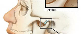
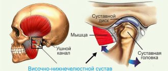
From an anatomical point of view, the temporomandibular joint is a connection between the lower jaw and the temporal bone of the skull, formed by the ellipsoidal fossa of the temporal bone of the skull and the head of the condylar process of the mandible.
Often, diseases of this area are based on pathologies of the articular disc, or destabilization of the intra-articular ligament.
No less often, the primary source of all problems is the inability to perform its functional duties as a capsule. Even arthritis does not bypass this joint.
Despite the fact that radiography is not always able to provide a 100% clear image of the area being examined, it is the most popular way to find the root cause of the disease.
Pathogenesis
The study of the mechanisms of development of pathology allowed us to come to conclusions about their progression associated with a lack of physiological endurance of the joint.
The resulting injuries and the appearance of inflammation lead to improper distribution or excessive load on the tissue, which causes disruption of the synchronization of the joints, causing loss of functionality of the muscular system. The result of the development of such unpleasant processes is a disruption of the nutrition of cartilage tissue, as well as a gradual loss of their elasticity, which inevitably leads to destruction.
Simultaneously with all of the above, serious transformations of bone tissue develop, which provokes the formation of growths that modify the elements of the joint: the head of the lower jaw changes shape, and signs of osteoporosis are visible.
As a result of the uncontrolled course of pathological processes, severe deformation and serious disruption of the tissue structure occur.
Indications for use
There is no clear list of symptoms when one should sound the alarm due to the individual characteristics of the human body. But, if problems begin with limited functional mobility of the lower jaw and connecting joint, then this is a reason to seek help.
Among other generally accepted signs, pain syndrome in a given zone is distinguished. Moreover, this can be either an acute pain that occurs when performing a certain movement, or an aching pain syndrome.
You also need to pay attention to:
- facial asymmetry;
- clicking and other sounds when chewing food;
- tinnitus;
- dizziness.
If you notice at least one of the above symptoms along with joint pain, you should immediately contact a traumatologist or maxillofacial surgeon for advice.
X-rays are also taken to determine the extent of damage to functional deviations that are caused by:
- dislocations;
- subluxations;
- fractures.
When a patient undergoes surgery, even if it is an ordinary plastic surgery, it is still impossible to do without an image. More serious reasons for visiting an x-ray office may be the following diseases at different stages of development:
- rheumatism;
- Bekhterev's disease;
- malocclusion.
Moreover, the sooner the victim contacts the attending physician, the more effective subsequent treatment will be, based on the detailed information received.
Is x-ray dangerous for health?
X-ray examination is a very useful method of studying the area of interest; the doctor can see the real bone picture and make conclusions that are important for health! At the same time, modern digital (not film) devices can significantly reduce the dose load.
Radiation safety standards are strictly regulated by the relevant SANPIN, which indicates the standard radiation load for humans, which corresponds to 1 mSv per year for 5 years, but not more than 5 mSv per year. While a snapshot of the jaw joint gives a load of 0.006 mSv, so this study is relatively harmless.
Diagnosed anomalies
Separately, situations are considered when the doctor has every reason to suspect deforming arthrosis of the joint for a long time. Such a serious pathology is a consequence of the degenerative process in the joint. And, if initially it was common to believe that inflammation was the trigger for damage to the mandibular joint, today the range of causes has expanded. Hormonal imbalances, physical injuries and changes in the tone of the masticatory muscles for any reason, including genetic diseases - all this contributes to the destructive process of arthrosis.
A black and white image of the bone structure will provide visualization of the joint space. If even a slight narrowing is visualized, this will be the beginning of painstakingly constructed restorative therapy. Consolidation of the articular tubercle will also indicate a possible deviation. Occasionally, patients even experience exophytic formations, which are characterized by growth inside the joint, aggravating the condition.
Another common ailment that affects both young and old patients is temporomandibular arthritis. The underlying reasons for the development of a serious illness may be:
- childhood infections;
- mumps;
- otitis.
The course of the disease is less than a month to trigger destructive mechanisms inside the joint. Already in the third week of advanced disease, the image will demonstrate a narrowing of the joint space. Since this imaging point is also characteristic of arthrosis, doctors will definitely consider additional parameters to establish an accurate diagnosis.
We are talking about the clarity of the end plates and marginal usurative changes. If the former have become less expressive in the image, and erosion is visible on the bone elements, then effective treatment cannot be avoided.
Periodically, X-rays are prescribed for those victims who have suffered fractures and dislocations, but are already on the mend. Regular monitoring is necessary in order to adjust the previously established course of treatment, taking into account intermediate indicators of effectiveness. Monitoring the condition will allow you to track how quickly and correctly the bones grow together.
Usually arthrologists are sent for analysis, but since not all public medical institutions have such a professional on staff, appointments are mostly issued by:
- surgeons, including plastic ones;
- traumatologists;
- dentists.
Even old devices that produce examination results on film are suitable for almost all types of diagnosis confirmation. But occasionally doctors insist that the patient do a test on a digital machine. It is believed to be able to provide clearer images.
The most likely causes of arthrosis of the jaw joint
Before trying to determine the causes of arthrosis of the jaw joint, it is necessary to understand that the pathology is primarily multifactorial, which makes it likely that several prerequisites for its development exist at once.
The group of common causes of arthrosis of the jaw joint usually includes genetic factors and various diseases. Among the local ones there are:
- the presence of a chronic form of arthritis;
- partial absence of molars;
- pathologies of dental tissues (abrasion, etc.);
- violation of prosthetic technology/placement of fillings, especially on chewing surfaces;
- injuries and surgical interventions.
In addition, it is worth noting that among women, the risks of the causes of arthrosis of the jaw joint include a natural decrease and instability of hormonal levels, on which the effectiveness of metabolic processes occurring in tissues largely depends.
A combination of factors provokes difficulties in treatment.
Procedure algorithm
Despite the widespread use of X-ray equipment, everyone knows that exposure to radiation has a negative impact on overall well-being. This is especially true for people from specific risk groups, as well as those who have managed to be exposed to radiation several times in a row in a short period of time.
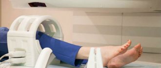
For adults, the dosage of radiation exposure used is considered within the normal range, but only on the condition that in the next few months he will avoid such diagnostics. But the same dose of rays has a more pronounced effect on children. Hence the desire of doctors to protect babies by sending them from X-ray to computed tomography, where the radiation exposure is much less.
In addition to babies, pregnant women at any stage are at risk. Some women in an interesting position naively believe that when examining the jaw, radiation will not reach the abdomen, but this is a mistaken opinion. Even with the slightest fluctuations in background radiation, the normal development of the fetus may be at risk. We are talking about increased risks of mental and physical retardation of the unborn child. To reduce the chances of a negative scenario, it is better to turn to an ultrasound as a more gentle alternative.
But even issuing a referral to an adult should be carried out under the strict supervision of the attending physician. The increased vigilance is explained by the fact that the ionizing effect negatively affects the condition of the salivary glands located nearby.
Despite a number of specific contraindications, the technique does not require significant preparatory measures. Typically, adults are prescribed a survey version of the study, even if the dental structure of the lower jaw and joint are to be examined.
But for a child, doctors limit themselves to close-up contact photography, if the functional features of the innovative version of the X-ray machine allow. Then even a dental examination can be carried out without a tube for generating radiation, which will not frighten the youngest patients of the dental clinic.
To get a reliable picture, the laboratory technician can ask the patient to fix the oral cavity in two positions in turn:
- open;
- closed
If serious complications or rare pathologies are suspected, the doctor may suggest additionally administering a contrast agent, which increases information content by increasing the clarity of visualization of the lesion.
But here it is worth being double vigilant, since at the contrast stage there is a possibility of an allergic reaction. To avoid anaphylactic shock, you need to ask for an allergy test in advance, and only then go for diagnostics.
Classification
Based on the results of an X-ray examination, in accordance with the diagnosed changes, it is customary to distinguish between two types of arthrosis:
- sclerosing - changes suggest sclerotic changes in bone surfaces, accompanied by a significant narrowing of the gap between the heads of the bones;
- deforming – there is a significant loss of texture and inherent forms of bone tissue, as well as a significant proliferation of osteophytes.
According to their origin, there are two types of pathology:
- primary – forms independently, without previous diseases/damages, mainly in old age;
- secondary - has a close connection with previous trauma, inflammation or metabolic disorders.