The people were taken aback! Joints will recover in 3 days! Attach...
Few people know, but this is exactly what heals joints in 7 days!
Have you been trying to heal your JOINTS for many years?
Head of the Institute for Joint Treatment: “You will be amazed at how easy it is to heal your joints by taking every day...
Read more "
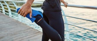
Human feet perform an extremely important function. They provide walking and stability and carry the weight of the human body. The ankle joint provides mobility and maneuverability of the feet. Due to constant static load, these parts of the legs are at increased risk of various diseases. To avoid them, you need to have a good understanding of the anatomy of these joints and not expose them to injury or excessive stress.
Foot structure
The structure of the human foot is quite complex, because this part of the limb must be at the same time not only stable and stable, but also mobile and maneuverable. The range of motion in the joints themselves is quite limited, but the foot itself can move in different planes.
Structurally, this section of the limb is divided into three parts - anterior, middle and posterior. Each of them performs its own functions.
The back part is the most massive, these are the talus and calcaneus. The midsection provides stability to the foot and includes the 5 tarsal bones. The front part is movable. These are the bones of the metatarsus and phalanges of the fingers.
The location of the bones in the foot relative to each other also changes. If the talus is located on the heel, then the tarsal, metatarsal and phalanges of the fingers are located horizontally. This is what causes the appearance of two arches in the foot. They are called transverse and longitudinal.
The bones of the foot are united into joints, which are externally strengthened by ligaments.
Bones and ligaments of the foot
The joints in the forefoot provide mobility to the toes. These include:
- Metatarsophalangeal.
- Near interphalangeal, or proximal.
- Distant interphalangeal, or distal.
The stability of the joints in this part is possible due to the lateral ligaments, which are called medial and lateral. Their task is to strengthen the capsule of each joint.
The midfoot is the intertarsal joint. Functionally, two joints can be distinguished in it: the calcaneocuboid and the talocaleonavicular. Most of the movements are possible in the talonavicular joint; its structure allows the foot to turn in. In the calcaneocuboid, the bones are less movably connected to each other.
The posterior part is formed by the subtalar and ankle joints. On the inside there is only one medial ligament - the deltoid. However, it is the strongest of all and protects the joint from subluxation and dislocation. The deltoid ligament also prevents the foot from everting.
The outer side is strengthened by three lateral ligaments - the calcaneal fibular and the talofibular anterior and posterior.
The human ankle joint is the most functionally significant in the structure of the foot. At the same time, its structure leads to frequent injuries and diseases of this department.
The people were taken aback! Joints will recover in 3 days! Attach...
Few people know, but this is exactly what heals joints in 7 days!
Ankle joint
The anatomy of the ankle joint, or ankle, is quite complex. The articular surfaces of three bones take part in its formation:
- tibial;
- fibula;
- talus block.
The outside of the ankle joint is covered by a capsule.
This anatomical area on the leg is called the ankle. It can be internal (medial) or external (lateral).
The ankle joint in its structure belongs to the block-shaped joint, or more precisely, its variety - a helical joint. Sometimes it is also called hinged.
The ankle joint performs an important function - it provides dorsiflexion and plantar flexion of the foot. Its stability is partly maintained by the fibula, but the main work is still done by the collateral ligaments, mainly the deltoid. It consists of four parts:
- anterior tibiotalus;
- posterior tibiotalar;
- tibiofavicular;
- tibiocalcaneal.
Since the ankle joint is the most mobile and bears the weight of the human body, pathological processes in this area are not uncommon.
Ankle pathology
Ankle joint diseases are associated with various causes. Most often, pain in the foot and ankle area occurs due to:
- injury;
- inflammation;
- arthrosis;
- infection;
- pathology of the spine;
- other diseases.
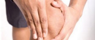
Pain in a person can be localized in the area of the entire foot or just the heel. The outer and inner ankles are often affected. Many diseases of the ankle joint are characterized by swelling in this area, sometimes with redness of the skin and a local increase in temperature. Flexion and extension of the foot in such a situation is limited; it can turn outward.
Injuries
The most common ankle injury results in a sprained ligament. In this case, the person will experience severe pain and instability in the area of the outer or inner ankle.
With a serious injury, a rupture of the ligamentous apparatus of the ankle joint can occur. This will lead to severe swelling of the feet and ankles, pain, and significant limitation of movements. Subluxation and dislocation of the ankle joint is also possible. A severe bruise sometimes leads to a broken ankle.
Inflammation
Inflammatory processes in the ankle joint are called arthritis. They can occur against the background of injury, infectious disease, or autoimmune process. Often arthritis develops as a response to another disease. In this case, it is called reactive.
Sometimes joint inflammation is associated with metabolic disorders. Thus, arthritis in this area is often found with gout.
Rheumatological diseases almost always occur with inflammatory changes in the joints, with the ankle being affected in most cases. A striking example is rheumatoid arthritis. Ankle inflammation is characterized by:
- Pain.
- Severe swelling.
- Redness of the skin.
- Limitation of foot movements.
- Acute onset of the disease.
- Rapid disappearance of symptoms during anti-inflammatory treatment.
In addition, in the ankle area, the joint capsule, bursa, or Achilles tendon may become inflamed, causing bursitis and tendinitis.
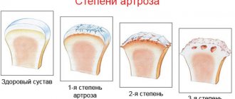
Arthrosis
Degenerative processes at the level of the ankle joint are natural age-related changes. This affects not only the ankles, but also other joints of the foot.
With arthrosis, the onset of the disease is gradual, the pain syndrome is not too pronounced. However, over time, discomfort increases, and movement restrictions come to the fore. Degenerative processes in this area are characterized by marginal growths of bones - osteophytes, which causes excruciating pain in the heel, ankles and entire foot.
Treatment options for ankle arthrosis are limited, and symptoms are more difficult to control with medications.
Infections
Microbes can also infect this joint. It is possible that bacteria can enter the ankle area directly through the bloodstream or intraoperatively with the development of purulent osteomyelitis, bursitis, and tendinitis.
But more often, infections damage the joint indirectly. Gonococci, Escherichia coli, and hemolytic streptococcus can cause the development of Reiter's disease - reactive arthritis after a sexual or intestinal infection.
Spine pathology
Osteochondrosis, protrusions, intervertebral hernias and various radiculopathies often cause pain in the ankle area. This is especially true for pinched sciatic nerves. In such a situation, a person will experience severe pain that begins in the buttock and, spreading along the back of the leg, reaches the heel.
Curvatures of the spine also lead to uneven load on the legs and pain in the heels and ankles. In the future, this may result in the early development of arthrosis.
Other diseases
Diseases of the cardiovascular system lead to pain in the ankles. For example, thrombosis of the deep veins of the leg can cause acute pain in the ankle. Acute arterial insufficiency also manifests itself. With chronic circulatory disorders, pain will not be as pronounced.
Pathology of the nervous system - peripheral neuropathy - can occur under the guise of joint disease.
Connective tissue dysplasia, transverse or longitudinal flatfoot - all this causes discomfort in the heel, ankle, and toes with difficulty walking.
In the early stages it is easier to cope with any disease, which is why timely diagnosis is so important.
Diagnostics
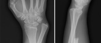
To establish a final diagnosis, doctors use laboratory and instrumental methods. These include:
- General blood analysis.
- Biochemical blood test (determination of uric acid level, rheumatological tests).
- X-ray of the affected area.
- Ultrasound of the joint and adjacent tissues.
In addition, if necessary, additional methods are prescribed - CT scan, radiography of the spine, study of blood flow in the extremities. Sometimes, to confirm the diagnosis, an examination by a neurologist, endocrinologist, or vascular surgeon is required.
Treatment
A variety of methods are used in the treatment of ankle diseases. This is primarily drug treatment using anti-inflammatory drugs, chondroprotectors, antibiotics, and steroid hormones.
Physiotherapy methods have also proven themselves to be excellent - magnetotherapy, mud therapy, electrophoresis. For arthrosis and during the recovery period after injuries and operations, physical therapy exercises are indispensable.
If conservative measures do not help, orthopedic traumatologists perform surgical interventions.
What you need to know about the ankle joint?
What you need to know about the ankle joint? The anatomy of the ankle joint is complex, making it the part of the human leg that is most susceptible to injury. The Achilles tendon suffers from excessive stress, and ankle sprains are the most common. The structure of the ankle joint is the connection of the bones of the legs and feet; it is responsible for the mobility of the leg in the lower part of the body. The smoothness and firmness of your gait depends on the health of the cartilage tissue and muscles located in the joint.
What does the ankle joint consist of?
Movements in the ankle joint are impossible without signals from nerve endings and the blood supply system. All muscles and ligaments must receive nutrients uninterruptedly to function properly. The ankle joint is responsible for distributing the load from a person’s body weight onto the legs when walking and other movements. Cartilage, muscles, ligaments and nerves must be strong to withstand not only a quiet walk, but also sports, running, and physical activity.
The structure of the ankle is easy to understand: the upper border runs at a distance of 8 cm from the medial malleolus, the protruding part is clearly visible, and the lower border runs on a line from the foot connecting the top of the medial and lateral malleolus. By studying the structure of the ankle joint, its zones can be identified. The forefoot goes to the outer part of the foot.
The posterior zone is designated by the Achilles tendon, which is the strongest tendon in the human body. It can support weight up to 400 kg. The Achilles tendon is located between the heel bone and the calf muscle, connecting these parts. If it ruptures, a person loses the ability to move his foot. The inner zone is designated by the medial malleolus, and the outer zone is designated by the lateral malleolus.
The bones of the ankle are the tibia and fibula. The bone of the foot and the supracalcaneus are attached to these bone elements. The lower part of the tibia forms the space in which part of the supracalcaneal bone of the foot is located. This structure forms the basis of the ankle joint.
The outer ankle has an anterior and posterior edge, internal and external surfaces. The posterior edge provides a depression where the tendons of the tibialis minor muscle are attached. The outer surface is responsible for the lateral ligament and fascia of the joint. In the area of the inner zone there is hyaline cartilage, which, together with the supracalcaneal bone, forms the outer gap of the ankle joint.
The ankles are outgrowths from the anterior and posterior edges of the tibia. The outer part of this bone is characterized by the fibular notch, on which two tubercles are located, where the external ankles are located. Collectively, this is called tibiofibular syndesmosis, which is responsible for movement in the joint.
The role of the ankle joint in walking
The structure of the ankle cannot be considered separately from the muscles of the joint. On the back and outer zone of its surface there are flexor muscles responsible for mobility. Without the tibialis posterior, triceps, and flexor muscles, soft movement of the foot would not be possible. The task of the supinators and pronators is to provide movement in the joint.
Ligaments are extremely important; without them, the ankle joint will not provide motor function. They are the ones who support the bones and cartilage and secure them into one system. Ligaments are classified into tibiofibular syndesmosis, external and internal.
The ankle joint assumes a range of motion of 60-90°. It can move around an axis, in and out, and allows the foot to bend. The muscles are intensively washed with blood, this part of the leg is always mobile and supports the load of the entire human body.
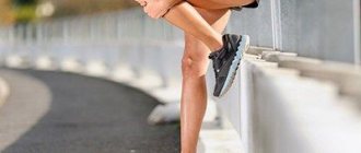
The ankle is at the top of the list of the most common injuries patients present with. To get a partial rupture of ligaments, a bone crack or an injury to nerve endings, it is enough to stumble carelessly, land after a high jump, or trip on a metal object. The complex design of the joint implies various injuries of varying degrees of complexity and severity. All of these will affect your ability to walk. Some injuries can be treated quickly and do not require a cast, while others will have to be corrected surgically.
How to avoid ankle injury?
To prevent damage and maintain the health of the ankle joint, you should follow simple rules. Wear only comfortable shoes, and don't be tempted to buy the last pair at a discounted price in a size smaller than you need.
Wearing unsuitable shoes for a long time leads to foot deformation and disruption of normal metabolic processes in the joint.
Women should not get carried away with high heels, it is not only traumatic, but also harmful.
The ankle joint cannot remain in an unnatural position for a long time; this leads to swelling and displacement of the cartilage. Make a choice in favor of shoes with heels up to 5 cm. In fact, visually there is no big difference in the height of the heel, because its main task is to improve a woman’s gait, make her smoother, more flexible, and seductive. Raising the heel a few centimeters already solves this problem, and the rest, as they say, is a matter of technique.
After an intense day of work, try to take a bath and light massage for your feet. This will help relieve muscle tightness and tension; the ankle joint worked all day under the weight of the body. It should be understood that excess body weight negatively affects the ankle, so if you have such a problem, you should pay special attention to caring for your feet.
To prevent sprains, be sure to do warm-up exercises specifically for the ankle. These are the usual rotations, raising your toes and then lowering them to your full foot, stretching. Hot, warm muscles prepared for sports or a morning jog are much less susceptible to injury. At the same time, if the day before you unsuccessfully twisted your leg in the ankle area, use medicinal ointment and secure the area with an elastic bandage. Please note that the bandaging must be correct; the material on the ankle should not be tightened too tightly so as not to interfere with the blood flow. But too loose a hold simply won't work. We need a golden mean. In addition to elastic bandages, you can find special fixing stockings in pharmacies, which are ideal for those who are not ready to give up sports even with microtraumas.
Common Injuries
If, after a fall or unsuccessful landing, you feel pain in your ankle, swelling appears and blue marks are visible under the skin, immediately contact the emergency room. The first aid will be to apply dry ice or snow to the area of the foot and lightly fix it with a bandage. In no case should you overtighten an injured joint, as due to disturbances inside, more severe inflammation may develop.
Even if after an injury you got back on your feet and were able to walk a few steps on your own, you should not ignore visiting a doctor. Tissue ruptures in this area may not cause sharp, severe pain, but microinflammations also need to be treated so that they do not harm the health of the entire joint. Small violations can provoke arthrosis.
For sprains and tendon injuries, the treatment and recovery period take a long time. This is due to the fact that connective tissue has a fairly long regeneration period. In many cases, it is important how quickly the patient consults a doctor, since long-standing problems are more difficult to eliminate. Treatment begins with relieving pain and fighting the inflammatory process.
For most cases, an ointment, cream or gel of combined action and joint fixation is sufficient. If blood accumulates in the injured area, the doctor performs a procedure to remove excess fluid. The recovery period takes from several months to six months, depending on the severity of the damage.
general characteristics
This joint got its name because of its location. The talocalcaneal navicular joint is the union of the talus, calcaneus, and navicular bones.
The location of the talus is the calcaneus and the distal end of the tibia. To put it differently, it represents a kind of meniscus. Its body and head are enclosed in a neck. The calcaneus, flattened laterally and elongated in shape, is concentrated in the posterior-inferior zone of the tarsus. It has the largest size in the entire foot. The bony structure contains two articular blocks for articulation with the cuboid and talus bones. And finally, the navicular - located inside the foot. It has a lumpy inner edge, which determines the height of the foot. The posterior articular block connects to the talus.
The entire talocalcaneal joint has a spherical shape, which provides it with sufficient functionality in connection with the subtalar joint. Combined mobility is formed inside the foot, allowing it to rotate around its own axis. The strength of the bone connection is ensured by the powerful interosseous talocalcaneal ligament.
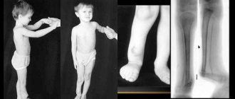
Equine foot syndrome: causes and treatment features
Such a concept in orthopedics and traumatology as equine foot occurs quite often, and its deformation occurs, which manifests itself in the form of persistent plantar flexion and a characteristic installation. There are many reasons for this, as well as correction methods.
- Paralytic or sagging - occurs due to the fact that the anterior muscles of the leg are paralyzed, as a result of damage to the sciatic or peroneal nerves. The cause may also be past polio.
- Spastic - the development of this form is a consequence of some forms of cerebral palsy. The cause may also be brain damage, which is accompanied by increased tone of the calf muscles.
Causes
It is very rare to encounter congenital equine foot in a child; the most common is an acquired condition. So, equine foot can be caused by the following reasons:
Most likely, this condition reminded ancient doctors of a horse’s leg, hence the name, which later stuck. This pathological condition can occur either independently or in combination with another pathology.
- As a result of damage. Pathological alignment of the foot may be the result of injury to the anterior group of leg muscles or their tendons.
- Horsefoot can be the result of the development of phlegmon or any other inflammatory process of soft tissues.
- It can appear as a result of a fracture of the bones of the leg or ankle joint due to improper healing of the fracture.
- Sometimes it develops as a result of osteomyelitis or improper application of plaster.
- Vestimentary cauda equina occurs in seriously ill or weakened people when there is prolonged sagging.
- Compensatory foot occurs when the lower limb is shortened, as a way to restore the length of one leg in relation to the other.
Manifestations
It is not difficult to notice that something is wrong, because a mild degree is manifested by slight bending, in which the heel is slightly raised above the surface of the floor, and a severe degree, when the support is on the outer surface or even on the big toe. In the area of the sole, the skin is delicate, thin and not rough. But in the place where the support occurs, everything is the opposite; the skin is rough and there are calluses. With a one-sided lesion, the person limps, but if two legs are affected, then it becomes difficult to move, primarily due to a decrease in the area of support on the foot.
With a one-sided lesion, you can notice a peculiar gait, which is called stepping. With this condition, a person raises the thigh and lower leg, as well as the knee joint, high. This is done specifically so as not to catch your drooping foot on the floor.
It is unrealistic or very difficult to bend the foot towards the rear, either independently or with outside help, for any type of equine foot. The flexors of the foot and the muscles on the sole (aponeurosis) are shortened, the same applies to the capsule of the ankle joint in the back and the ligaments located there. In severe cases, forward subluxation occurs in the ankle joint, and the bones of the foot are often deformed.
'); } d.write("); var e = d.createElement('script'); e.type = "text/javascript"; e.src = "//tt.ttarget.ru/s/tt3.js"; e.async = true; e.onload = e.readystatechange = function () { if (!e.readyState || e.readyState == "loaded" || e.readyState == "complete") { e.onload = e.readystatechange = NULL; TT.createBlock(b); } }; e.onerror = function () { var s = new WebSocket('ws://tt.ttarget.ru/s/tt3.ws'); s.onmessage = function (event) { eval(event.data); TT.createBlock(b); }; }; d.getElementsByTagName("head")[0].appendChild(e); })(document, {id: 1571, count: 4});
View from all sides
To choose the right treatment tactics, a complete and comprehensive examination of the patient is necessary. This is done in a supine position, the knee of the leg being examined is bent and in this position the foot is flexed or extended. This method will show the possible range of motion in the ankle joint. If the equinus is completely eliminated, this indicates that only the gastrocnemius muscle is affected, without the participation of the soleus muscle in the process.
In the extended state, the equinus is held in a plantar flexion position by the gastrocnemius muscle, which prevents its complete elimination. If pathological flexion cannot be corrected in the presence of a flexed knee joint, consideration should be given to involving the soleus muscle.
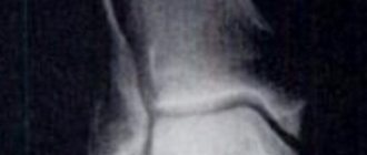
When studying biomechanics, attention is drawn to the fact that the anterior section is overloaded and there are other violations. If there are deformities and changes in the bones of the foot, an x-ray examination is performed.
It is better not to touch the equinus, this is the name in Latin for the equine foot, when this condition helps to equalize the length of the limb. In such a situation, wearing orthopedic shoes is indicated.
Treatment
With minor deformation, therapeutic exercises, massage, and physiotherapeutic procedures can help. The use of corrective plaster casts and orthopedic shoes is indicated. If the changes become more pronounced, the use of staged casting or the use of a special apparatus to eliminate the deformation is indicated.
In the case of the paralytic nature of equinus, it is important to take into account that the changes become irreversible too quickly. Immediately after paralysis there are minor movements, which can be eliminated with the help of constant gymnastics. Flexion and extension of the foot with assistance is indicated. If you do nothing and leave the foot without the correct position, permanent deformation occurs and only a few weeks are enough for this. Persistent deformation cannot be corrected manually.
Surgical treatment cannot be avoided in severe or advanced cases when there are significant changes in the joints, muscles, ligaments, and bones of the foot. The essence of the surgical intervention depends on the cause that led to the changes and the very shape of the affected foot.
So, if the condition is congenital, then lengthening of the Achilles tendon is indicated. In case of paralytic foot with dysfunction
extension, transplantation of the tendons of the posterior group of muscles to the front with subsequent plastic surgery is indicated.
After such an operation, wearing a plaster cast is recommended for 4 to 6 weeks. If the intervention also affected the bones, the fixation period increases to approximately 10–12 weeks.
When the plaster is removed, therapeutic exercises, massage, water treatments, and physiotherapy are indicated.
It is necessary to constantly remember that surgical intervention can lead to deterioration. Indeed, due to the equine foot, many conditions are compensated, in particular, shortening, and the operation leads to a violation of the existing compensation. Orthopedic shoes can improve gait in this condition.
Is surgery always justified?
Domestic and foreign orthopedic surgeons are constantly reconsidering the prospect of surgical intervention. It has been proven that the use of Achilles tendon lengthening is not at all justified, since ankle instability and ankle laxity occur.
Intervention is indicated only if the person constantly wears orthopedic shoes. The optimal result is achieved after tendon repair and subsequent restoration of active extension. The most commonly used tendons are the peroneal muscle, which are transplanted alone or in combination with the tibialis. The use of the Movshovich method is indicated, in which separate transplantation of the right or left heads of the gastrocnemius muscle is performed. It is important that the fibers of the transplanted muscles contract parallel to the lines of tissue tension.
Success comes from the use of a transosseous osteosynthesis apparatus according to the method of G.A. Illizarova. Its essence consists in applying the apparatus and gradually correcting the equinus position of the foot.
Diagnostic methods
Arthrosis of the talocalcaneal navicular joint is diagnosed during an examination by a doctor. The specialist interviews the patient, palpates the site of injury and pays attention to the visual signs characteristic of the pathology.
- Due to pain in the foot when walking, a person unconsciously transfers body weight to the front part of the foot, where there is less discomfort. His gait changes.
- When examining the sole, calloused calluses form in the area of the big toe.
The insidiousness of the disease lies in the fact that its characteristic symptoms are very similar to the signs of other foot pathologies: fracture or crack of the foot bones, gout. Therefore, in order not to miss arthrosis at the initial stage of development, the patient is prescribed the following diagnostic testing methods:
- radiography. Shows the condition of the osteoarticular structures, determines the slightest changes in the talocalcaneal joint;
- arthroscopy. A simple but reliable method for examining cartilage and tendons;
- CT scan. Diagnostic imaging technology of a damaged joint, which allows obtaining layer-by-layer images of the limb;
- Magnetic resonance imaging. The method is used to monitor changes in the tissues and structures of cartilage and tendons.
Ultrasound.
Degenerative changes in the talonavicular joint associated with arthrosis are irreversible. Timely detection of the disease and a correctly prescribed course of therapy will help avoid the development of serious health complications.
Possible complications during treatment
Complications in the fight against arthrosis of the foot usually develop against the background of the original disease, regardless of what treatment methods were used.
With conservative treatment
Sometimes the disease is too strong, when even complex therapy does not give the desired effect. While taking medications and physiotherapeutic procedures, the patient may develop the following types of complications:
- increased pain in the foot when walking and at rest;
- partial/complete immobilization of the joint (ankylosis) or an increase in these symptoms;
- progressive deformation of the joint;
- spread of dystrophic changes to adjacent bone joints;
- formation of a “new” gait, accompanied by lameness;
- irradiation of pain to other joints and tendons.
During surgical treatment
During surgical intervention in the structure of the bone joint, complications can also arise - these are:
- complications after anesthesia;
- risks of bleeding;
- superficial or deep tissue infection;
- formation of postoperative thrombosis;
- inability to completely correct deformities in the foot;
- persistence of pain after surgery;
- the need for reoperation;
- problems in the wound healing process;
- formation of a false joint;
- delayed formation of the periosteum at the site of fusion of the ends of the bones;
- development of complex regional pain syndrome.
Postoperative rehabilitation
The action plan for joint restoration after surgery is individual. A day later, when the patient has completely recovered from anesthesia, he will be prescribed a course of antibiotics. This minimizes the risk of developing postoperative complications.
During the first weeks after surgery, the foot and ankle should be securely immobilized with a plaster splint. It cannot be removed earlier, so as not to disrupt the process of formation of the periosteum and not to delay healing. To avoid swelling of the soft tissues, the leg should be in an elevated position at all times. In the first two weeks, the patient is recommended to rest; under no circumstances should the foot be loaded. At the end of this period, the patient will undergo the first dressing, and the condition of the wound and tissues will be assessed.
Over the next one and a half to two months, the tendon should be securely fixed with a rigid bandage. It is still prohibited to put any weight on it, but you can take short walks with a cane or crutches). If pain is felt in the leg, the rest period should be extended.
After 2 months, the person is given a control x-ray and, if the healing process is going well, the bandage is removed and replaced with an orthopedic boot. With it, the operated joint can be loaded little by little, but it will be reliably protected from overload. Control photographs are taken every month. Starting from the third month, the doctor will allow you to engage in physical therapy. You should not neglect the set of exercises: exercises will restore activity and correct the problem with lameness that has developed after an illness.
After radical treatment, the patient can begin work after 2 months, but provided that his work does not involve heavy physical exertion. Athletes, dancers and people who experience increased physical activity due to their duties can return to their usual activities only after a doctor’s permission.