September 24, 2019
54149
0
4.1 out of 5
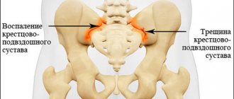
Arthrosis of the sacroiliac joint is an inflammatory degenerative-dystrophic disease that affects not only the joints connecting the pelvic bones and the sacrum, but also all nearby tissues. Its development can provoke severe pain and significant limitation of mobility, since it is the area of the sacromovable joint that bears a large load when walking. Therefore, it is important to detect the disease as early as possible and carry out proper treatment, which will prevent the loss of a person’s ability to move freely.
“SL Clinic” invites you to undergo diagnostics of diseases of the musculoskeletal system using modern equipment and receive a detailed consultation with a doctor. You will be able to ask any questions you are interested in, receive a carefully written treatment regimen, an objective assessment of the situation, and, if necessary, solve the problem surgically.
Reasons for development
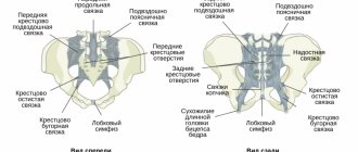
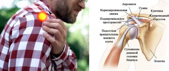
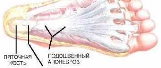
The sacroiliac joint is a paired joint that is the junction of the spinal column and the pelvis. In the sacral region, the vertebrae are firmly fused to each other, forming an almost monolithic bone. Both of its lateral surfaces are covered with hyaline cartilage. The ilium is tightly adjacent to them.
The joint or articulation thus formed is strengthened by numerous rigid, practically immovable ligaments. It has a slit-like shape, and the articular cavity itself is filled with synovial fluid and acts as a shock absorber during movement.
The sacroiliac joint is practically motionless, but at the same time provides stability when standing, is responsible for stabilizing the body position when performing certain movements, when sitting, and distributes the load on the pelvis and legs when walking. Therefore, the slightest disturbances in its functioning affect the condition of the spinal column and can provoke the development of a wide variety of complications.
Over the years, the sacroiliac joint gradually changes and becomes prone to degenerative and dystrophic changes. The following can provoke the development of arthrosis:
- various types of injuries to the lower back and tailbone;
- multiple pregnancies and births, especially when carrying a large fetus;
- abnormalities of bone tissue development;
- infectious diseases affecting bone tissue;
- obesity;
- metabolic disorders that provoke calcium deficiency in the body;
- sedentary lifestyle;
- autoimmune disorders, in particular ankylosing spondylitis;
- excessive physical activity.
A genetic predisposition to it significantly increases the risk of pathology. Most people diagnosed with this condition are over 55 years of age. Although recently there has been a tendency towards rejuvenation of diseases of the musculoskeletal system, therefore today it is no longer uncommon to find arthrosis of the sacroiliac joint in people from 25 to 35 years old. Most often, the pathology is diagnosed in women, due to the peculiarities of the anatomy.
Is contrast necessary?
If it is necessary to confirm or refute the presence of tumors, a CT scan of the coccyx is performed with the use of additional radiocontrast agents - special preparations containing iodine, which, when administered intravenously, stain the vessels. Tumors have their own blood supply network, so pathologically altered areas begin to stand out clearly in the photographs. Thanks to this, the doctor can see the location, size of the space-occupying lesion, the degree of destruction of bone tissue and other parameters.
A CT scan of the sacrococcygeal region with contrast is performed for the purpose of differential diagnosis of tumors and cysts.
Computed tomography of the sacrum and pelvis with contrast
After injection of an iodine-containing substance, side effects may appear:
- pain at the injection site;
- metallic taste in the mouth;
- flushes of heat in the body.
These symptoms are not life-threatening and do not require medical attention.
Indications for stopping the procedure are:
- nausea, vomiting;
- itching;
- hives;
- swelling of the ears, face;
- difficulty breathing;
- bronchospasm;
- lowering blood pressure.
Any sign of a complication should be reported to the radiologist via a voice communication device.
Sacral osteosarcoma on CT scan
Symptoms of arthrosis of the sacroiliac joint
In the early stages of development, the disease practically does not manifest itself. Signs of a degenerative process occur when cartilage tissue is destroyed. This may be accompanied by:
- discomfort in the lumbosacral spine and buttocks, aggravated by physical work, bending, turning, prolonged sitting or walking;
- increased muscle tone in the sacral area;
- limitation of motor activity and range of motion, feeling of stiffness;
- the appearance of a characteristic crunch when bending or turning the body;
- gait disturbance;
- increased urge to urinate;
- decreased libido.
The pain can initially usually be described as pulling, aching. They tend to radiate to the groin, legs, perineum, and buttocks.
The inflammatory process quickly worsens, which is accompanied by redness, increased sensitivity and swelling of the soft tissues in the projection of the diseased joint, impaired circulation in the affected area and a decrease in the amount of nutrients supplied. As a result, osteophytes are formed. As arthrosis progresses, the symptoms intensify, and in advanced cases the person almost completely loses the ability to move independently.
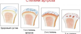
Stages of the disease
The intensity of signs of arthrosis of the sacroiliac joint depends on the stage of its development. There are 4 stages:
- At first, the disease practically does not manifest itself. After physical work, long walking or sitting, minor short-term discomfort may occur in the sacrum and buttocks, which quickly disappears after rest. At the same time, the functions of the joints are completely preserved, so the person does not notice the limitation of mobility. Attacks of acute pain that interfere with standing are rare.
- The pain intensifies and occurs more frequently, and can only be relieved by taking painkillers. This is triggered by the appearance of irreversible changes in the state of the cartilage tissue of the joint.
- Due to severe deformation of the cartilage, osteophytes are formed along the entire surface of the joint, and the bone surfaces are exposed. In some cases, separation of the sacrum is observed. This is accompanied by severe pain, completely incapacitating the person.
- Degenerative-dystrophic processes lead to complete stiffness and constant severe pain.
Clinical picture
Symptoms of SIJ syndrome are often difficult to distinguish from other types of low back pain. The most common symptoms include:
- Lower back pain.
- Pain in the hips.
- Discomfort when sitting for long periods of time.
- Local tenderness of the posterior surface of the SIJ (next to the posterior superior iliac spine).
- Pain occurs when there is mechanical stress on the joint, for example, when bending forward.
- No signs of neurological deficits/nerve root tension.
- Aberrant movements of the SIJ.
- The joint may be hyper- or hypomobile, which can cause pain.
- The pain is usually localized directly above the SIJ.
- Patients may complain of sharp, stabbing and/or shooting pain that extends down the back of the thigh, not below the knee.
- The pain may mimic radicular pain and therefore be misdiagnosed as radicular pain.
- Patients often complain of pain when sitting, lying ipsilaterally, or climbing stairs.
Differential diagnosis
SIJ syndrome is a controversial diagnosis, so SIJ pain and injury are commonly overlooked. This condition is often defined as dysfunction (a term that serves as an umbrella term for various conditions).
Differential diagnosis should include:
- Radicular pain.
- Piriformis syndrome.
- Ankylosing spondylitis.
- Lumbar facet syndrome.
- Spondyloatropathy.
- Greater trochanteric bursitis.
- Fracture of the femur.
- Hip overload syndrome.
Diagnostics
It is impossible to establish the cause of pain only on the basis of the existing clinical picture, since more than 15 different diseases are accompanied by symptoms typical for arthrosis of the sacroiliac joint. In particular, ankylosing spondylosis, L5-S1 hernias, spinal canal stenosis, scleroderma and other diseases manifest themselves in a similar way.
To diagnose the disease, the following are prescribed:
- CBC - used to detect signs of an inflammatory process (with arthrosis, the number of leukocytes in the blood and ESR are usually not much higher than normal);
- biochemical blood test - necessary to assess the functioning of internal organs;
- X-ray or CT - each method allows you to detect narrowing of the joint space, signs of destruction of cartilage tissue and osteophytes;
- MRI is the most informative diagnostic procedure, with which you can detect even minimal deviations from the norm in the condition of ligaments, muscles and joints;
- Osteoporosis screening – prescribed to identify bone weakness.
At SL Clinic you can get advice from a specialist who has been treating this problem for many years. Therefore, when undergoing treatment with us, you will definitely not miss the progression and development of the disease when you contact immediately after the first symptoms appear.
Preparation for CT scan of the sacrococcygeal region
No special preparation is required for the procedure without contrast. On the eve of a CT scan of the sacrococcygeal region, you can take medications; you do not need to refuse food. You should arrive for diagnostics approximately fifteen minutes before the appointed time to sign the contract and fill out the form. You must have your passport, a doctor’s referral, and the results of previous examinations with you.
If, according to indications, a computed tomography scan of the sacrococcygeal region with contrast is prescribed, the patient must donate blood for creatinine. Some centers do a rapid test on site. Exceeding the permissible limits is a contraindication for the procedure.
Before the examination, mothers with infants are advised to prepare food for their babies: after contrasting, the milk will have to be expressed twice and poured out.
Intraosseous pneumocyst of the sacrum on a computed tomogram
Treatment of arthrosis of the iliosacral joint
The effectiveness of treatment largely depends on how early it was started. Along with conservative therapy, radiofrequency ablation of the joint is prescribed, in a set of measures that together help eliminate the inflammatory process and stop the destruction of the joint. It includes:
- drug therapy;
- physiotherapy;
- manual therapy;
- Exercise therapy.
In case of exacerbation of the disease, bed rest is recommended until the severity of pain decreases. In the future, patients are advised to give up heavy physical work, sports and long running. But moderate physical activity is an integral part of the fight against arthrosis of the sacroiliac joint. When working in a sedentary or standing position, it is important to take regular breaks and walk around.
A vertebrologist may recommend that the patient wear a special orthopedic corset. It will help reduce stress on the back muscles and pressure on the affected joints. The bandage is selected individually by the doctor. It should be worn for several hours during the day.
If conservative treatment is completely ineffective or advanced forms of arthrosis have led to the formation of osteophytes, patients can be helped surgically. This will protect the person from pain, preserve his ability to work and avoid disability.
Drug therapy
In order to relieve pain and eliminate the inflammatory process, patients are prescribed drugs from different groups. They, as well as the route of administration (oral, intramuscular, intravenous) and doses are selected individually, based on the stage of development of arthrosis of the sacroiliac joint. In this case, existing concomitant diseases must be taken into account.
Patients are advised to use:
- NSAIDs – used for moderate pain. In addition to the analgesic effect, they have anti-inflammatory properties. They are most often used in the form of drugs for oral use, but if they are ineffective, intramuscular injections can be prescribed. The negative side of drugs in this group is their negative effect on the condition of the mucous membranes of the gastrointestinal tract with long-term use.
- Corticosteroids - indicated for severe inflammatory processes that cannot be treated with NSAIDs. They are prescribed in short courses and have a powerful anti-inflammatory effect.
- Muscle relaxants are drugs that relieve muscle spasms. They are used to eliminate reflex spasms caused by pain. This reduces the intensity of pain and improves blood circulation in the area.
- Chondroprotectors - drugs in this group are designed to stop the destruction of cartilage tissue and improve its structure. They are intended for long-term use.
- Vitamin complexes – help increase the effectiveness of drugs from other groups and normalize metabolic processes.
- Local remedies in the form of ointments, creams or gels most often contain NSAIDs and are used to relieve mild pain.
Lidocaine or novocaine blockades can quickly relieve unbearable pain. They are performed exclusively under completely sterile conditions, i.e. in a medical facility. The essence of the procedure is to inject anesthetic solutions into precisely defined points in the sacral area. The manipulation brings relief within 2–5 minutes. It can also be carried out at the SL Clinic.
But the blockade cannot be performed during pregnancy or the presence of pustular rashes on the skin in the projection of the affected joint.
Physiotherapy
Physiotherapeutic procedures that are correctly selected in terms of duration and frequency of implementation significantly increase the effectiveness of drug therapy and can reduce the intensity of pain. Patients are recommended:
- electrophoresis with the introduction of drugs from the NSAID group - the procedure involves the introduction of drugs through a weak electric current directly into the lesion;
- laser therapy – the thermal effect of the laser activates the processes of regeneration of cartilage tissue cells;
- reflexology - influencing biologically active points helps improve blood circulation and reduce pain;
- Magnetic therapy - a method that helps reduce the intensity of pain and reduce the rate of degenerative processes.
Usually a course of procedures is prescribed, consisting of 10–12 sessions. They can only be carried out during remission. Contraindications to physiotherapeutic treatment are serious diseases of the cardiovascular system, severe renal or respiratory failure, febrile conditions, and epilepsy.
Manual therapy
Manual therapy sessions conducted by a specialist, taking into account the characteristics of the patient’s condition, can not only activate blood circulation in the area of the sacroiliac joint and thereby improve tissue nutrition, but also slow down the course of degenerative processes.
The procedures are carried out in courses. They should be started only after the acute phase of arthrosis is completed.
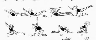
Exercise therapy
Although rest helps relieve pain, a specific set of exercises can be even more helpful. Moreover, physical therapy plays an important role in the conservative treatment of arthrosis of the sacroiliac joint. Depending on the stage and general condition of the patient, a training schedule and workload are individually developed for him. It is usually necessary to do therapeutic exercises daily for 20–30 minutes.
The first sessions of exercise therapy are recommended to be carried out under the supervision of a specialist. This will help not only to master the necessary set of exercises, but also to maintain an optimal rhythm when performing it. In most cases, patients are prescribed body turns, bends, rotations, etc. When performing any exercise, it is important to avoid sudden movements and overexertion, and if pain occurs, you should definitely consult a doctor.
Swimming and yoga have a beneficial effect on the patient's condition. But they are only permissible outside of exacerbation of arthrosis.
Surgical treatment of arthrosis of the sacroiliac joint
In some cases, the only way for patients to get rid of excruciating pain and avoid long-term back pain and joint pain is surgical treatment of arthrosis. It is indicated when attempts to cope with the disease using conservative methods are unsuccessful, as well as in severely advanced cases, i.e., stage 3 arthrosis of the sacroiliac joint.
The essence of surgical intervention depends on the nature of the existing changes. Radiofrequency ablation of nerve endings can be used to eliminate pain. Its essence consists in introducing a special electrode through a pinpoint puncture of soft tissue directly to the pain-causing nerve and its destruction by the generated thermal energy.
The procedure in most cases leads to immediate pain relief. In other situations, there is a progressive decrease in their intensity over 6-8 weeks. After it is completed, the patient can almost immediately move independently and return home on the same day.
If irreversible changes have occurred in the sacroiliac joint, the surgeon may recommend arthrodesis to the patient. The operation is performed on both sides in 2 stages:
- With the patient lying on his stomach after treatment of the surgical field, an incision about 2 cm long is made and a channel is formed into the articulation cavity through the posterior portion of the sacroiliac ligament. Using special instruments, a thorough curettage is carried out, i.e., cleaning the articular surfaces. The cavity is washed with solutions of antiseptics and antibiotics and the wound is sutured. Drainage is inserted into it and an antiseptic bandage is applied.
- The patient is turned over onto his back and a cushion is placed under the lumbar curve. An incision up to 4–5 cm long is made along the iliac crest, through which a bone canal is formed in the crest using an awl. Also, 2 more channels are created at a distance of 1–2 cm from the first. 3 rods are inserted into them and the rigidity of the installation is carefully controlled. An external fixation device is mounted on them and the necessary compression parameters are set.
During the rehabilitation process, surgeons change compression modes, thereby achieving effective arthrodesis of the sacroiliac joint. Patients can get up already on the second day after surgery. In the absence of adverse events, he can stand up independently and begin learning to walk. A control x-ray is performed on day 5 and, if there are no complications, the patient can return home, receiving detailed recommendations on the specifics of recovery.
Every 2–3 days, the patient must independently or with the help of relatives carry out dressings and take prescribed medications. After 8–10 weeks, a repeat X-ray examination is performed and part of the external fixation device is removed. After this, the patient needs to walk for about an hour, leaning on crutches and without them. In the absence of pain, final dismantling is performed, which leads to normalization of range of motion.
You can undergo conservative and surgical treatment for arthrosis of the sacroiliac joint at the SL Clinic. Our highly qualified vertebrologists are able to accurately determine the causes of pain and stiffness of movement, and select the optimal treatment tactics, which are sure to give positive results.
In severe cases, you can receive professional surgical care from us and regain the ability to move calmly. We provide medical services at the level of well-known clinics in Germany, Israel and the Czech Republic, but at the same time make them accessible to a wide range of patients. The cost of all types of services, including specialist consultations, blockades, surgical treatment, is given in the price list. Entrust your health to us, and we will do everything possible so that pain does not bother you anymore.
Disease prevention
Since arthrosis is an age-related disease, it is impossible to guarantee its absence, especially if there is a hereditary predisposition. But it is possible to reduce the risk of its development. For this purpose it is recommended:
- regular moderate physical activity;
- avoid prolonged sitting or standing;
- promptly treat any infectious disease;
- avoid stress and nervous tension;
- do not lift too heavy objects;
- maintain normal weight.
Cost of treatment for arthrosis of the sacroiliac joint at SL Clinic
Treatment of SI joint arthrosis involves inserting needles into the projections of the diseased joint and treating them in a temperature regime. The procedure is minimally invasive and outpatient i.e. you can go home the same day.
The cost of radiofrequency treatment of arthrosis of the SIJ is 68,000 rubles and depends on: - The cost of needles for radiofrequency ablation; — Clinics and class wards. The price includes: — Stay at the clinic before and after surgery; — Operation; — Cost of needles for radiofrequency ablation; — Observation and consultation during the rehabilitation period. All clinic services and costs are listed in the price list.