The sternoclavicular joint (SJJ) is formed by the articulation of the sternal end of the clavicle and the clavicular notch of the manubrium of the sternum. It is one of the four joints that make up the shoulder complex. The RCL is classified as a low-moving, flat-type joint that has a fibrocartilaginous articular disc. The ligamentous apparatus of this joint is so strong that the collarbone is more likely to break than the joint itself to dislocate.
Sternoclavicular joint
The purpose of the joint is to connect the upper limbs with the chest by combining the bones of the clavicle and shoulder girdle with the torso. By its origin, the sternoclavicular joint is a rudiment, which is a connection of the upper or forelimbs not only in humans, but also in animals, starting with reptiles.
The sternoclavicular joint is shaped like a saddle joint. In its structure, it has a communicating shape, having concavities and convexities corresponding to each other. This joint, having two axes and freely making movements along them, from the point of view of simple mechanics is a universal joint. Its structure includes the following cartilaginous tissues:
- cartilaginous covering of the clavicular bone;
- cartilaginous covering of the sternocostal cavity;
- cartilage disc;
- cartilage tissue covering the joint.
Thus, the structure of the joint includes:
- the medial end of the clavicle with its main surface;
- superior ligament;
- anterior ligament;
- costoclavicular ligament;
- posterior ligament;
- concave arches of the sternocostal surface.
The sternoclavicular joint is also supported by:
- The intervertebral ligament stretches over the notch of the jugular cavity of the sternum between the ends of the clavicular bones.
- The sternoclavicular ligament complex. According to their location, they converge on the front, back and top surfaces of the joint, strengthening its strength.
- The most powerful and durable ligament in the sternum is the costoclavicular ligament. It runs from the very top edge at the first rib and rises to the collarbone. Controls the maximum upward elevation of the collarbone.
The sternoclavicular joint, having a saddle-shaped structure in shape, resembles a spherical one in terms of its movement capabilities.
The sternoclavicular joint is one of the key joints of the shoulder girdle. Thanks to it, a strong contact of the shoulder joint is created, which ensures the attachment of the ends of the bones of the upper limbs to the chest. The strength of the connection is quite high, and the joint is able to withstand powerful functional loads. This allows a person to make rotational movements with his hand in three planes.
The sternoclavicular joint makes it possible to raise the arm and place it behind the head, and rotate the shoulder. A person performs all these movements hundreds of times a day, thanks to the characteristics of the sternoclavicular joint, which, on the one hand, is highly stable, but at the same time allows the upper limb to make free movements in different directions.
The sternoclavicular joint includes the sternal end of the clavicle and the clavicular notch located on the sternum. The adjacent planes of the sternoclavicular joint are covered with hyaline cartilage. Its main role is to ensure complete compliance of one joint part with another so that movements are as comfortable as possible.
But this feature does not prevent the joint from performing its functions, since for mutual correspondence there is a special intra-articular disc in the sternoclavicular joint. It is located between the bone surfaces, but does not connect to them. Only along the perimeter of the intraarticular disc there are ligaments connecting it to the capsule.
The intra-articular disc divides the cavity into two parts: the upper lateral and lower middle. If the disk has holes in the middle of its body, then the cavities can be connected to each other. This is not a pathology, it is only a characteristic of the sternoclavicular joint, and this feature does not prevent it from performing its functions.
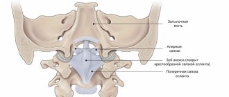
A complex diagram of the sternoclavicular joint (Fig. 82, according to Rouviere) shows the following. The right half of the figure shows a vertical frontal section.
- Costoclavicular ligament 1, attached to the upper surface of the first rib and running upward and outward towards the lower surface of the clavicle.
- Very often, the two articular surfaces have different radii of curvature, and their congruence is ensured by the meniscus 3, like a saddle between horse and rider. This meniscus divides the joint into two secondary cavities, which may or may not articulate with each other depending on the presence or absence of a perforation in the center of the meniscus.
- The sternoclavicular ligament 4, lining the upper part of the joint, is strengthened on top by the interclavicular ligament 5.
The left half of the picture shows a front view of the joint.
- Costoclavicular ligament 1 and subclavian muscle 2.
- The X axis runs horizontally and slightly obliquely anteriorly and outward, which corresponds to the movements of the clavicle in the vertical plane within the following limits: upward by 10 cm and downward by 3 cm.
- The Y axis, running in the vertical plane obliquely downward and slightly outward, crosses the middle part of the costoclavicular ligament and corresponds, according to traditional concepts, to the movements of the clavicle in the horizontal plane. The amplitude of these movements is as follows: the outer end of the clavicle can move 10 cm anteriorly and 3 cm posteriorly. From a purely mechanical point of view, the true axis (Y′) is parallel to the (Y) axis, but is located inside the joint.
Another, third type of movement occurs in this joint, namely axial rotation of the clavicle by 30°. This is only possible when the ligaments are relaxed. Since the sternoclavicular joint is biaxial, during voluntary rotation around its two axes, automatic (combined) rotation occurs. Observations in practice show that this automatic rotation always accompanies voluntary movements in a given joint.
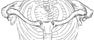
Movements of the clavicle in the horizontal plane (Fig. 83, top view).
- The thick line indicates the position of the clavicle at rest.
- Movements are carried out in relation to point Y′.
- Two crosses indicate the extreme positions of the clavicular attachment of the costoclavicular ligament.
In the inset, section A is taken at the level of the costoclavicular ligament to demonstrate the tension developed in the ligament in extreme positions.
- Anterior movement is controlled by tension on the costoclavicular ligament and the anterior capsule ligament 1 .
- Posterior movement is limited by the tension of the costoclavicular ligament and the posterior capsule ligament 2.
Movements of the clavicle in the frontal plane (Fig. 84, front view). The cross corresponds to the X axis of motion. As the outer end of the clavicle rises (shown by the thick line), its inner end slides downward and outward (red arrow). This movement is controlled by the tension of the costoclavicular ligament (shaded line) and the tension of the subclavian muscle 2.
As the clavicle descends, its inner end rises. This movement is limited by the tension of the superior ligament of capsule 4 and the contact between the clavicle and the superior surface of the first rib.
"Upper limb. Physiology of joints” A.I. Kapandji
The sternoclavicular joint, articulatio sternoclavicularis, is formed by the clavicular notch of the sternum and the sternal end of the clavicle. The joint is simple. The articular surfaces are covered with connective tissue cartilage, incongruent and most often saddle-shaped. The discrepancy between the articular surfaces is leveled by a composite disc located in the joint cavity.
The articular capsule is strong and attached to the edges of the articular surfaces of the bones. The joint cavity is divided by the articular disc into two parts that do not communicate with each other - inferomedial and superolateral. Sometimes the articular disc has a hole in the middle; in these cases, both joint cavities communicate with each other.
1. Anterior and posterior sternoclavicular ligaments, ligg. sternoclaviculare anterius et posterius, which are located on the anterior, superior and posterior surfaces of the articular capsule, strengthening the latter,
2. Costoclavicular ligament, lig. costoclaviculare. which is a powerful ligament running from the upper edge of the first rib upward to the collarbone and inhibits its upward movement.
3. Interclavicular ligament, lig. irtterclavicure, stretched between the sternal ends of the clavicles above the jugular notch of the manubrium of the sternum; inhibits the downward movement of the collarbone.
In terms of the range of movements, the sternoclavicular joint approaches the spherical type, articulatio spheroidea.
The sternoclavicular joint (articulatiostemoclavicularis).
Front view. On the left side of the specimen, the joint is opened with a frontal incision.
1-clavicle (right); 2-anterior sternoclavicular ligament; 3-interclumbar ligament; 4-sternal end of the clavicle; 5-intra-articular disc (fudinoclavicular joint); 6-first (I) rib; 7-costoclavicular ligament; 8-fudinocostal joint (11th rib); 9-intra-articular sternocostal ligament; 10th cartilage of the 11th rib; 11-synchondrosis of the handle of fudina; 12-radiate fudinocostal ligament.
The sternoclavicular joint. Front view. On the left side of the specimen, the joint is opened with a frontal incision. 1-clavicula (dextra); 2-ligamentum sternoclaviculare anterius; 3-ligamentum interclaviculare; 4-extremitas sternalis claviculae; 5-discus articularis (articulatio sternoclavicularis); 6-costa (I); 7-ligamentum costoclaviculare;
Sternoclavicular joint. Anterior aspect. Frontal section of joint at left. 1-clavicle (right); 2-anterior Sternoclavicular ligament; 3-interclavicular ligament; 4-sternal end ot'clavicle; 5-articular disc (ol'sternoclavicular joint); 6-1 rib; 7-costoclavicular ligament; 8-sternocostal joint (of the II rib); 9-intra-articular sternocostal ligament; 10-cartilage of the II rib; 11-manubriosternal synchondrosis; 12-radiate sternocostal ligament
The sternoclavicular joint (articulatio sternoclavicularis), formed by the connection of the sternal end of the clavicle with the clavicular notch on the manubrium of the sternum, is the only joint connecting the axial skeleton with the skeleton of the upper limb. The shape of both articular surfaces is close to saddle-shaped. The powerful articular capsule is strengthened by the interclavicular (lig.
Clinical manifestations of degenerative pathology
Acromioclavicular arthrosis first manifests itself as pain in the shoulder joint. As the disease progresses, the pain increases, becomes more acute, and spreads to the cervical and thoracic region. Many people initially attribute shoulder pain and stiffness to lack of exercise or aging, but this is far from the truth. If you experience any pain in the musculoskeletal system, you should immediately contact a rheumatologist or arthrologist for further examination. Arthrosis of the acromioclavicular and sternoclavicular joints can progress, exacerbating the patient's condition several times. Early detection of the disease will help begin timely treatment. Symptoms of the disease:
- crepitus and clicking when moving the shoulder;
- weakness and muscle atrophy;
- the shoulder may become stiff after a long period of rest;
- it is difficult for the patient to raise his arms above his head or place them behind his back;
- swelling of the shoulder and partially of the collarbone appears (depending on which side of the joint is affected by the disease).
With arthrosis of the sternoclavicular joint, the patient complains of severe difficulty breathing, increased heart rate, and pain in the thoracic region. Inaction when primary and further signs appear leads to deformation, the joint becomes immobilized, and growths and protrusions may appear. The symptoms of the disease worsen every day, causing the patient hellish suffering. It is important to report the symptoms in time and treatment will begin faster; it will help restore the mobility and functionality of this joint.
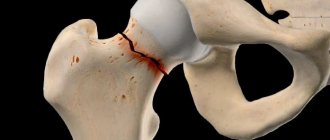
Damage
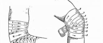
Due to its superficial location and role in the movements between the bones and joints of the shoulder girdle and torso, the clavicle itself and the joints attached to it are often subject to fractures and dislocations. Dislocation occurs as a result of sudden movements of the shoulder girdle backwards or downwards and backwards. In this case, the anterior ligament is torn, forming a subluxation.
With a stronger impact on this joint, all ligaments are torn, releasing the collarbone from the articular fossa, forming a dislocation of this joint, which is easily recognized by external signs. Another type of dislocation occurs if the impact on the collarbone and joint is direct, that is, through a direct blow or strong pressure when the posterior ligament is torn.
Because the sternoclavicular joint is one of the most mobile joints in the body, it is susceptible to dislocations and fractures. Mechanical impact in the form of a blow, jerk or strong compression can lead to partial or complete dislocation of the joint, causing sharp, severe pain. Although the injury occurs in the thoracic region and shoulder girdle, pain is felt by the rib and its muscles.
Of the diseases, the most common and dangerous is osteochondrosis of the clavicle. In its advanced form, it leads to the formation of osteophytes on the clavicular head and partial immobilization of the limbs. A person loses the ability to freely extend and contract, raise and lower his arms, perform body turns and shoulder movements. Untreated rheumatoid arthritis also leads to limited mobility of the upper limbs, progressing to ankylosis of the clavicular joint.
The joint node is susceptible to Friedrich's disease - aseptic necrosis of the tip of the clavicle attached to the sternum. The pathology is characterized by swelling and changes in texture, color of tissue and skin. With a large number of osteophytes, marble disease occurs, which is characteristic of congenital syphilis disease. Only a doctor can determine the disease and prescribe treatment; self-diagnosis is dangerous and is not recommended.
Due to its superficial location and role in the movements between the bones and joints of the shoulder girdle and torso, the clavicle itself and the joints attached to it are often subject to fractures and dislocations. Dislocation occurs as a result of sudden movements of the shoulder girdle backwards or downwards and backwards. In this case, the anterior ligament is torn, forming a subluxation.
With a stronger impact on this joint, all ligaments are torn, releasing the collarbone from the articular fossa, forming a dislocation of this joint, which is easily recognized by external signs. Another type of dislocation occurs if the impact on the collarbone and joint is direct, that is, through a direct blow or strong pressure when the posterior ligament is torn.
Causes
As with deforming arthrosis of other localizations, the factors causing the pathology of the described joints may vary significantly in different patients. Possible causes of osteoarthritis:
- Elderly age.
- Female.
- Menopause period.
- Hereditary predisposition.
- High loads on the upper limb.
- Injuries to the corresponding joint.
- Metabolic diseases.
- Arthritis of infectious nature.
The listed factors can cause pathology either individually or in combination with each other.
Diseases
This joint is characterized by diseases such as ankylosis, which is a consequence of gonococcal or rheumatoid arthritis. After the age of forty, arthrosis often appears, which, as it progresses, forms marginal osteophytes on the head of the clavicle. Pain caused by impact on the sternoclavicular joint, crunching, swelling should be a reason for a visit to an osteopathic doctor.
Aseptic necrosis of the end of the clavicle attached to the sternum, which is better known as Friedrich's syndrome, is determined by palpation. Causes painful swelling of the tissue around the joint, swelling and redness of the skin. Hyperostotic changes in the attached end of the clavicular bone appear in marble disease (Paget's disease). The manifestation of hyperostosis is typical for congenital syphilis.
Development mechanism
The mechanisms of degeneration development are not fully understood. Under the influence of the cause, the following changes occur in the joint:
- The density of the bones that form the joint decreases.
- Intra-articular cartilage is destroyed.
- A sluggish inflammatory process occurs in the synovial cavity.
In response to such mechanisms, compensatory mechanisms are activated in bone tissue. This leads to the formation of bone outgrowths - osteophytes. Such formations only worsen the course of the disease.
Possible collarbone injuries
- The intervertebral ligament stretches over the notch of the jugular cavity of the sternum between the ends of the clavicular bones.
- The sternoclavicular ligament complex. According to their location, they converge on the front, back and top surfaces of the joint, strengthening its strength.
- The most powerful and durable ligament in the sternum is the costoclavicular ligament. It runs from the very top edge at the first rib and rises to the collarbone. Controls the maximum upward elevation of the collarbone.
Blood flow and lymph flow
The general health and well-being of a person depends on the movement of two fluids in the body: blood and lymph. Lymphatic and blood channels run along the lines of the clavicular muscles, exiting the armpits. Through them, the tissues of the joint node are enriched with oxygen and nutrients, and the removal of decay products and muscle acid is ensured after intense physical activity of the shoulder girdle and arms.
Functions of the joint
Having evolved as a result of upright walking, this node in humans has become less functional than in animals. Its purpose remains the same - to ensure free mobility of the upper limbs, but the degree of involvement and load has decreased. Thanks to the clavicular articular and muscular ligament, people swing freely and move their arms along three axes:
- frontal,
- vertical,
- sagittal.
The condition of the joint determines the ability to raise and lower the shoulders, bring the shoulder blades together, and also perform complex movements that involve muscle ligaments in several planes at once. The elasticity of the muscle and cartilage tissue of the joint joint regulates the flexibility of the upper torso. It is responsible for the strength of attachment of the upper limbs to the thoracic skeleton. Injuries to parts of the node lead to impaired motor functions of the arms and torso.
For a long time, there was no consensus among scientists on the classification of the sternoclavicular joint. In various literature on anatomy, one can find various classifications, according to which the sternoclavicular joint is classified as flat joints, and in function – as spherical. Scientists also say that it is more reminiscent of a saddle-shaped one in its characteristics.
The most common belief is that the sternoclavicular joint is a simple joint, because it is formed by only two surfaces. It can also be called complex, because, in addition to the main elements that provide movement, it contains an intra-articular disc. In this case, the shape of the element can be called saddle-shaped, since the articular surfaces are located as if on top of each other. This classification is optimal and best reflects the characteristics of the joint.
It is covered with strong ligaments that secure it, as well as cartilage. There are four ligaments in total in the sternoclavicular joint:
- sternoclavicular - there are two such ligaments (anterior and posterior), they serve to strengthen the articular articulation along three surfaces - upper, anterior and posterior. The ligaments themselves are quite short, but wide, therefore strong and can intertwine with the connective tissue formations of the capsule;
- costoclavicular ligament - this element of the connection originates from the upper edge of the 1st rib, and it is attached to the clavicular bone. This ligament is also wide and short, its main purpose is to slow down upward movements and provide stability to the upper limb;
- interclavicular ligament - stretched between the ends of the clavicle above the jugular notch. The main function of this ligament is to hold the articulation elements during sudden downward movements.
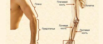
The clavicle is the only bone formation in the human body that connects the skeletons of the upper limb and torso. This paired bone is located directly above the first rib and is part of the shoulder girdle. Its length varies from 12 to 16 centimeters.
It is a long curved bone located between the clavicular notch of the sternum and the acromion process of the scapula. This bone formation is attached to the manubrium of the sternum by the sterno-clavicular joint, and to the acromion (humeral process of the scapula) by the acromioclavicular joint.
The clavicle is the first bone of the human skeleton, the ossification of which begins as early as 5–6 weeks of embryonic development, but its complete ossification ends only by the age of 25.
In its shape, the clavicle is a tubular bone, and in structure it is a spongy bone (its cavity contains spongy substance, and not bone marrow, as in most other bones of the human skeleton).
The bone has a rounded body and two ends: the sternal and acromial. At its sternal end there is a saddle-shaped sternal articular surface designed for articulation with the sternum. The upper surface of the clavicle is completely smooth, and on the lower there are two tubercles: cone-shaped and elongated (trapezoidal line). Muscles and ligaments are attached to these formations, allowing movement of both the collarbone itself and the bones of the upper limbs.
Muscles attached to the clavicle and their places of attachment:
- Deltoid – deltoid tuberosity;
- Sternum - hyoid - medial third of the bone;
- Trapezius - lateral third of the clavicle;
- The sternum major is the rounded edge of the medial third;
- Subclavian – subclavian groove;
- Sternocleidoid – mastoid – medial third.
Ligaments that have a connection to the bone and the place of attachment:
- Trapezoidal – trapezoidal line.
- Conical - a cone-shaped tubercle.
There are many blood vessels and nerves around the collarbone, including the brachial plexus, which is responsible for normal innervation and movement of the upper limb.
- Conductive: acts as a conductor through which physical impulses from the torso travel to the upper extremities.
- Protective: provides protection for lymphatic and blood vessels, nerves located between the neck and arm, from various mechanical damage.
- Supporting: it is this that acts as a kind of support for the upper limb and the scapula, which is also attached to it. In addition, the clavicle provides active mobility of the upper limb and is a direct connection between the arm and torso.
Possible mechanical damage to the collarbone:
- Dislocation: there are two types - acromial or sternal (depending on which end of the bone was damaged). The main symptoms of this injury are: swelling and pain in the collarbone area; inability to move your hand; protrusion of one of the ends of the bone;
- Fracture: In most cases, a clavicle fracture occurs in the middle of the bone's body (at a place called the "diaphysis"). Symptoms: lengthening of the arm that is directly connected to the broken collarbone; difficulties when trying to lift the upper limb; hematomas at the site of injury; swelling and severe pain in the fracture area;
- Bruise: distinguish between direct and indirect. A direct bruise occurs when there is a direct mechanical impact on the bone, while an indirect bruise can be caused by damage to the chest, arm or shoulder. The only sign of a collarbone injury is the appearance of hematomas. In the event of a severe injury of this type, the sensitivity of the hand is lost and its motor ability is reduced.
Non-mechanical pathologies affecting the collarbone:
- Osteolysis: this pathological condition is extremely rare and involves the resorption of bone tissue. This process is associated with the production by the human body of antibodies that have a detrimental effect on it. There are no special signs of this disease. It manifests itself only in the poor ability of the bone to heal during fractures and cracks.
- Arthrosis: this disease most often appears after 40 years in people whose professional activities involve heavy physical activity on their hands. For a long time, arthrosis may not manifest itself in any way, but later a person experiences increased fatigue, limitation in the movement of the upper limbs, pain in the shoulder area and sometimes the crunching of bones in this place is heard.
- Chondroma: This is one of the types of benign neoplasms of the collarbone. The only symptoms of chondroma in humans are pain when moving the bone. Most often, this disease appears in adolescence, since it is during this period that the human skeleton is actively developing, and the increased growth of cartilage tissue can transform into this tumor.
- Neuroma: This is another type of benign tumor that affects the collarbone. Unlike chondroma, this formation does not grow inside the bone, but outside, as a result of which it can be easily felt by palpation. When you press on this swelling, pain impulses are sent to the elbow or shoulder. In addition, in the presence of this pathology, pain may also occur when turning the head sharply.
- Osteomyelitis: is an inflammation of bone tissue that occurs when the human body is exposed to infectious agents. Osteomyelitis is of two types: hematogenous and traumatic. In hematogenous osteomyelitis, pathogenic bacteria penetrate to the collarbone through the circulatory system, and in traumatic osteomyelitis, the development of the inflammatory process is a consequence of suppuration, which can occur with a fracture of the clavicle.
The clavicle is an undeniably necessary bone for a person, which ensures the movement of the upper limbs, without which a person’s life cannot be complete. Therefore, any pathologies associated with this bone can cause irreparable harm to human life and health.
6.Functional classification of the joint (by form and function)
___________________________________________________
muscles:
- The intervertebral ligament stretches over the notch of the jugular cavity of the sternum between the ends of the clavicular bones.
- The sternoclavicular ligament complex. According to their location, they converge on the front, back and top surfaces of the joint, strengthening its strength.
- The most powerful and durable ligament in the sternum is the costoclavicular ligament. It runs from the very top edge at the first rib and rises to the collarbone. Controls the maximum upward elevation of the collarbone.
Why does pathology occur?
Acromioclavicular arthrosis has several causes. First of all, heredity influences the development of degenerative pathology. The likelihood of developing arthrosis depends on the diligence of the parents; in order to prevent the occurrence of pathology, the child should be examined regularly (1 or 2 times a year). The exact extent of the participation of genetic factors in the formation of acromioclavicular arthrosis is unknown, but scientists suggest that heredity still plays an important role in its development. Other causes of the disease:
- Injury. Most often, the disease occurs in loaders, athletes, bodybuilders, mechanics and miners. Broken bones, dislocations, sprains and repeated contusions lead to damage to this joint, which ultimately leads to arthrosis.
- The disease is most often found among people over 50 years of age. The prevalence of degenerative pathology increases with age, because over time all joints of the musculoskeletal system become less flexible and elastic.
- Previous diseases, for example, gout, septic arthritis, and metabolic disorders, can increase the risk of developing acromioclavicular arthrosis.
Diagnosis of changes in the joint
Methods for diagnosing diseases and disorders in the sternoclavicular joint are inspection and palpation, x-ray of the chest bones. All studies are carried out by a traumatologist or osteopath. The presence of any asymmetry or deformation, redness or pain during movements in the sternoclavicular joint, or the appearance of a crunching sound during movement indicate the presence of one of the above diseases or injuries.
Palpation is carried out with the second and third fingers of the right hand, with the doctor positioned behind or to the side of the patient. The fingers are placed in the middle of the sternum and, focusing on the notch under the patient’s neck, they feel the joint. For better detection, the patient is asked to raise his arms in a horizontal plane, which greatly facilitates the search.
The sternoclavicular joint is simple in structure. But at the same time it is quite strong, keeping the limbs attached to the body. When this joint is damaged, hand movements become very limited and cause pain.
The sternoclavicular joint, articulatio sternoclavicularis, is formed by the clavicular notch of the sternum and the sternal end of the clavicle. The joint is simple.
The articular surfaces are covered with connective tissue cartilage, incongruent and most often saddle-shaped. The discrepancy between the articular surfaces is leveled by a composite disc located in the joint cavity.
Sternoclavicular joint (articulatiostemoclavicularis)
| The sternoclavicular joint (articulatiostemoclavicularis). Front view. On the left side of the specimen, the joint is opened with a frontal incision. 1-clavicle (right); 2-anterior sternoclavicular ligament; 3-interclumbar ligament; 4-sternal end of the clavicle; 5-intra-articular disc (fudinoclavicular joint); 6-first (I) rib; 7-costoclavicular ligament; 8-fudinocostal joint (11th rib); 9-intra-articular sternocostal ligament; 10th cartilage of the 11th rib; 11-synchondrosis of the handle of fudina; 12-radiate fudinocostal ligament. The sternoclavicular joint. Front view. On the left side of the specimen, the joint is opened with a frontal incision. 1-clavicula (dextra); 2-ligamentum sternoclaviculare anterius; 3-ligamentum interclaviculare; 4-extremitas sternalis claviculae; 5-discus articularis (articulatio sternoclavicularis); 6-costa (I); 7-ligamentum costoclaviculare; 8-articulatio sternocostalis (II); 9-ligamentum sternocostalis intraarticulare; 10-cartilago costae (II); ll-synchondrosis manubrii sterni; 12-ligamentum sternocostale radiatum. Sternoclavicular joint. Anterior aspect. Frontal section of joint at left. 1-clavicle (right); 2-anterior Sternoclavicular ligament; 3-interclavicular ligament; 4-sternal end ot'clavicle; 5-articular disc (ol'sternoclavicular joint); 6-1 rib; 7-costoclavicular ligament; 8-sternocostal joint (of the II rib); 9-intra-articular sternocostal ligament; 10-cartilage of the II rib; 11-manubriosternal synchondrosis; 12-radiate sternocostal ligament |
Atlas of human anatomy. Akademik.ru. 2011.
Author of the article: Vasily Shevchenko
Let me introduce myself. My name is Vasiliy. I have been working as a massage therapist and chiropractor for over 8 years. I believe that I am a professional in my field and want to help all site visitors solve their problems. All data for the site has been collected and carefully processed in order to convey all the required information in an accessible form. Before using anything described on the site, a MANDATORY consultation with your specialist is always necessary.
✔
About me
✉
Feedback
To make a diagnosis and determine changes in the clavicular joint that cause pain and other unpleasant symptoms, a comprehensive examination of the node is performed. Depending on the severity of the injury or suspicion of degenerative disease processes within the body, diagnostics are carried out using traditional therapeutic and instrumental methods.
Visual inspection
Since the anterior sternoclavicular ligament is clearly visible only in people who do not have sufficient fat, the diagnosis of damage is carried out as follows: the patient is asked to raise his shoulders synchronously. In a healthy state, the clavicular protrusions are symmetrical, the skin is smooth, without swelling or discoloration, and there is no pain. If changes are noticeable, an additional puncture of the area is performed.
Palpation of the area
The doctor performs manual palpation of the clavicular node by standing to the side or behind the patient. The manipulation is performed with the thumb and index finger of one hand. Palpation is carried out from the notch under the neck where the joint is located and to the area that begins to hurt when an injury or pathology is diagnosed. To facilitate examination through palpation, the patient should raise and extend his arms forward.
The most traditional method of hardware diagnosis of the clavicular region is chest radiography. The method allows for comprehensive monitoring of the clavicular joint and the surrounding area. The picture clearly shows the characteristics of deviations, if any. Complex cases of arthrosis diseases require additional diagnosis using an MRI machine.
Pathology treatment methods
It is essential that the patient and doctor have a good relationship and talk openly about the hope of recovery, expectations and risks of treatment. When arthrosis of the acromioclavicular joint is treated, various methods are used to control pain and slow down the progression of the disease. If the patient responds well to non-surgical treatment, the degenerative process may slow down. If conservative therapy does not bring the desired result, surgical intervention is necessary.
Non-surgical therapies
The ability to perform exercises will be limited at first and will be supervised by a physiotherapist. The physical therapy program is developed based on the general condition of the patient, the course of the disease and the person’s age. The exercises are focused on stretching and strengthening muscles. The training complex changes every day; it is necessary to develop it for all muscle groups and joints affected by degenerative pathology. Conservative treatment is carried out with the use of medications. To relieve symptoms and slow the progression of arthrosis, use:
- analgesics, drugs have few side effects and relieve pain well;
- nonsteroidal anti-inflammatory drugs such as Indomethacin, Aceclofenac, Nimesulide or COX-2 inhibitors, for example, Celebrex;
- Doctors prescribe topical anti-inflammatory drugs such as Voltaren.
Topical analgesics (creams) contain counteracting agents, such as eucalyptus, which stimulate nerve endings and help distract the brain from joint pain. Examples of brand names include "Ben-Gay", "Icy-Hot" and "Zostrix". If necessary, steroid injections are given to relieve swelling, reduce stiffness and pain.
Surgical intervention for the last stages of the disease
After diagnosing the third or fourth stage of the disease, the patient is offered surgery. Conservative treatment, physiotherapy and folk remedies are ineffective at the last stage of the pathology. Doctors offer the following types of operations:
- Arthroscopy to remove loose fragments of damaged cartilage.
- Osteotomy reduces friction between bones and eliminates formed osteophytes.
- Endoprosthetics or complete replacement of the acromioclavicular joint.
Arthroplasty and hemiarthroplasty can reduce or completely eliminate pain. However, such operations require long-term recovery and rehabilitation. In addition, the formed false connecting joint will have to be replaced again after some time. Surgery is a serious step in which it is necessary to weigh the positive aspects and study possible complications after the operation. It is also necessary to approach the choice of a surgeon seriously; the operation cannot be trusted to a novice who has just received a diploma.
If you find an error, please select a piece of text and press Ctrl+Enter.