Why does flat feet occur?
Foot deformity can be congenital or acquired. There are many reasons for this:
- fractures, injuries, ankle sprains;
- injuries of the foot, heel and tarsal bones;
- rickets, against which the bones lose density and suffer from excess load;
- weakness of muscles, ligaments and bones;
- metabolic disorders;
- excess body weight;
- incorrect, narrow, uncomfortable shoes.
The percentage of congenital flat feet is not high - only 3%. The foot is deformed due to joint dysplasia. Regardless of the source of the problem, it must be addressed, since the pathology disrupts the normal biomechanics of movement and contributes to diseases of the musculoskeletal system.
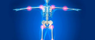
Flat feet combined with excess weight are a huge threat to your joints.
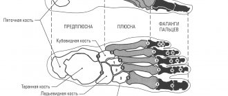
Anatomy of the joints of the human foot - information:
In the articulations between the tarsal bones, articulationes intertarseae, there are 4 joints:
- Subtalar joint , art. subtalaris, is formed by the posterior articular surfaces of the talus and calcaneus, generally representing segments of a cylindrical surface.
- Talocaleonavicular joint , art. talocalcaneonavicularis, lies anterior to the subtalar and is made up of the almost spherical head of the talus, the corresponding articular cavity formed by the scaphoid bone, the articular facet on the sustentaculum tali of the calcaneus and lig. calcaneonaviculare plantare, filling the gap between the sustentaculum and the posterior edge of the os naviculare and containing in its thickness a layer of fibrocartilage, fibrocartilago navicularis. The articular capsule is reinforced on the back side with lig. talonavicular and from the plantar side lig. canacaneonaviculare plantare. Between both of these joints there passes a bone canal - sinus tarsi, in which lies a strong ligament, lig. talocalcaneum interosseum, stretching between the talus and calcaneus.
- Calcaneocuboid joint , art. calcaneocuboidea, formed by the articular surfaces of the calcaneus and cuboid bones facing each other. It takes part in the movements of the subtalar and talocalcaneal-navicular joints, increasing their volume. Art. calcaneocuboidea together with its neighboring art. talonaviculare is also described under the general name of the transverse tarsal joint, art. tarsi transversa. In addition to the ligaments that strengthen the art. calcaneocuboidea and art. talonavicularis separately, the transverse joint also has a ligament common to both joints, which is very important in its practical significance. This is lig. bifurcatum - a ligament whose posterior end originates on the upper edge of the calcaneus and then divides into two parts, one of which is lig. calcaneonaviculare, is attached to the posterolateral edge of the scaphoid, and the other, lig. calcaneocuboideum, grows onto the dorsal surface of the cuboid bone. This short but strong ligament is the “key” of the transverse joint, since only by cutting it can a wide divergence of the articular surfaces be achieved during the operation of isolating the foot in the named joint.
- Wedge-scaphoid joint , art. cuneonavicular, formed by articulation of the posterior articular platforms of the sphenoid bones with three facets of the distal articular surface of the scaphoid bone. As for the movements in artt. intertarseae, here, first of all, the calcaneus rotates together with the navicular and the anterior end of the foot around the sagittal axis with a range of movements of 55° (this axis goes obliquely, entering the head of the talus on the back side and exiting from the side of the sole on the lateral surface of the calcaneus) . When the foot rotates inward (pronation), its lateral edge is raised, and the back of the foot turns medially; on the contrary, when rotating outward (supination), the medial edge is raised with the dorsum of the foot turning to the lateral side. In addition, adduction and abduction around a vertical axis are possible here, when the tip of the foot deviates from the midline medially and laterally. Finally, there may also be extension and flexion around the frontal axis. Movements around three axes are also performed in art. talocalcaneonavicularis, which is a complex spherical joint. All these movements are small and usually combined together, so that at the same time as supination there is adduction of the forefoot and slight flexion, or vice versa: pronation is accompanied by abduction and extension. In general, the ankle joint in combination with artt. intertarseae allows great freedom of movement of the foot like a multi-axis joint.
Tarsometatarsal joints , artt. tarsometatarseae, connect the bones of the second row of the tarsus with the metatarsal bones. Artt. tarsometatarseae - typical tight joints, slight mobility in which serves to give elasticity to the arch of the foot. Separate articular capsules have articulations of the first metatarsal bone and the medial cuneiform, and articulations of the second and third metatarsals with the cuboid. The tarsometatarsal joints are supported by the dorsal, plantar and interosseous ligaments, ligg. tarsometatarsea dorsalia, plantaria et cuneometatarsea interossea. Intermetatarsal joints, artt. intermetatarseae, are formed by the surfaces of the metatarsal bones facing each other; their joint spaces often communicate with the artt cavity. tarsometatarseae. The joints are strengthened by transversely running ligg. metatarsea dorsalia, plantaria et interossea. Articulations of the finger bones:
- Metatarsophalangeal joints , artt. metatarsophalangeae, between the heads of the metatarsal bones and the bases of the proximal phalanges, in the nature of the structure and ligamentous apparatus are similar to similar articulations of the hand. Movements in the joints are generally the same as in the hand in the corresponding joints, but limited. Apart from the slight abduction of the fingers to the side and the reverse movement (adduction), then there is only extension and flexion of all fingers, and extension occurs to a greater extent than flexion, in contrast to what we have on the hand.
- Interphalangeal joints , artt. interphalangeae pedis, do not differ in their structure from similar joints on the hand. It should be noted that often the distal and middle phalanges on the fifth finger are fused together boney. The joints of the foot are vascularized from the branches of the arcus plantaris u r. plantaris profundus a. dorsalis pedis. Venous outflow occurs into the deep veins of the lower limb - vv. tibiales anterior et posterior, v. peronea. The outflow of lymph is carried out through deep lymphatic vessels in the nodi lymphatici poplitei. The innervation of the joint capsules is provided by the branches of the nn. plantares medialis et lateralis u nn. peronei superficialis et profundus.
Why is this problem more pressing for women?
Most often, knee arthrosis and flat feet are diagnosed simultaneously in women. The reason is wearing narrow shoes with high heels. Shoes compress the toes and reduce the permeability of blood vessels. There is a deficiency of vitamins and microelements. The cartilage begins to deform and is not restored to its original shape. The next step is treatment of osteoarthritis.
Women are more susceptible to arthrosis along with flat feet, since endocrine changes in the female body occur more often and more intensely than in the male body. This problem is especially acute during menopause.
Women are more susceptible to arthrosis and flat feet
What symptoms indicate that you have arthrosis and flat feet?
The problem can be recognized by the following signs:
- foot pain;
- swelling;
- tired legs;
- change in gait;
- curvature of the bones of the foot.
At first, the pain appears sporadically. Then it intensifies and manifests itself even at rest. Pain from the foot is transmitted upward - to the lower back and hip joint. After a night's rest, a person experiences relief.
After active movement, swelling occurs on the foot. To get rid of pain and discomfort, you have to rest for several hours. It becomes increasingly difficult to squat and maintain balance as the midfoot gradually expands.
If flat feet and arthrosis of the ankle or knee progress, the gait changes. From the side, a heavy gait and club feet are noticeable. Posture is disturbed - problems with blood circulation and new diseases of other systems and organs arise.
Flat feet lead to a whole chain of problems
Pain in the joints of the foot
Very often, doctors hear complaints from patients about foot pain when walking. This symptom for various diseases can be of a different nature: “aching”, sharp, cutting, burning, etc. Depending on the place of manifestation, it can be localized or diffuse. Therefore, before self-medicating and taking painkillers, you need to consult a doctor and find out the cause.
Flat feet
- a change in the shape of the foot, characterized by drooping of its longitudinal and transverse arches. There are primary, transverse and longitudinal flat feet, a combination of both forms is possible.
Signs:
• Pain in feet, knees, hips, back
• Unnatural gait and posture
• It is easier to bend over than to squat; crouching down, it’s hard to maintain balance
• “Heavy” gait
• Clubfoot when walking
• Deformed feet (flat feet, crooked, disproportionately long toes, bunions on the big toe, overly wide feet), deformation of the knee joints, disproportionate development of the muscles of the legs and lower legs
• Flat feet predisposes to the development of ingrown toenails
Foot pain can also be caused by:
Diabetic foot syndrome
- a complex of anatomical and functional changes that develop against the background of diabetic neuropathy, micro- and macroangiopathy, osteoarthropathy, contributing to increased trauma and infection of the soft tissues of the foot, the development of a purulent-necrotic process and, in advanced cases, leading to amputation.
Clubfoot
- deformation of the foot, in which it deviates inward from the longitudinal axis of the lower leg. Clubfoot can be equinovarus, when the patient’s foot is turned down and inward, varus, in which the heel is turned inward, and valgus, in which the heel is turned outward.
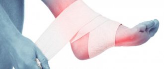
Metatarsal fracture
- fracture of the metatarsal bone of the foot. The fracture is often located in the midfoot. Patients with a fracture experience pain in the area, swelling, and difficulty walking
Neuritis
- an inflammatory disease of the peripheral nerves, in which, along with pain, symptoms of so-called loss are detected, i.e. loss or decrease in sensitivity, as well as paralysis and paresis.
Gout
is a metabolic disease characterized by the deposition of urate crystals in various tissues of the body in the form of monosodium urate or uric acid.
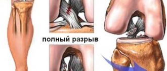
Dislocation
- violation of the congruence of the articular surfaces of bones, both with and without violation of the integrity of the articular capsule, under the influence of mechanical forces or destructive processes in the joint
Treatment at the Naran Clinic and results
Experienced and qualified doctors of the Naran clinic, first of all, will identify the cause of such symptoms using oriental diagnostic methods. A thorough survey, examination, pulse examination and palpation will together give an overall picture of the patient’s body condition and indicate what is causing pain in the foot area.
The Naran Clinic uses in its medical practice a set of methods that are combined with each other depending on the general condition of the patient, the course of the disease, age status, constitutional affiliation, the presence of concomitant pathological disorders and other factors.
The methods of Tibetan medicine are divided into two types - external effects on the body and internal ones. The first type refers to procedures using acupuncture, acupressure, moxotherapy (warming with wormwood cigars), vacuum therapy (cupping), cupping massage, the Mongolian massage method “khorme” using medicinal herbs and oils, herbal compresses, etc. . By internal action we mean Tibetan herbal remedies - unique medicinal products, compiled according to the recipes of ancient Tibetan emchi monks, the naturalness and power of which cannot be doubted. The most advanced Westerners have long appreciated the power of precious Tibetan herbal medicines.
The combined effect of all methods shows
excellent medical results:
- Removes congestion and blockages in the flow of qi
and blood - Improves blood flow and lymph flow
- “Removes” the resulting stagnation, allowing internal organs to function normally
- Nourishes all systems and tissues of the human body
- Affects the immediate cause of the disease and eliminates it
- Has a beneficial healing effect on the entire body
- Strengthens immunity
How is arthrosis treated for flat feet?
Since the patient has a history of two dangerous diseases of the musculoskeletal system, treatment must be comprehensive. Depending on the severity, clinical manifestations, and intensity of pain, the following measures and medications are prescribed:
- painkillers and non-steroidal anti-inflammatory drugs, for example diclofenac, ibuprofen, ketoprofen;
- for muscle spasms of the foot - muscle relaxants to relax muscle tissue;
- chondroprotectors – to stimulate the growth of cartilage tissue and partially reduce pain;
- in difficult cases - surgical intervention (surgical correction of bone deformities, cleaning bones from osteophytes or endoprosthetics of a damaged joint).
What is the real danger of flat feet and can it be cured?
Symptoms of arthrosis of the foot
The symptoms of the disease are varied. The degree of its severity may vary depending on the stage of the disease: from mild pain during prolonged standing work to curvature of the fingers and a noticeable change in gait. The most common symptoms of arthrosis of the feet:
- Crunching or squeaking noises when moving (crepitus).
- Swelling, redness of the skin at the site of pathology;
- Painful sensations during physical activity, which lose their severity during rest.
- Weather dependence: dull pain in the joints when the weather changes.
- Deformation of the joints of the feet, increase in their size.
- A change in gait caused by the patient’s natural desire to unload the affected joint.
- Fever.
- Deterioration in performance, increased fatigue, formation of characteristic calluses.
- When the disease is advanced, there is significant deformation of the joints, which becomes noticeable even during an external examination of the leg.
If you notice these symptoms, we recommend that you make an appointment with a doctor to undergo an examination and appropriate treatment for arthrosis of the joints of the foot or heel .
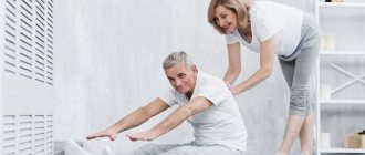
Will physiotherapy help in such cases?
Physiotherapy is also effective in reducing pain. After a course of procedures, there is no need to take large doses of medications, therefore, along with drug treatment, physiotherapy is often prescribed.
- Manual therapy is aimed at correcting the position of the foot joints, correcting the heel and metatarsal bones, and toes. Chiropractors effectively restore blood circulation in the tissues around the damaged joint, especially if their sessions are combined with massage.
- Mud baths and clay wraps will improve muscle tone, increase blood circulation, and activate metabolic processes.
- The same problems are solved by other physical procedures, for example magnetic or laser therapy.
Physiotherapy does not treat flat feet or arthrosis: it is an auxiliary method
How to avoid arthrosis if you have flat feet
Flat feet today are easily corrected with the help of orthopedic insoles and special shoes. With arthrosis, the situation is more complicated: the disease has no cure and tends to progress. Therefore, if you have a deformed foot, remain vigilant so as not to trigger the disease. What can you do?
- Avoid wearing tight shoes or high heels except occasionally.
- It is no coincidence that orthopedists and rheumatologists recommend losing weight. If you have extra pounds, the heel bone deforms faster, as do the joints of your toes. Heart function deteriorates, coxarthrosis and gonarthrosis, spondyloarthrosis, and other diseases develop. Therefore, reconsider your diet and physical activity.
- Try not to overexert your feet. Change positions more often, do not squat, do not cross your legs.
- Take part in safe exercise, such as walking or swimming.
- Walk barefoot on grass or sand more often - this is an excellent prevention of arthrosis with flat feet.
Arthrosis is a serious and dangerous disease. If there is a history of flat feet, the situation becomes more complicated, but by no means becomes hopeless. Reconsider your lifestyle and habits, take care of your joints - and they will serve you faithfully for many years!
The harm and benefits of heels
Many of us women love heels. And this is not surprising, because putting on such shoes, you immediately feel slimmer and more beautiful. Unfortunately, when worn for a long time, high heels pose a health risk.
Features of the anatomy of the foot
The main function of the foot is shock absorption, i.e., distributing the load when running and walking on two legs. Thanks to the movable bone connections (joints) and the special shape of the arches of the feet, the body weight is distributed to the lower spine, pelvic bones and joints of the lower extremities so that we can walk and run without experiencing pain or discomfort.
If even a small part of this well-functioning system of body weight distribution changes, then problems begin. For example, flat feet or arthrosis of the knee joint can lead to impaired gait biomechanics, improper weight redistribution and increased load on the hip joints or lower spine with subsequent damage.
Damage from high heels
You may ask, what does the heel have to do with it? It has been proven that a heel height of only 2-3 cm increases the load on the forefoot by 22%, and a heel of 6 cm increases the load on the forefoot by 60%! This means that with prolonged loading (for example, for several months), the forefoot (toe joints, metatarsal joints) will begin to deform under the influence of excess load, and pathological bone growths (osteophytes) will appear.