Tarsal joints
Connections of the bones of the foot.
The ankle joint, articulatio talocruralis, is formed by the articular surfaces of the distal ends of the tibia and fibula.
On the fibula there is an articular surface of the ankle, fades articularis malleolaris.
The articular surface of the talus from above has the shape of a block, trochlea, and on the sides it is represented by flat articular platforms - the lateral and medial malleolus surface, fades malleolaris lateralis
et
medialis.
The fork-shaped bones of the lower leg cover the block of the talus.
Articular capsule, capsula articularis,
over a long stretch, it is attached along the edge of the articular cartilage and only in front of it, on the surface of the body of the talus, it recedes somewhat, attaching to the neck of the talus. The anterior and posterior sections of the articular capsule are weakly stretched.
Ankle ligaments
lie on its lateral surfaces.
Medial (deltoid) ligament, lig. mediale (deltoideum) is divided into the following parts:
• anterior tibiotalar part, pars tibiotalaris anterio
extends from the anterior edge of the medial malleolus downward and attaches anteriorly to the posteromedial surface of the talus.
• tibiofacial part, pars tibionavicularis,
longer than the previous one, starting from the medial malleolus and reaching the dorsum of the scaphoid bone;
• tibiocalcaneal part, pars tibiocalcanea,
stretched between the end of the medial malleolus and the sustentaculum tali;
• posterior tibiotalar part, pars tibiotalaris posterior
runs from the posterior edge of the medial malleolus downwards and is laterally attached to the posteromedial parts of the body of the talus.
The following ligaments lie on the lateral surface of the ankle joint:
• anterior talofibular ligament, lig. taloflbulare anteriusM
follows from the anterior edge of the lateral malleolus to the lateral surface of the neck of the talus;
• calcaneofibular ligament, lig. calcaneoflbulare,
begins on the outer surface of the lateral malleolus and, going down and back, attaches to the lateral surface of the exact bone;
• posterior talofibular ligament, lig. taloflbulare posteriu
runs from the posterior edge of the lateral malleolus almost horizontally to the lateral tubercle of the posterior process of the talus.
The ankle joint is a type of trochlear joint - a helical joint.
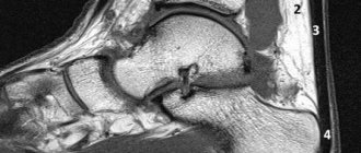
The subtalar joint, articulatio subtalaris, is formed by the posterior articular surface of the calcaneus, fades articularis posterior calcanei, and the posterior calcaneal articular surface of the talus, fades articularis calcanea posterior tali.
Articular capsule, capsula articularis,
weakly stretched, attached over a long distance along the edge of the articular cartilages, and only in front - on the talus and behind - on the calcaneus, it recedes somewhat from the edge of the articular surfaces.
The ligaments that strengthen this joint include.
• interosseous talocalcaneal ligament, lig. talocalcaneum interosseum,
located in the sinus tarsi, attaching its ends to the sulcus tali et sulcus calcanei;
• lateral talocalcaneal ligament, lig. talocalcaneum laterale,
stretched between the upper surface of the neck of the talus and the superolateral surface of the calcaneus;
• medial talocalcaneal ligament, lig. talocalcaneum mediale,
goes from the posterior process of the talus to the suspensory process of the calcaneus.
The talocalcaneal-navicular joint, articulatio talocalcaneo-navicularis, is formed by the articular surfaces of the talus, calcaneus and navicular bones. The talus forms the articular head,
and the calcaneus and navicular bones are
the glenoid fossa.
The articular capsule, capsula articularis, is attached along the edge of the articular cartilage.
The joint is strengthened by the following ligaments:
• talonavicular ligament, lig. talonaviculare,
stretched between the neck of the talus and the scaphoid;
• plantar calcaneonavicular ligament, lig. calcaneonaviculare plantare,
follows from the sustentaculum tali to the plantar surface of the scaphoid. The upper part of this ligament passes into the scaphoid fibrous cartilage, which takes part in the formation of the articular fossa of the joint.
Talocaleonavicular joint
in shape it belongs to
the spherical joints, articulatio spheroidea, but movements
The calcaneocuboid joint, articulatio calcaneocuboidea, is formed by the posterior articular surface of the cuboid bone, fades articularis posterior ossis cuboidei, and the cuboid articular surface of the calcaneus, fades articularis cuboidea
The articular surfaces of the calcaneocuboid joint are saddle-shaped. Articular capsule, capsula articularis,
in the medial section it is attached to the edge of the articular cartilage and stretched tightly, and in the lateral section it is attached slightly from the edge of the articular cartilage.
The joint is strengthened by a number of ligaments, which are more developed on the plantar side:
• long plantar ligament, lig. plantare longum,
the most powerful It begins on the lower surface of the tubercle of the calcaneus and, moving forward, is thrown through the sulcus ossi cuboidei, forming an osteofibrous canal; reaches the bases of the II-V metatarsal bones. The deep bundles of this ligament, which are shorter, are attached to the tuberosity of the cuboid bone
• plantar calcaneocuboid ligament, lig. calcaneocuboideui, plantare,
is deeper than the previous ligament.
Its bundles are adjacent directly to the articular capsule and connect the plantar surfaces of the calcaneus and cuboid bones. The calcaneocuboid joint is similar in the sella sella, articulatio sellaris, but
a uniaxial joint
The transverse tarsal joint, articulatio tarsi transversa, combines two joints:
• talocalcaneonavicularis, articulatio talocalcaneonavicularis,
• calcaneocuboid, articulatio calcaneocuboidea.
The joint line is curved in an S-shape.
• the medial section is convexly facing forward;
• lateral - back.
The joints are anatomically separate, but have a common bifurcated ligament, lig. bifurcation. This ligament begins on the surface of the calcaneus v
its
anterior edge and is immediately divided into two ligaments: • lateral calcaneocuboid ligament, lig. calcaneocuboideun
heading towards the dorsum of the cuboid bone;
• medial - calcaneonavicular ligament, lig. calcaneonavicular
going to the scaphoid bone.
Bifurcated ligament, lig. bifurcatum, also called the key of the transverse tarsal joint,
since after cutting all the ligaments located in the circumference of this joint, it holds the bones in the described joint, and only after cutting the lig. bifurcatum, it is possible to isolate the foot in this joint during surgery.
The wedge-scaphoid joint, articulatio cuneonavicularis, is a complex joint in the formation of which the scaphoid, cuboid and three sphenoid bones take part.
The following joints are formed here:
• wedge-navicular joint, articulatio cuneonavicularis,
between the anterior articular surfaces of the scaphoid and the posterior articular surfaces of the medial, intermediate and lateral sphenoid bones;
• joints between the facing surfaces of the cuboid, scaphoid and sphenoid bones.
Articular cavity
between the scaphoid and sphenoid bones is located in the frontal plane, and from it three
• between the medial, intermediate and lateral sphenoid bones;
• lateral wedge-shaped and cuboid;
• one joint space back - between the scaphoid and cuboid. Articular capsule, capsula articularis,
attached to the edge of the articular cartilage. The joint cavity communicates through the gap between the medial intermediate and lateral cuneiform bones with the cavity of the tarsometatarsal joint, articulatio tarsometatarsea, in the region of the second metatarsal bone.
8. Tarsometatarsal joints, articulationes tarsometatarseae,
connect the tarsal bones to the metatarsal bones. There are three tarsometatarsal joints:
• between the medial cuneiform and first metatarsal bones;
• between the intermediate and lateral cuneiform and II-III metatarsal bones;
• between the cuboid and IV-V metatarsal bones.
The joint between the medial cuneiform and first metatarsal bones is formed The line of the joint space of the tarsometatarsal joints is uneven, since the second metatarsal bone is longer than the others, and the lateral cuneiform bone protrudes somewhat forward compared to the anterior part of the cuboid bone.
Articular capsule, capsula articularis,
each of the tarsometatarsal joints is attached along the edge of the articular cartilages and
is supported by the following ligaments: • dorsal
tarsometatarset dorsalia,
located on the dorsum of the joints;
• plantar tarsometatarsal ligaments, ligg. tarsometatarsee plantaria,
located on the plantar surface;
• interosseous metatarsal ligaments, ligg. metatarsea interossea,
located between the bases of the metatarsal bones;
• interosseous wedge-metatarsal ligaments, ligg. cuneometatarseq interossea,
connect the sphenoid bones to the metatarsal bones. The middle (medial) of them connects the medial sphenoid bone with the base of the second metatarsal bone and is the “key” of the tarsometatarsal joints. These joints are classified as low-moving joints.
The intermetatarsal joints are located the direction of the ligaments that strengthen these joints is basically the same as on the hand. Articular capsules, capsulae afticulares,
strengthened by the following
• interosseous metatarsal ligaments, ligg. metatarsea interossea;
• dorsal metatarsal ligaments, ligg. metatarsea dorsalia;
• plantar metatarsal ligaments, ligg. metatarsea plantaria.
The intermediate spaces between the individual metatarsal bones are called interosseous spaces of the metatarsus spatia interossea metatarsi.
Metatarsophalangeal joints, articulationes metatarsophalangeae,
formed by the articular surfaces of the heads of the metatarsal bones and the bases of the proximal phalanges. The heads of the third metatarsal bones have an irregularly spherical shape, their dorsal part is somewhat narrowed.
Articular capsules, capsulae articulares,
attached along the edge of the articular cartilages, weakly stretched.
The dorsal part of the articular capsules is thinned on the side of the plantar surface; they are strengthened by plantar plantaria,
and on the sides by collateral
collateralia.
In addition, a deep
metatarseum transversum profundum
Metatarsophalangeal joints belong to the type of spherical joints, articulatio spheroidea.
Interphalangeal joints of the foot, articulationes interphalangeae pedis,
connect the proximal phalanges with the middle ones, and the middle ones with the distal ones.
Articular capsules, capsulae articulares,
These joints are thin.
Their lateral sections are supported by collateral collateralia,
and on the plantar side -
by the plantar ligaments, ligg.
plantaria Interphalangeal joints belong to the type of trochlear joints, ginglymus.
Metatarsal fractures
Dancer's fracture (avulsion fracture of the base of the 5th metatarsal)
Avulsion fractures occur at the base of the 5th metatarsal (zone 1), where the peroneus brevis tendon and plantar fascia attach.
This injury is often called a “dancer’s fracture” because it occurs when landing poorly after a jump or twisting the foot after a jump. In such a situation, the ankle joint twists while the peroneus brevis muscle contracts, which leads to a separation of the base of the 5th metatarsal bone. The original description of this term belongs to the legendary orthopedist Sir Robert Jones, who in 1902 diagnosed himself with such an avulsion fracture as a result of an injury received while dancing.
Rice. X-ray of a dancer's fracture (avulsion fracture) in zone 1 of the 5th metatarsal bone.
Jones' fracture (stress fracture of the metadiaphysis of the 5th metatarsal)
A true Jones fracture is a fracture in the 2nd zone of the 5th metatarsal. The line of such a fracture extends to the area of the articulation of the 5th metatarsal bone with the fourth. The fracture occurs due to tensile loads along the outer 5th metatarsal bone when the foot rolls in. This situation often occurs in patients with high arches. Most Jones fractures are stress fractures associated with repetitive stress, although they can result from a single injury. In an athlete, such an injury may result from a sudden change in running direction, when the heel bone leaves the ground.
Rice. Jones fracture in the metadiaphyseal zone of the 5th metatarsal bone.
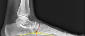
Metatarsal base fractures and Lisfranc injuries
Fractures in the area of the bases of the metatarsal bones are often accompanied by damage to the tarsometatarsal joints - Lisfranc injuries. To detect such injuries, the doctor must evaluate the radiographs very carefully. Signs of Lisfranc injury may include an increase in the interval between the 1st and 2nd metatarsals, small fractures in the area of the bases of the 1st and 2nd metatarsals, and disruption of the normal relationship between the edge of the sphenoid and the base of the 2nd metatarsal. To exclude this damage, computed tomography (CT) is the most informative.
If a Lisfranc injury is suspected, even if nothing is visible on radiographs, an MRI may also be indicated.
Metatarsal stress fractures
Metatarsal stress fractures are rarely visible on x-rays at first and only become visible 5 to 6 weeks after the onset of symptoms when callus appears. Before this period, the diagnosis can be established on the basis of MRI or scintigraphy. Stress fractures of the 2nd and 3rd metatarsals usually occur at the level of the diaphysis or neck. Often such fractures occur with a sudden increase in physical stress, for example, among army recruits during long marches. Therefore, such fractures are also called “marching” fractures. Ballet dancers who frequently stand on their toes may experience stress fractures at the base of the 2nd metatarsal.
Rice. Stress fracture of the 2nd metatarsal bone.
Neuropathic metatarsal fractures
Patients with decreased sensation in the foot, for example due to diabetic neuropathy, may also develop stress fractures of the metatarsals. A common location for such fractures, especially in patients with high arches or varus deformity of the lower extremity, is the metadiaphyseal region of the 5th metatarsal bone (Jones fracture).
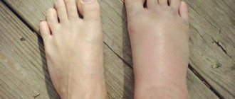
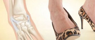
Important muscles and ligaments of the foot. Treatment of flat feet.
Stability and mobility in joints depends on three components - the shape of the bones, the condition of the capsule and ligamentous apparatus that connects the bones, forming the joint, and the muscles that surround it.
Acquired flatfoot in adults is associated with weakening of the tibialis posterior muscle (m. tibialis posterior), which in turn leads to degradation of the plantar calcaneonavicular ligament (lig. calcaneona-viculare plantare) and destruction of the bony arch of the foot.
The basis of acquired flatfoot in adults is a muscle imbalance - a weakened posterior tibialis muscle (m.tibialis posterior) on the medial side, the tendons of which pass to the sole and an overstrained short peroneus muscle (m.peronaeus brevis), belonging to the lateral group, which is an antagonist. This imbalance puts undue stress on the plantar calcaneonavicular ligament (lig. calcaneona-viculare plantare), which ultimately leads to collapse of the medial arch of the foot and pain.
The true cause of the disease is unknown, but it should be assumed that the cause of flat feet lies in several factors, one of which, scientifically based, is muscle imbalance. In addition, there is stiffness of the Achilles tendon and the muscles associated with it - the gastrocnemius and soleus (stretching these muscles can be used to prevent and treat acquired flat feet). Women are more susceptible to this disease than men, especially in their sixth decade.
The plantar calcaneonavicular ligament (lig. calcaneona-viculare plantare) connects the calcaneus (heel) to the navicular. The shape of the navicular bone, which is located on the medial side in the center of the foot, resembles the shape of a boat. This ligament is an important stabilizer of the medial longitudinal arch of the foot.
The key muscle supporting the plantar calcaneonavicular ligament is the tibialis posterior. This muscle originates from the interosseous membrane at the level of 2/3 of the height of the posterior parts of the fibula and tibia. Having passed under the plantar calcaneonavicular ligament, the tibialis posterior muscle is divided into two bundles; one part is attached to the tuberosity of the scaphoid, and the other is again divided into bundles, which are attached to the second through fourth metatarsal bones and to the second sphenoid bone.
The main function of the tibialis posterior muscle is to supinate the foot (turn the sole inward), and also helps to adduct the leg and flex the ankle joint.
This muscle is an important stabilizer of the midfoot during walking when the heel is up. The anterior tibialis muscle (m.tibialis anterior), which runs medially in the area of the middle of the foot, helps to turn the sole inward (supinate).
The antagonist of the posterior tibial muscle is the short peroneus muscle (m.peronaeus brevis), which starts from the lower half of the lateral surface of the fibula and from the intermuscular septum of the leg, goes down and runs near the tendon of the long peroneus muscle. Having wrapped around the lateral malleolus at the back, the tendon runs forward along the outside of the calcaneus and attaches to the tuberosity of the fifth metatarsal bone. Its function is to flex and pronate the foot (turn the sole outward), lowering its medial edge and raising the lateral edge, as well as abduct the foot. It helps stabilize the transverse arch of the foot.