3 February 2020
118711
0
4.1 out of 5
Spinal fractures account for about 2–2.5% of injuries. Among them, the most common are compression fractures, which are a serious test for the human body and psyche. They can occur due to a wide variety of reasons, but always require immediate treatment with conservative, and in severe cases, surgical treatment.
What is a compression fracture?
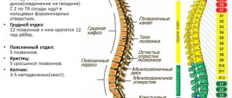
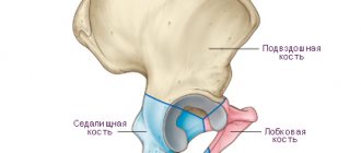
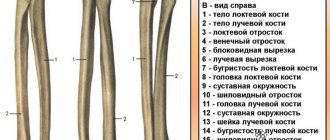
The spine is formed by 33 vertebrae, each of which has a body and 2 arches with processes of different sizes. The processes of the arches of adjacent vertebrae are connected to each other, forming intervertebral joints, and intervertebral discs are located between the surfaces of the vertebral bodies to prevent friction and soften impacts when walking and other loads.
The spine of a healthy person can withstand a load of up to 400 kg.
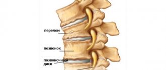
With severe compression, compression of one or an entire group of vertebrae is observed, as a result of which their shape changes, and cracks or even separation of bone fragments may occur. Therefore, a compression fracture is a back injury in which flattening occurs, i.e., a decrease in the height of one or more vertebrae under the influence of a perpendicularly directed load on the spine. In most cases, they are observed in the thoracic and lumbar region. Compression fractures of the cervical vertebrae practically do not occur.
Causes of compression fractures
Most often, compression fractures occur in older people. This is due to the fact that they often suffer from osteoporosis, i.e. a disease of bone tissue that causes a decrease in density and thinning of bones. As a result, bones, and vertebrae in particular, become fragile and easily break even under the influence of minor traumatic factors. Sometimes a patient with advanced osteoporosis only needs to make a careless movement, bend over, or even cough for a compression fracture of the spine to occur.
But even in young people and even children without bone pathologies, compression fractures of the spine are diagnosed. In most cases, they are the result of a powerful external mechanical impact on the back, the force of which exceeds the physiological potential of bone tissue strength.
Among the main reasons leading to disruption of the integrity of the vertebrae are:
- falls from a height onto the back, buttocks and especially legs;
- lifting weights with a jerk with the load on the back, not on the legs;
- sports injuries;
- car crashes;
- diving with your head hitting the bottom or any object;
- strong blows to the back;
- squats;
- the formation of metastases of malignant tumors in the spine or bone tuberculosis.
Among older people, women are significantly more likely to suffer from osteoporosis than men. Therefore, compression fractures are observed much more often in them. At the same time, among young people, men are more likely to experience spinal fractures.
A compression fracture can occur in any of the 33 vertebrae that form the spine. But injuries to the lumbar and thoracic regions are more common. Therefore, in the vast majority of cases, fractures of the 11th and 12th thoracic vertebrae, as well as 1st lumbar vertebrae, are diagnosed.
Kinds
Compression fractures differ in the degree of compression of the vertebrae, their modification and the presence of complications.
Based on the degree of compression of the vertebral bodies, there are 3 types of spinal compression fractures:
- Grade 1 – the height of the vertebral body has decreased by less than 20–30% of its original value;
- 2nd degree – compression of the vertebra reaches 50%;
- Grade 3 – vertebral height decreases by more than 50%.
Depending on the severity and direction of the load applied to the spine, as well as its initial strength, the vertebrae can be deformed in different ways. Therefore they distinguish:
- wedge-shaped fractures, in which the vertebral body is compressed on only one side, as a result of which it takes the shape of a wedge, with its sharp end facing the inside of the body;
- compression-tearing, accompanied by separation and movement of the anterior-superior part of the vertebra forward and downward, which is accompanied by the formation of uneven marginal surfaces and injury to the ligaments;
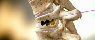
- comminuted - the most severe type of fracture, in which the vertebra is crushed into several fragments that can damage the spinal cord and provoke severe neurological symptoms.
Thus, a distinction is made between uncomplicated and complicated fractures. In the first case, patients are only concerned about pain in the area of the affected vertebra, sometimes radiating to the limbs or chest. In the second case, damage to the nerve roots occurs, which leads to the appearance of neurological symptoms and their gradual increase.
The main danger of compression fractures is that in the first days after receiving an injury, sometimes a person can move independently without serious pain in the back. But after a few hours or even days, partial or complete paralysis may occur.
Compression fractures of the spine that occur due to osteoporosis are often asymptomatic or with minimal impairment.
In the absence of timely medical care, an uncomplicated compression fracture can develop into a complicated one. This will require more complex and lengthy treatment.
Medicines
Most patients in the acute stage of fractures receive analgesics. With pain control, the patient can get up and move around easily and avoid problems caused by immobilization.
Corseting
Most patients are prescribed the use of a rigid corset, which is selected individually. A corset can dramatically reduce the range of motion and fix a broken vertebra. Patients who wear a special brace are allowed to move, but strenuous activities such as lifting and bending are significantly limited.
Motor activity control
. Bed rest is usually recommended for several days, since prolonged immobilization leads to weakening of the muscular corset, and in the presence of osteoporosis, to further bone loss and progression of osteoporosis.
Correction of osteoporosis
. Bone-strengthening drugs such as bisphosphonates (Actonel, Boniva, and Fosamax) help stabilize or reverse bone loss. This is a critical part of the treatment and helps prevent further fractures when axial loads are placed on the vertebrae.
Symptoms
Compression fractures are characterized by:
- acute pain at the site of injury, which can radiate to the arms (in case of damage to the thoracic region) and legs (in case of injury to the lumbar region), and also intensify when pressed;
- increased skin temperature and swelling of soft tissues in the projection of the affected vertebra;
- limited mobility, muscle weakness;
- general weakness, confusion;
- nausea and vomiting;
- spinal asymmetry;
- bulge formation.
If a compression fracture is caused by a blow to something, abrasions, bruises, or at least redness of the skin may be present on the skin of the back at the site of the projection of the affected vertebra.
In complicated fractures, the nerve roots passing in close proximity to the spinal column are affected. In such cases, you may experience:
- decreased sensitivity in the arms or legs;
- paresis and paralysis of the limbs;
- disruption of the pelvic organs, which is manifested by involuntary diversion of urine and feces; in men, the sensitivity of the genital organs may decrease and erectile dysfunction may occur.
A characteristic sign of a spinal fracture is a sharp increase in pain at the site of injury when gentle pressure is applied to the head.
The nature of the symptoms also greatly depends on the location of the fracture. So, if the thoracic vertebrae are damaged, it is possible:
- difficulty breathing;
- pain in the chest and heart;
- numbness of the skin of the legs;
- disruption of the gastrointestinal tract;
- disturbance of urination and defecation.
At the same time, lumbar injuries are often accompanied by:
- girdling pain or radiating to the buttocks and legs;
- disorders of the genitourinary system;
- difficulty turning over and lifting legs;
- loss of consciousness;
- passing lameness;
- paralysis of the legs.
Cervical spine injuries are caused by:
- pain in the neck, spreading to the back of the head, shoulder girdle, arms and increasing when turning the head and pressing on the affected area;
- dizziness;
- discomfort when swallowing;
- noise in ears;
- reflex hypertonicity of the neck muscles;
- discomfort when inhaling.
Thoracic region
The main tasks of rehabilitation for compression fractures of the thoracic spine are solved through exercise therapy. By performing certain exercises, physiologically normal mobility is ensured not only of all central segments, but also of the entire back, muscle tone is restored, pain in the scapulothoracic zone is eliminated, and posture is normalized. Here are examples of some useful exercises. The average repetition frequency is 10 times.
- Take a standing position. Place your feet hip-width apart, hands on your waist. As you inhale, bend back, slightly raising your shoulders and pushing them back; as you exhale, round your back, pointing your shoulders forward.
- Now we complicate the task a little by doing the same thing, but with the body turning to the right/left: we turn to one side and spread the shoulder girdle, return to the starting position and round the back, turning the shoulders forward.
- Let's move on to a new task. The position is the same, only the arms are extended along the body. Perform lateral bends: bend to one side, the arm of the bending part slides down the side of the limb, while simultaneously pulling the opposite arm towards the armpit. Similarly, we tilt the body in the other direction. Each side – 10 repetitions.
- Get on all fours with your straightened upper limbs resting on your palms. As you inhale, we bend your back, your pelvis moves back, your head drops down. As you exhale, arch your back upward (like a cat), smoothly throwing back your head, while tightening your stomach.
- We partially change the previous pose - the support in the hands now falls on the forearm area. We lift our right hand off the floor, take hold of the shoulder, slightly turn our chest to the right and make 5 springy movements with this glenohumeral part with the emphasis “up”. We work in a similar way with the left side.
- Lie on the floor with your stomach down, resting on your forearms, chest open. When the thoracic fracture has healed, the “sphinx” pose has a beneficial effect on this line of the spine; ask your orthopedist whether your rehabilitation can include this type of warm-up. Method of execution: pull your head up towards the ceiling (feel your ribs straightening), stay in this position for a few seconds, then relax, lowering your head to the floor.
- Lie on your side (head on the floor), bend your knees (knees together), stretch both arms forward in front of you, clasping your palms. Next, with a sliding movement of the upper hand, as if opening, we pass it along the inner surface of the adjacent arm, then along the chest, and finally place it on the floor. At the moment of the so-called “opening”, turn your head in the direction of movement of the limb, as a result the shoulder blades lie on the plane. The pelvis and legs should remain in position at all times. p. Then “get together” again and repeat the task. After five times, turn to the other side and repeat the exercise by analogy.
- Lumbar part
Rehabilitation for compression fractures of the lumbar spine has similar goals. This is to restore the full functionality of the musculoskeletal system, make it resilient and strong, and balance your posture. What kind of exercise do physical therapy instructors and orthopedic doctors suggest their clients do?
- Lying on your back, perform simultaneous flexion of the knee and hip joints. Thus, the lumbosacral region and abdominal muscles are excellently trained.
- We do not change the IP. Raise your legs straight up. Extending the limbs to the sides followed by bringing them together. When your legs come together, you need to cross them slightly.
- The well-known exercise called “bicycle” brings considerable benefits. We won’t dwell on the technique; we think everyone knows how to simulate riding a bicycle in a recumbent position.
- Roll over onto your stomach, spread your arms to the sides. Lift your chest off the surface along with your arms, hold in this position for as long as your physical fitness allows, then lower and relax. Note that the feet do not come off the floor. If it is more convenient for someone, you can stretch your arms in front of you.
- The position remains the same, but your legs need to be spread slightly and your arms should be extended straight in front of you. Lifting the sternal complex (along with the arms) and legs from the support, lift them simultaneously with a bend in the back. Fix the pose and stay in it for as long as possible. Lower yourself, rest a bit, then repeat.
- Get on your knees. The pelvis and back are strictly vertical, hands on the belt. Walk on your knees in a straight line forward, then backward. We complicate the task: we begin to walk in a circle, first clockwise, and then in the opposite direction.
Diagnostics
Patients with back pain should be examined by a traumatologist or vertebrologist. Based on the examination, medical history, and test results, the doctor may suspect the presence of a fracture. To confirm the diagnosis, as well as clarify the type and degree of fracture, patients are prescribed:
- X-ray of the spine in direct and lateral projection is the main method for diagnosing fractures, allowing one to determine which vertebra is damaged and the degree of its destruction;
- CT scan – provides more complete data on the condition of the spine and allows you to assess the stability of the damaged segment;
- MRI is a highly informative research method that provides comprehensive information about the condition of the intervertebral discs, ligaments and the condition of the spinal canal.
To assess the degree of preservation of the spinal cord in compression fractures, myelography may be prescribed. In older people, densitometry is performed to assess bone density. Thanks to this, it is possible to diagnose osteochondrosis or discard this factor as the cause of a compression fracture of the spine.
MRI
MRI is necessary in cases where there are motor or sensory disturbances in the lower extremities. Radicular pain or suspected spinal cord compression is also an indication for MRI.
Magnetic resonance imaging is important because it provides the best visualization of the neural structures of the spine. In addition, MRI can be performed using contrast, which allows better diagnosis of conditions such as hemorrhage, tumor or infection with greater sensitivity.
Treatment of spinal compression fractures
Patients with such injuries become patients of traumatologists, vertebrologists or even neurosurgeons. It is these specialists who, from the very first day, must supervise the treatment of a compression fracture and select the optimal treatment tactics. It is selected strictly individually for each patient.
The selection of treatment is carried out taking into account many factors, including:
- location of the fracture;
- number of affected vertebrae;
- their degree of compression;
- the presence of neurological or other complications;
- presence of concomitant diseases, etc.
As a result, the doctor develops the most effective treatment strategy. It may involve the use of conservative methods or immediate surgical intervention, but is always aimed at eliminating pain, stimulating regeneration processes, eliminating compression of the nerve roots and restoring the tone of the back muscles.
Conservative
For mild, uncomplicated grade 1 compression fractures, treatment is possible without surgery. In such cases, skeletal traction is performed to restore the normal position of the vertebrae and the body is fixed to consolidate the bone using a plaster cast, a special bandage or an orthopedic corset. They help reduce the load on the spine, reduce the risk of complications and create suitable conditions for a favorable recovery.
Patients are prescribed bed rest for several weeks. In some cases, when the intensity of the recovery processes is weak, it must be adhered to for several months. During this time, hardware traction of the spine is periodically performed.
The mattress is replaced with a hard board. This is where you have to spend almost all your time, lying on your back.
Traditionally, you can get up and move independently after 2 months, and sit no earlier than after 4 months.
Patients are also prescribed:
- drug therapy;
- exercise therapy;
- physiotherapeutic procedures;
- massage.
Drug therapy
The use of medications is a mandatory component of therapy, even if surgery is planned and after it. Patients with compression fractures of the spine are prescribed:
- analgesics (often narcotic) – used to relieve severe pain;
- novocaine blockades – have a pronounced and long-lasting analgesic effect;
- antibiotics – used to eliminate the risk of developing infectious complications of injury;
- corticosteroids - used to eliminate the inflammatory process;
- chondroprotectors – help activate regeneration processes in the affected vertebrae and cartilaginous bodies;
- immunomodulators – necessary to strengthen weakened immunity;
- Calcium and vitamin D preparations help strengthen bone tissue.
The complex of medications, as well as the method of their administration to each patient, is selected individually by the doctor. During the hospital stay, the necessary medications can be administered intravenously and intramuscularly.
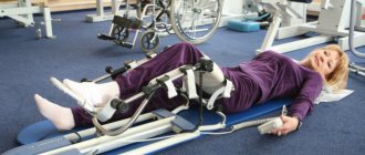
Exercise therapy
Patients, even in the first time after injury, are advised to undergo regular exercise therapy, since the muscles will inevitably weaken due to forced bed rest and immobilization of the spine. Initially, they are performed from a lying position and consist of performing simple breathing exercises and working out the joints. This allows you to avoid muscle atrophy, the development of congestion in the lungs and disruption of tissue trophism caused by poor circulation.
Gradually the difficulty of the classes increases. As the patient recovers, more active exercises are added. They allow you to work your back muscles. This will strengthen them and reduce the load on the spine.
Physiotherapeutic procedures
Physiotherapy sessions are indicated approximately 45 days after injury. The most effective are:
- Ural Federal District;
- electrophoresis with alternating calcium and phosphorus preparations;
- UHF;
- reflexology;
- paraffin or ozokerite wraps;
- electrical stimulation;
- Sollux;
- ultrasound therapy.
Typically, a course of physiotherapeutic treatment includes 10–12 procedures, each lasting 10–15 minutes.
Massage
The first massage sessions can be carried out no earlier than 2 months after the start of rehabilitation. At the same time, it is important to find a qualified specialist whose actions will contribute to the restoration of the spine, and not provoke the destruction of already achieved results.
Surgery for compression fracture of the spine
The patient’s condition and the nature of the injury do not always allow non-surgical treatment. Surgical intervention is indicated for:
- 2 and 3 degrees compression fracture;
- injuries complicated by displacement of the vertebrae, the formation of fragments;
- compression of nerve roots and spinal canal stenosis;
- lack of effect from conservative therapy.
Modern surgery and neurosurgery in particular have made great strides forward. Today, surgical treatment of compression fractures of the spine is possible in extremely gentle ways through a pinpoint puncture of soft tissue, which does not even require sutures. These include vertebroplasty and kyphoplasty. These microsurgical interventions are virtually free of intra- and postoperative risks, are highly effective and provide rapid recovery.
In more complicated situations, when kyphoplasty or vertebroplasty cannot be used, patients are prescribed surgical fixation of the vertebrae with special plates, meshes, laminar contractors, or transpedicular fixation.
The most difficult and traumatic is the complete replacement of a broken vertebra with an artificial implant or autograft. In such situations, it is possible to carry out all the necessary manipulations only during a laminectomy.
But surgery is only the first stage of treatment for a compression fracture of the spine. After it, the patient needs to receive full conservative therapy and rehabilitation.
Vertebroplasty
Vertebroplasty is a modern, gentle microsurgical operation that belongs to the group of percutaneous procedures. It involves increasing the strength of a broken vertebra by introducing a special polymer composition called bone cement into it. Since the bone tissue of the vertebrae is spongy, filling its voids with polymer allows us to achieve reliable consolidation of bone fragments and prevent compression fractures of this vertebra in the future.
Bone cement is a material specially developed for these purposes, which is based on polymethyl methacrylate. It is characterized by high safety and biocompatibility with high viscosity and strength.
To administer it, a special puncture needle is used, which is inserted into the damaged vertebra under X-ray control. As a rule, a CT scanner or image intensifier is used for this purpose.
Bone cement is prepared only after placing the cannula in the desired position, as it hardens very quickly. It comes into the hands of a neurosurgeon in the form of two separate components: a liquid monomer and a powdered polymer. At the moment of their connection, a chemical transformation reaction occurs, accompanied by the active release of thermal energy. As a result, a creamy mass is formed, which spreads well throughout all cavities of the vertebral body and hardens in 8–10 minutes. As a result, a high-strength conglomerate is formed that is not subject to compression fractures in the future.
The composition of bone cement specifically includes antibacterial and X-ray contrast agents. The former are necessary to eliminate the risk of developing infectious and inflammatory complications after surgery, and the latter to assess the quality of filling the vertebral body with bone cement.
Vertebroplasty is an operation with proven high efficiency. After it, 85% of patients experience improvement almost instantly, and the remaining 15% experience improvement after some time. At the same time, the total duration of vertebroplasty does not exceed 40 minutes, it does not require general anesthesia, which eliminates the risks associated with it, and avoids long-term hospitalization.
But restoring the integrity of the spine using this technique is impossible if:
- compression fractures accompanied by a decrease in vertebral height by more than 70%;
- individual intolerance to substances included in bone cement;
- damage to the spine by metastases of malignant tumors.
After vertebroplasty, only a tiny needle wound remains, which does not require stitches. Therefore, after the operation there are no scars left, and the wound is covered with a sterile plaster.
Kyphoplasty
For compression fractures, kyphoplasty, which can be called a more advanced version of vertebroplasty, is often used. This is due to the fact that it is capable of not only restoring the integrity of the vertebra and strengthening it, but also returning it to normal size. Therefore, kyphoplasty is performed for compression fractures with a decrease in vertebral height by more than 70%.
This becomes possible thanks to the preliminary introduction of a special balloon into the vertebral body. A saline solution in which an X-ray contrast agent is dissolved is injected into it. This allows you to monitor, using an X-ray machine, the nature of the restoration of the size and position of the injured vertebra and, if necessary, correct it.
After the anatomically normal position of the vertebra has been restored, the balloon is removed, bone cement is immediately prepared and the resulting space is immediately filled with it. After 10 minutes it hardens, after which the cannula is removed and the wound is covered with a sterile dressing.
Kyphoplasty, unlike vertebroplasty, has a minimal risk of bone cement spreading beyond the operated vertebra, and also allows for immediate elimination of kyphotic spinal deformity.
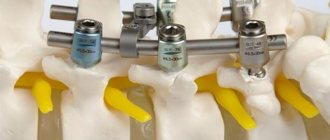
Transpedicular fixation
For unstable fractures, microsurgical interventions are not indicated. In such situations, it is possible to fix the vertebrae in the required positions using transpedicular fixation. This surgical technique has been used since the middle of the last century and has been well studied.
The operation involves making a soft tissue incision and exposing the spinous processes and vertebral arches. Each vertebra has a special point at which the transverse and articular processes intersect. A hole is made in it with a special probe, into which titanium screws of the required type and size are screwed.
Titanium is a durable metal with high strength. Therefore, the screws installed in the spine reliably fix the bone structures and eliminate the risk of their deformation. There are caps on the surface of the screws, which eliminate the risk of their distortion and the development of undesirable consequences.
After all the screws are installed at the required points, springy rods are inserted into them. As a result, the load on the structure is distributed evenly.
When transpedicular fixation is performed by an experienced neurosurgeon, the risk of damage to nerve fibers and blood vessels is minimal. But this operation is not recommended for severe concomitant diseases or pregnancy.
Laminectomy
For severe fractures requiring removal of bone fragments, laminectomy is indicated. This operation is the most traumatic for the body and involves making a large incision. But it gives the surgeon good access to the injured vertebra, which makes his work easier and allows him to remove sharp fragments.
During a laminectomy, both the spinous processes, vertebral arches and its body can be removed. This makes it possible to eliminate compression of nerve fibers and eliminate bone fragments that threaten the spinal cord or vertebrae that cannot be restored by other methods. During laminectomy, intervertebral discs can also be removed, and an implant of a suitable type can be inserted into the resulting space. These can be bone grafts formed from the patient's own bones, and implants made from artificial inert materials.
Thus, during the operation, the height of the vertebra increases due to the opening of its arches and the removal of bone fragments. But it implies a long and sometimes difficult recovery period.
Vertebroplasty
This procedure is most useful for reducing pain. It also strengthens damaged bone, allowing patients to recover faster. Using a needle, under X-ray control, a cementing substance, polymethyl methacrylate (PMMA), is injected into the area of the vertebral fracture. The cement hardens within 15 minutes. This fixes the bone and prevents further destruction and reduces pain in more than 80% of patients.
Rehabilitation
Even achieving complete bone consolidation is not a reason to stop rehabilitation measures. In total, it can take 6 months or more, up to several years. At the same time, complete recovery is not always possible, but the completeness of recovery is directly affected by how accurately the patient follows medical recommendations.
In the first 2 months, bed rest is indicated, and the patient should be provided with a hard orthopedic mattress, as well as wearing a corset. For severe compression fractures, rehabilitation is carried out in specialized centers.
Patients are advised to enrich their diet as much as possible with foods containing calcium, phosphorus, B vitamins, zinc and magnesium. A sufficient amount of these microelements in the body will contribute to the strengthening of bones and rapid tissue regeneration. Therefore, enter into the menu:
- dairy products;
- nuts, in particular almonds;
- seafood;
- bananas, lettuce, beets, cabbage;
- oatmeal and buckwheat porridge;
- liver;
- legumes
At the same time, you should avoid foods and dishes that interfere with the absorption of calcium, in particular fatty foods, alcohol, processed foods, strong tea and coffee. It is also worth limiting the consumption of yeast baked goods, since patients are forced to lead a sedentary lifestyle, which requires fewer calories to maintain normal functioning.
Following nutritional rules is important. This will not only optimize regeneration processes, but also avoid digestive disorders, stomach pain and constipation associated with a sedentary lifestyle.
Rehabilitation also includes:
- Breathing exercises are an effective method for preventing joint stiffness, which can be practiced from the first days of recovery. Classes are conducted together with a doctor who can correctly select the required load and take into account the characteristics of compression fractures of the cervical and thoracic spine.
- Exercise therapy - the first classes are conducted under the supervision of a specialist, and the intensity and duration of classes are increased at a slow pace. A set of exercises is always developed by a doctor; amateur performance in this matter is absolutely inappropriate. The nature of the exercises is selected depending on the location of the injury and the general physical fitness of the patient.
- Physiotherapy – complex physiotherapeutic treatment using the above methods in combination with properly performed massage accelerates recovery and the patient’s return to a normal lifestyle.
After completing the main stage of rehabilitation, patients are recommended to undergo sanatorium-resort treatment, which will include hydrotherapy, paraffin, ozokerite or mud wraps and other procedures.
Cervical area
After removing the plaster, rehabilitation after a fracture of the cervical spine involves restoring the mobility of the corresponding segment, strengthening the muscular-ligamentous apparatus of the neck and upper shoulder girdle. At the same time, coordination abilities are improved, and complete adaptation to social and living conditions occurs.
Surprisingly, all of the above methods are optimally suited after such a spinal-motor body injury as a fracture of the cervical spine; rehabilitation with the inclusion of these techniques in the exercise therapy complex, naturally, must be approved by a specialist. As for the fundamental exercises, they are outlined below.
- First you need to warm up the glenohumeral part, which will increase blood flow in the neck. To do this, take a vertical position, place your hands on your shoulders. Perform calm rotational movements in the shoulder joints - 10 times back, and then the same amount forward. When finished, lower your hands and shake them.
- I. p. is the same as in the first exercise. No. 1. Now spread your upper limbs in different directions, straightening them, and place them on your shoulders again. Make sure that at the moment when you spread your arms, they remain on the same horizontal plane with your shoulders, that is, do not lift them up or lower them. After 10-15 repetitions, lower the limbs that worked hard and shake them a little, and at the same time rest.
- Cross your hands with your fingers, place them on your forehead (with the inside of your palms facing the frontal area). Apply resistance to your head with your upper limbs, continuously pressing your forehead on your palms for 5 seconds. Simply put, imagine that you want to lower your head, but an obstacle prevents you from performing this action. Please note that the head does not drop anywhere, but remains in place, but you feel tension in the back of the neck. On the count of five, relax and remove your hands from your forehead. We do 5-7 approaches.
- The next method of warming up is based on the same principle, but we place the “lock” on the back of the head, pressing the head backwards. Make sure that the spine does not bend and the head does not fall back. Work with this type of exercise, observing the time and frequency of sets according to method No. 3.
- Both for stretching, balance, strength of the neck muscles, and for a beneficial effect on the respiratory center and heart function, shoulder abduction is used. The person stands with his arms along his body. In practice, it looks like this: we tense the shoulder muscles and turn our shoulders forward as if you want to close them, then we open our shoulders, straighten up and try to bring our shoulder blades towards each other.
- We save the original position. Now simultaneously move your right hand along the side of the body to the armpit and turn your head to the right, while the shoulder rises and touches the chin. Now we will carry out an identical manipulation with the reverse side.
Such fruitful rehabilitation after a fracture of the cervical spine, or rather its biomechanics, will lead to an amazing state. Of course, training should be implemented systematically, and not just during this difficult period, but preferably throughout life.
Possible complications
If you receive a back injury or just sudden pain, you should immediately consult a doctor, especially older people and those who are forced to regularly lift heavy objects as part of their job. Only timely, well-chosen treatment for a compression fracture can minimize the risks of complications and subsequent disability.
Since the spinal cord passes between the vertebral bodies and their arches, damage to it is the most dangerous complication of a compression fracture. It is he who is responsible for a person’s ability to fully move.
With compression fractures, especially when combined with the separation of fragments, it is possible that the nerve roots of the spinal cord, the blood vessels that feed it, and also the spinal cord itself may be pinched. In more severe cases, contusion or even rupture of the spinal cord is possible, leading to irreversible paralysis.
If treatment is not started in time, injury can provoke secondary degenerative and neurological disorders:
- segmental vertebral instability;
- curvature of the spine or the formation of a hump (kyphotic deformity), which is especially typical for older people;
- persistent movement disorders;
- protrusion and herniation of intervertebral discs;
- scoliosis, radiculitis;
- formation of bone structure calluses;
- disruption of the functioning of internal organs, including the genitourinary system, which can provoke urinary and fecal incontinence, and in men, additionally, erectile dysfunction;
- osteochondrosis and the formation of intervertebral hernias.
They tend to progress gradually and also lead to paralysis. Therefore, it is better not to hesitate and immediately contact a traumatologist or vertebrologist.
Forecasts
For grade 1 fractures, with timely, correct treatment and strict adherence to all medical recommendations, the prognosis is favorable, especially if the injury occurred in a young person. In such situations, the ability to work is fully restored and in the future the risk of intervertebral hernias is within the average range.
With compression fractures of degrees 2 and 3, the appearance of pain in the future cannot be ruled out; the risk of developing osteochondrosis, radiculitis, protrusions and herniated intervertebral discs increases significantly.
Thus, compression fractures of the spine are quite common, especially in older people, and at the same time a dangerous injury that can lead to disability or even death. Treatment started from the first days allows you to avoid such sad consequences and return a person to normal life. Therefore, if there are indications for surgery, you should not be afraid or refuse, because modern microsurgical methods are highly effective and safe.