Muscles are anatomical formations that have the ability to contract, while ensuring movement in the joints, performing certain work and maintaining the position of the body in space. Tendons are structures through which muscles are attached to bones. In the area of the foot and ankle, the tendons, with the exception of the Achilles tendon, are named after their corresponding muscles.
The muscles responsible for the functioning of the foot and ankle joint can be divided into external ones, i.e. those located on the back or front surface of the lower leg, and those located on the dorsal (upper) or plantar (lower) surface of the foot.
An exception is the gastrocnemius muscle, which starts on the back of the lower third of the thigh just above the knee joint and attaches to the heel bone.
Muscles and tendons of the leg
Calf muscle
This powerful calf muscle consists of two heads, medial and lateral, which originate on the posterior surface of the distal end of the thigh and are attached by the Achilles tendon to the calcaneus.
The gastrocnemius muscle is involved in running, jumping, and all types of activities that involve high-intensity stress on the lower extremities.
Together with the soleus muscle, it forms the calf muscle, called the triceps surae muscle. The function of the gastrocnemius muscle is to flex the foot and ankle downward (plantar flexion).
Forceful dorsiflexion of the foot can cause damage to this muscle.
Soleus muscle
This muscle starts from the tibia below the level of the knee joint and is located under the gastrocnemius muscle. Distally, its tendon unites with the gastrocnemius tendon to form the Achilles tendon. Like the gastrocnemius muscle, the main function of this muscle is plantar flexion of the foot.
The calf muscle is involved in walking, dancing, and maintaining an upright body position when we stand. Also, one of its important functions is to ensure blood flow through the veins from the lower limb to the heart.
Plantaris muscle
It is a small muscle that originates along the lateral head of the gastrocnemius muscle. The tendon of this muscle is the longest tendon in the human body. It is a weak but still plantar flexor of the foot. Damage to this muscle can occur when playing sports.
Achilles tendon
The Achilles tendon is formed at the mid-calf level by the gastrocnemius and soleus muscles and is attached to the heel bone. This is the most powerful and durable tendon in the human body.
It is subjected to the most significant loads compared to all other tendons. When running and jumping, the tendon is subjected to loads that are 8 times greater than body weight, and when walking - 4 times.
Through the Achilles tendon, the gastrocnemius and soleus muscles perform plantar flexion of the foot and ankle joint.
The tendon consists of three parts:
- Musculotendinous part (proximal part of the tendon, at the level of which muscle fibers turn into tendon fibers)
- Non-insertional part (body) of the Achilles tendon
- Insertion part of the Achilles tendon
The blood supply to the Achilles tendon is quite poor compared to other anatomical structures. The tendon in its upper section receives blood supply from the muscles that form the tendon, and below - from the heel bone to which it is attached. The middle part of the tendon is supplied with blood by the branches of the peroneal artery and this blood supply is the poorest, so it is not surprising that this part of the tendon is most susceptible to damage. The Achilles tendon is surrounded by a soft tissue sheath called the paratenon. The middle part of the tendon receives its blood supply precisely through this sheath. The paratenon allows the Achilles tendon to glide relative to surrounding tissues for up to 1.5 cm.
Anterior to the Achilles tendon is the Kager fat pad, which performs an important function of protecting the Achilles tendon.
MR anatomy of the Achilles tendon
- Musculotendinous part
- Kager fat body
- Non-insertional part of the Achilles tendon
- Insertion part of the Achilles tendon
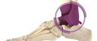
The structure of the ankle joint
Let's look at the structure of the ankle. It is a knot that is connected by bones. There are four main bones in the ankle joint. It also contains fibers called ligaments. They should hold the bones, but not hinder their movement. It is the ligaments that allow you to make movements of different amplitudes. The ligaments must be elastic.
There are definitely blood vessels in the joint. They are needed for normal blood circulation. They are not considered components of the ankle joint, but without them it will not fulfill its purpose.
Structure of the ankle joint and ligaments
It is possible to compare the anatomy of the ankle joint with a bag, which has 2 layers. It is where the bones are connected. The main purpose of the bag is to create a tightness and reproduce special synovial fluid. It will fill all the cavities.
External muscles and tendons of the foot
Tibialis posterior muscle
The tibialis posterior muscle starts from the posterior surface of the tibia and fibula (under the gastrocnemius muscle in the posterior muscle sheath of the lower leg). The tendon of this muscle on its way to the foot bends around the back of the inner ankle.
The main point of attachment of the muscle is the tuberosity of the scaphoid and the medial sphenoid bone. Also from the tendon there are bundles that attach to the bases of the 2nd, 3rd and 4th metatarsal bones, the intermediate and lateral cuneiform bones and the cuboid bone.
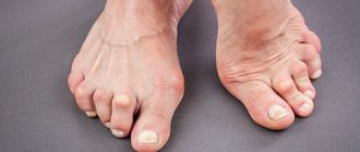
The muscle and its tendon play an important role in the formation and maintenance of the inner arch of the foot.
Contraction of the tibialis posterior muscle causes inversion (inward rotation) of the foot and plantar flexion of the foot and ankle.
Dysfunction of the tibialis posterior muscle, incl. rupture of its tendon may cause acquired flat feet.
Tibialis anterior muscle
The tibialis anterior muscle originates from the upper two-thirds of the outer surface of the tibia. Its tendon attaches to the medial cuneiform and 1st metatarsal bones of the foot.
The muscle performs dorsiflexion and inversion of the foot.
Damage to the common peroneal nerve, which innervates the muscle, or the tendon of this muscle leads to foot drop.
Peroneus brevis muscle
The peroneus brevis muscle originates from the lower two-thirds of the outer surface of the fibula. Its tendon passes behind the lateral malleolus, runs along the outer surface of the calcaneus, located above the tendon of the long peroneal muscle, and is attached to the tuberosity of the base of the 5th metatarsal bone.
The muscle performs eversion (outward rotation) of the foot and provides dynamic stabilization of the outer part of the foot and ankle joint. Trauma to the foot accompanied by inversion can lead to damage to the tendon of this muscle.
A – peroneus brevis tendon, B – peroneus longus tendon
Peroneus longus muscle
The peroneus longus muscle originates from the fibula above the peroneus brevis muscle. Its tendon also passes behind the lateral malleolus, continues onto the foot and attaches to the medial cuneiform and 1st metatarsal bones.
The main function of the muscle is plantar flexion of the 1st ray of the foot. It also performs plantar flexion and eversion of the foot. The muscle is involved in maintaining the transverse arch of the foot and provides lateral dynamic stability of the ankle joint.
Flexor digitorum longus 1 (FHL)
The muscle begins on the back surface of the leg (posterior muscle sheath) and attaches to the lower (plantar) surface of the distal phalanx of the 1st finger.
The muscle performs flexion (plantar flexion) and inversion of the foot. She also bends the 1st finger.
Extensor digitorum longus 1 (EHL)
This muscle is located between the tibialis anterior muscle and the extensor digitorum longus muscle in the anterior muscle compartment of the lower leg. It is attached to the base of the distal phalanx of the 1st finger. The long extensor of the 1st toe extends (straightens and raises) the first toe, performs dorsiflexion of the foot and is involved in eversion and inversion of the foot.
Flexor digitorum longus (FDL)
It is one of three muscles that originate on the back of the lower leg (the posterior muscle sheath), the other two being the flexor digitorum longus and the tibialis posterior. The flexor digitorum longus attaches to the inferior (plantar) surface of the distal phalanges of the small toes.
The muscle flexes the small toes.
Extensor digitorum longus (EDL)
The muscle begins with a wide base on the anterior surface of the tibia and fibula and the interosseous membrane. On the foot it is divided into 4 tendons, which are attached to the 4 small toes. Each tendon at the level of the MCP joint is divided into 3 bundles, the central bundle is attached to the base of the middle phalanx, the two lateral bundles are united and attached to the distal phalanx.
The main function of the extensor digitorum longus is to straighten the fingers. However, it is also involved in dorsiflexion of the foot and ankle.
Ankle ligament diseases
Other ligament diseases:
- inflammation . It can begin to develop with weakened immunity or a sedentary lifestyle;
- Ligament distortion is a sprain or tear of a ligament. The disease can begin with sudden exertion. This may damage the articular cartilage;
- calcification of the ligaments in the ankle joint occurs when injured and manifests itself in the form of bone shadows;
- foot tendinitis is a pathological process that promotes damage to ligament tissue and the development of inflammation;
- With prolonged overstrain of the ligaments, it is almost impossible to avoid their damage. It can lead to the development of inflammation, chronic pain;
- With ligamentitis of the ankle ligaments, the longest plantar ligament is damaged. Ligamentosis occurs when there is an infection or injury.
Own muscles and tendons of the foot
Flexor digitorum brevis (FDB)
The muscle starts from the internal (medial) process of the calcaneus and the central part of the plantar fascia. It is attached to all 4 small toes. At the level of the PFJ, each muscle tendon is divided into 2 bundles, each of which goes around the tendon of the long flexor of the finger and is attached to the middle phalanges of 2-5 fingers.
The muscle performs flexion (plantar flexion) of the middle phalanges of the fingers in the PIPJ. As the muscle continues to contract, the proximal phalanges flex at the MCP joint.
Vermiform muscles
These are 4 small muscles starting from the 4 flexor tendons on the foot. The tendon of each lumbrical muscle is attached to the tendon extension of the long extensor muscles on the dorsum of the proximal phalanges of the fingers. Contraction of the lumbrical muscles leads to extension of the fingers in the PIPJ and DIPJ. Because the tendons are located below the point of rotation of the MCP joint, they also perform flexion at these joints.
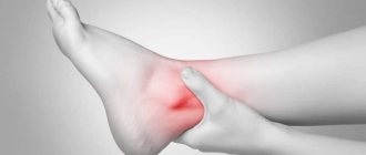
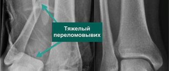
Bruised ankle
Bruises can have varying degrees of severity:
- In the first case, the skin is almost intact. There may be scratches. It usually goes away in a short period of time.
- In the second degree, muscle rupture occurs, swelling appears and a hematoma may appear. The pain is sharp and severe.
- At grade 3, tendon and muscle damage occurs . Sometimes it comes to dislocations.
- At grade 4 severity of bruises, changes will occur that will affect the quality of life. The ankle will not be able to perform any functions.
Diagnostics
To establish the correct diagnosis, taking into account the origin of the disease, the doctor first asks the patient about the onset and duration of the disease, its symptoms, and diseases of close relatives. Then a thorough examination of the patient is carried out to identify painful areas, the state of the ankle and foot function. The diagnosis is confirmed by laboratory and instrumental studies:
- Laboratory tests
- blood, urine, joint fluid taken by joint puncture (puncture) or during arthroscopy. The following are revealed: the severity of inflammation, the presence of infection, metabolic and hormonal disorders, autoimmune processes. - Instrumental studies
:- Ultrasound
- an increase in the volume of the synovial membrane, the presence of a large volume of exudate; - X-ray of the ankle and foot
– bone changes: narrowing of the joint space, bone growths, deformities; - MRI and CT
are the most informative studies; they reveal any changes; - arthroscopy
- examination of the internal articular surface using optical equipment (arthroscope).
What to do if the disease worsens
Arthritis of the foot and ankle occurs with painful relapses. In some clinical forms of the disease, exacerbations can be very painful. How to help yourself, reduce pain before the doctor arrives? This can be done like this:
- calm down by taking valerian or motherwort;
- take a tablet of any pain reliever - Analgin, Diclofenac, Ibuprofen, Nise, Paracetamol, etc.; the analgesic effect occurs very quickly after using a rectal suppository with Diclofenac;
- apply pain-relieving ointment (gel, cream), for example, Fastum-gel, to the ankle and foot area;
- call a doctor at home;
- lie on your back on a flat surface and elevate your sore leg, placing a pillow under your shin and heel;
- calmly wait for the doctor to arrive.
Treatment of ankle ligaments at home
If the sprain is not very severe and the ankle has been examined, it can be treated at home with folk remedies:
- Grated raw potatoes are applied to the sore spot.
- A compress is made from grated onion and sugar; you can treat with this remedy often.
- Lard and garlic need to be ground. Add eucalyptus leaves and strain through cheesecloth and rub the resulting liquid into the sore spot.
- Pour several cloves of garlic with vinegar, preferably apple cider vinegar, and vodka. Leave for 14 days. Then add 20 drops of eucalyptus oil to it and use it as a compress on a sore ankle.
- Apply fresh elderberry leaf.
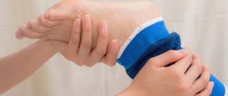
Step-by-step instructions for providing first aid to an injured person when injured:
- Take off your shoes.
- Apply a tight bandage to the leg so that it secures the ankle joint.
- Apply a cold compress to the sore joint.
- Call an ambulance or accompany the person to the hospital. Especially if a hematoma or severe swelling appears.
First aid for sprains
Sick leave for sprained ankle
Depending on the severity of the injury, the patient may receive sick leave. It is required to be given when performing surgery and diagnosing a ligament rupture. The duration of treatment depends on the outpatient condition of the patient and the opinion of the attending physician.
Types of surgery
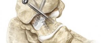
When the wear and deformation of the joint is too severe, this can become an obstacle to replacing the joint block with an endoprosthesis. Therefore, even with every desire, replacing a diseased segment with an artificial analogue is not always feasible. In this and all the situations described above, there is only one way out - to use arthrodesis surgery. It will stabilize the ankle and reduce pain symptoms to a minimum, thereby significantly improving the patient’s quality of life. There are several methods of surgical intervention.
- Intra-articular. During surgery, the joint capsule is opened, followed by removal of damaged hyaline cartilage from the surfaces of the bone elements. After repositioning the bones in an advantageous position, they are fixed with metal devices.
- Extra-articular. Fixation of the bones of the articulation only by placing a bone graft, while the cartilaginous covers are not subject to resection.
- Combined. This technique involves the combination of two methods in one surgical process: intra-articular and extra-articular. Thus, the cartilaginous structures from the joint are completely removed, an autograft is introduced, which is fixed with special metal plates.
- Compression. The operation consists of squeezing the articulating surfaces with a compression or compression-distraction type apparatus for their further fusion. Widely used designs are Ilizarov, Grishin, Volkov-Oganesyan apparatuses. Removal of cartilage is not excluded. Implantation of a bone graft is not required for the compression method.
Technique No. 1.